Not Available in Your Country
Sorry, this page is not
available in your country.
Overview
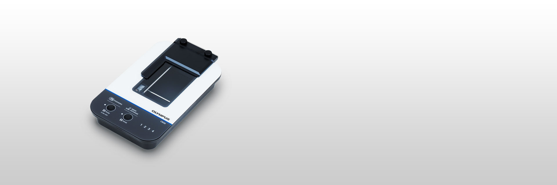 | Control Your Process with Smart Cell Culture MonitoringCulturing cells can be costly, complicated, and time-consuming. With the CM30 incubation monitoring system, there’s a simple way to improve your culture process. |
|---|
Improve the Cell Culture Process—
|    |
|---|
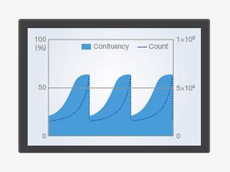
.jpg?rev=EEB6) 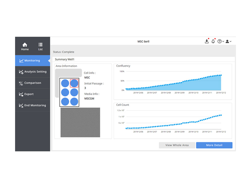 | Consistent Results Throughout Your LabConstant Analysis ParametersThe CM30 system uses image analysis technology based on machine learning to continuously measure and analyze the images acquired. Constantly visualizing the culture status as a quantitative value eliminates factors that cause variations in cell checks and contributes to the reproducibility and consistency of experiments. Compare Data Across Multiple SamplesUsing multiple heads helps you maintain a control sample while multiple test protocols run at the same time. Share your data with team members or compare it to past data. Customize the Analysis Parameters to Suit Your ExperimentThe CM30 system automatically performs confluency, cell counts, and colony counts from acquired images. You can configure the system’s analysis parameters to suit each cell culture’s conditions, such as cell type, culture conditions, or drug administered. Knowing the stepwise cell culture status at each timing improves the accuracy of the experiment. |
|---|
Cost EffectiveSave Time with AutomationImprove your traditional microscopy-based workflow and get more accurate results in less time. By automating cell culture monitoring using the CM30 system, you can expand your research and use your time more effectively No Need to Enter the Clean Room for MonitoringEvery time you enter the clean room, there is an operational cost for consumables and measurements. Now, you no longer constantly need to check cell culture status outside the incubator. You can reduce your costs by remotely checking the status of your cultures from outside the lab. Accurately Time Cell PassageTime cell passages consistently and without the subjectivity associated with manual counting. Based on your set standardized parameters, the software can indicate when your cells are ready for passage, helping prevent failures. | 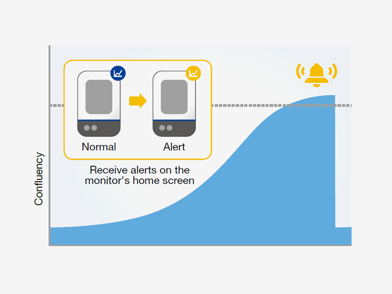 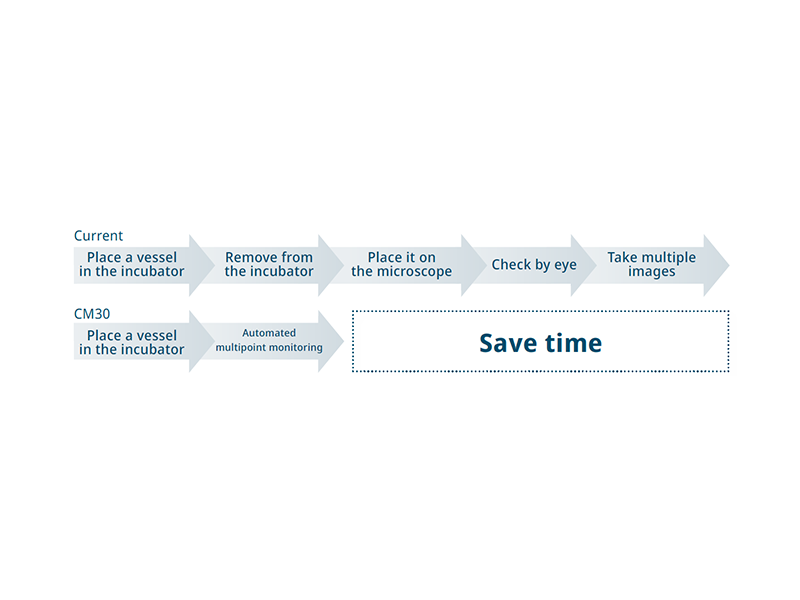 |
|---|
Testimonials |
Dr. Tadayuki Tsujita
| One of our research themes is developing novel hypoxic stress protection compounds. In order to validate the compound treatment, we were struggling to observe cell morphology and growth data without disturbing the continuous low oxygen environment. This is critical to analyzing super-short half-life proteins. We tried to several equipment combinations to solve this difficulty. In this time, we selected the CM30 system because it is compact and can be installed in our hypoxic incubator. It enables us to obtain experimental data such as serial dilution in multi-well plates in real time. The CM30 does not required mechanical moving to monitor all of the wells while maintaining hypoxic conditions, which had been difficult in the past. In addition, by connecting online, we can remotely observe the state of the cells from the outside, which we expect will be useful for troubleshooting. | |
Takanori Takebe, Dr.
| Numerous researchers may be curious about the biological mechanisms involved in the long-term culturing required for the formation of stem cells and organoids within an incubator. I was honestly surprised that such a simplistic microscope is poised to address these questions as this CM20* can visualize spheroid formation and organoids cultured in an extracellular matrix gel with amazing contrast. I believe that CM20 can be a transformative tool in a wide range of cell biology experiments. | |
Interstem Co., Ltd.
| I’m in charge of research and development of an autologous chondrocyte kit and conduct cell culture under various conditions when examining the culture protocol. I previously used a microscope to observe the conditions of cultured cells, but I can only make a qualitative evaluation with this method. Another challenge is that the analysis results vary depending on the experience and subjectivity of the worker. In contrast, the Olympus CM20* system can count cells and measure confluency with the same constant analysis parameters for highly reproducible and consistent analysis results. We can compare data between experiments when changing culture conditions, compare it with past data, and easily share it within the team— all of which can help us improve development efficiency. We’re also considering using it as an educational tool. By seeing the indicators, such as adhesion rate, uniformity, and proliferative ability, we can better evaluate the skills of workers. |
*The user comments above were made after using the CM20, but the same results can be obtained using the CM30. | |
Need assistance? |
Applied Technologies
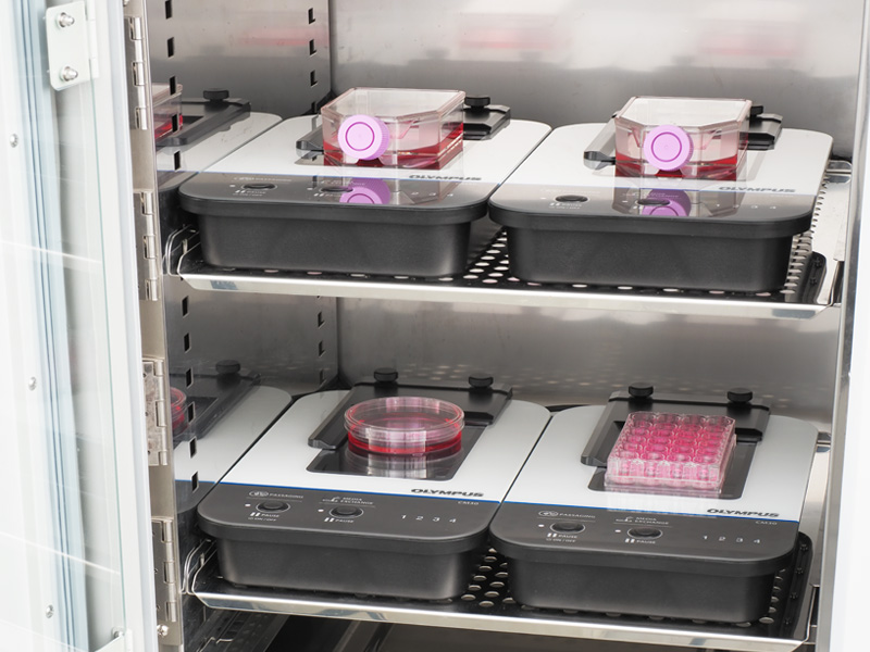
Leave Your Cultures in the IncubatorThe monitor lets you track the health of cell cultures without removing them from the incubator, reducing the risk of contamination or damage from temperature changes and vibration. Its unique design enables you to fit up to four head units inside a standard incubator for greater efficiency. | 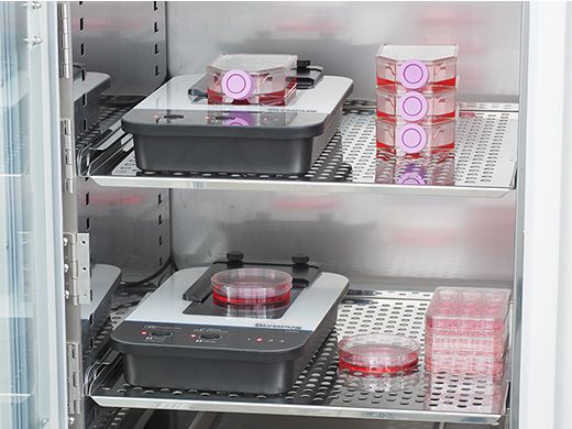 |
.jpg?rev=EEB6) 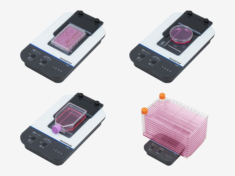 | Flat Design Accommodates Various VesselsEvident's epi-oblique optical system enables the CM30 incubation monitoring system to have a compact, flat design that accommodates most standard cell culture vessels. Simply place the culture vessel you normally use on the CM30. |
|---|
AI-Driven Automated Cell AnalysisBy training the system to distinguish between the cells and background using machine learning, it can automatically determine confluency. | 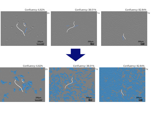 |
 | Reproducible, Comparable DataThe CM30 makes it easy to store and share detailed quantitative records of cell growth and health.
|
|---|
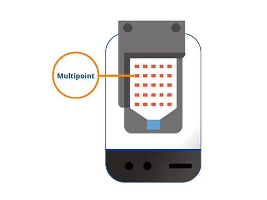
Customize the Settings to Meet Your NeedsMonitoring pointsSet the monitoring points in the vessel and well plate anywhere you would like, as shown in the vessel on the right. By specifying the rows and columns, you can set multiple points at once. Focus positionFreely set the focus position for each monitoring point. Monitoring timeFreely set the start time and interval time. |  |
|
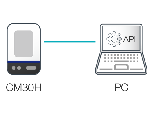
Application Programming Interface (API)
|
|---|
IQ/OQ Validation ServiceOur world-class support team can help you integrate the CM30 incubation monitoring system into your lab. IQ/OQ validation services are also available. |
|
Need assistance? |
Specifications
CM30H: Incubation Monitoring Head |
|
Installation environment
(inside the incubator) | Temperature: 37 °C (98.6 °F) + 0.3 °C (0.5 °F), humidity: 0—99% |
|---|---|
| Applicable vessels | Petri dish (90 mm (3.54 in.), 100 mm (3.94 in.)) |
| Microplate (6 well, 12 well, 24 well, 48 well, 96 well) | |
| Flask (T25, T75, T80, T150, T175, T225) | |
| Multi-layer flask | |
| Optical performance |
Field of view (H × V): 2.84 mm × 2.13 mm (0.11 in. × 0.08 in.);
(image size per one shooting) |
| Image size: 1280 × 960 pixels | |
| Illumination wavelength: λ = 630 nm (LED) | |
| Illumination method: epi-oblique illumination | |
| Cable length | Approx. 4.5 m (14.8 ft) |
| Sterilization resistance | Autoclave sterilization (for vessel holder and sponge rubber only) |
| UV ray sterilization | |
| Hydrogen peroxide (H₂O₂) gas sterilization (CM30H only) | |
| Disinfection resistance | Peracetic acid disinfection (cold sterilant) |
| Alcohol disinfection | |
| Weight | Approx. 3.1 kg (6.8 lb) |
Incubation Monitoring Station |
| Number of connectable CM30H | Max. 4 heads |
|---|---|
| HDD capacity | 4 TB or more |
Software |
| User management | 1000 user licenses (max) |
|---|---|
| Project setting | Project creation: new or load |
| Settings: standard or custom | |
| Culture conditions: vessel information, culture information etc. | |
| Cell analysis conditions: new or load | |
| Access authority: public or private | |
| Imaging interval: selection type | |
| Analysis | Cell analysis: cell confluency, cell count |
| iPS/ES cell analysis: colony confluency, colony count, colony size | |
| Data statistics: growth rate, doubling time | |
| Browsing | Image: entire area (tiling), fixed points |
| Analysis result: graph (time, passage) | |
| Export | Data export: image file, movie file* CSV file* *only for fixed points |
| Create report (PDF) |
Client PC (Recommended System Configuration for CM30 Software) |
| OS | Microsoft® Windows® 10 (64-bit) or higher |
|---|---|
| CPU | Intel® Core TM i3 (2.1 GHz) or more |
| RAM | 4 GB or more |
| HDD | Free space: 2 GB or more |
| Screen resolution | 1366 × 768 or more |
| Web browser | Google Chrome TM |
Operation Confirmed Incubator |
| Thermo Fisher Scientific | 51030388 |
|---|---|
| Panasonic | MCO-170AICUVH-PJ |
| ASTEC | SMA-165DRS |
| Eppendorf |
CellXpert® C170i (Operation confirmed by Eppendorf)
Reference data:Remote and quantitative cell culture monitoring: Vero cells |
Incubator conditions required for use of the CM30 system:
|
Resources
FAQ
FAQFunctions & SpecificationsCan the CM30 incubation monitoring head be installed in our current incubator?With its compact design (73 mm (2.9 in.) tall, 212 mm (8.3 in.) wide, and 360 mm (14.2 in.) long) and weighing only approximately 3.1 kg (6.8 lb), the CM30 fits easily in most standard-sized incubators. Is the head compatible with general cell culture containers?Yes. The CM30 is compatible with the following vessels:
What kinds of cells can the system observe?It’s designed to observe adherent cells, such as MSC and iPS cells. What can it measure?It estimates cell population and confluency from an acquired image. For samples that form a colony, it estimates the number of colonies and confluency. Will there be any damage to the cells?The system is designed to record quantitative data from inside an incubator, minimizing the chances of damaging your cell cultures. Can you observe fluorescence?It cannot observe fluorescence. We offer CKX53 inverted microscopes for fluorescence observation. How far can you remotely monitor?It depends on your router specifications and usage environment. If you want to extend the communication range, we recommend using a repeater. Can the CM30 incubation monitoring head be sterilized or disinfected?Yes. The head can be sterilized using:
Disinfected using:
SupportWhat is the support system after purchase?It depends on your region. Please contact your local sales representative. Is it possible to correspond when IQ / OQ validation is required?Yes. For details, please contact your local sales representative. Can the CM30 incubation monitoring system be connected to a network?Yes. It is possible after building the connection environment at the customer's responsibility. However, we do not guarantee the network connection in the customer's environment. When connecting to the network, it is necessary to set the station PC. Learn more from here. |
Release Notes
CM30 Version 2.2.3Improvements
|
CM30 Version 2.2.2Improvements
|
CM30 Version 2.2.1Improvements In this version, the cell count analysis function has been improved.
|
CM20H API Version 1.6.1 (Compatible with CM30H)Application Program Interface (API) which allows you to freely control the monitoring head CM20H/CM30H from your PC without the incubation monitoring station. Release note in the download package provides further details. New functions/Improvements
|
CM20H API for Linux Version 1.6.1 (Compatible with CM30H)Application Program Interface (API) which allows you to freely control the monitoring station CM20H/CM30H from your PC or microcomputer board equipped with Linux without the incubation monitoring station. Release note in the download package provides further details. New functions/Improvements
|


