Dr. Yosuke Yoneyama of the Graduate School of Medicine at the University of Osaka uses human iPS cell-derived liver organoids to conduct research on metabolic liver diseases, such as fatty liver disease, and regenerative medicine.
In this interview, we asked Dr. Yoneyama about his translational research with liver organoids, the applications of inverted microscopes in his work, and the utility of the IXplore™ IX85 motorized inverted microscope platform.
Dr. Yosuke Yoneyama
About Dr. Yoneyama
Dr. Yoneyama received his PhD from the Graduate School of Agricultural and Life Sciences at the University of Tokyo and currently works at the Graduate School of Medicine at the University of Osaka, where he conducts various studies using stem cells and organoids. His research is making significant contributions to the early diagnosis of liver diseases, the development of new treatments, and the advancement of regenerative medicine.
Q: What research are you currently focusing on?
Dr. Yoneyama: Our lab has the technology to generate liver organoids from human iPS cells, but we aim for translational research that considers medical applications, not just the generation itself. One example is our research into the mechanisms behind individual differences in the pathology of fatty liver disease. The number of patients with fatty liver disease is increasing globally, but the progression of symptoms varies among patients, and effective treatments have not yet been found. To overcome this, we are using organoid technology to reproduce the different liver conditions of individual patients and study the mechanisms that cause these individual differences in pathology.
Another approach is to use liver organoids for a new treatment: extracorporeal liver support device. We are developing a therapeutic device implemented with our liver organoids and continue to pursue research aimed at clinical applications.
Q: What led you to focus on organoid research?
Dr. Yoneyama: Organoids are a one-of-a-kind tool that enable us to directly study human organs. I started my involvement in liver organoid research around 2018. At that time, simply generating organoid models was enough for a paper. Even then, I wasn't content with just making organoids; as a scientist, I wanted to give back and contribute to clinical practice.
Another reason is my desire to elucidate how human organs are formed and how the dysregulation of those mechanisms is related to disease. For example, I am very interested in understanding the processes that lead to metabolic abnormalities and disease. Currently, I am focused on two things: as a scientist, elucidating the formation and functional mechanisms of human organs, and contributing to medical research that is useful in clinical practice.
Q: How do you primarily use microscopes in your experiments?
Dr. Yoneyama: For cell culture, I use an inverted microscope almost every day for observations, such as brightfield and phase contrast imaging. For high-end applications—cases where we need to know where and how much of a specific molecule is expressed, or what the intercellular and intracellular structures look like—we use high-resolution fluorescence microscopes like confocal microscopes. From the stage of creating organoids to the final evaluation of their function, the inverted microscope is an indispensable tool for organoid research.
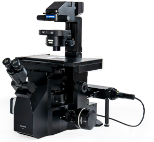
Evident inverted microscopes.
Left: CKX53 compact cell culture microscope.
Right: IXplore IX85 motorized inverted microscope platform.
Q: What aspects of the new IXplore™ IX85 inverted microscope were effective in your experiments?
Dr. Yoneyama: First, the throughput. The IXplore IX85 has a wide field of view, which reduces the number of images needed and enables me to efficiently collect data from the cells and specimens required for a single experiment. That is a huge benefit.
Improving throughput also helps when monitoring the homogeneity of the organoids we create or the frequency of abnormal formations. When we discuss data in the lab, the wide field of view allows us to monitor more organoids at once, which helps us have more accurate conversations about homogeneity and abnormalities.
Another improvement I genuinely appreciated was the image stitching function. I used it to acquire images of a relatively large rat liver specimen, about 20-30 mm, and it produced a high-resolution image with almost no visible seams. To obtain a similar image without these seams, we usually need to adjust conditions and make fine-tuned settings, but the IX85's automatic correction made it easy to capture uniform, high-resolution images. I was very impressed.
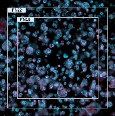
Organoids made from human iPS cells. Image courtesy of Dr. Yoneyama.
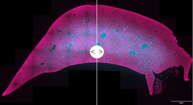
Rat liver tissue.
Left: Original stitched image.
Right: Stitched image with Intelligent Shading Correction applied.
Image courtesy of Dr. Yoneyama.
Q: How did the LUPLAPO25XS objective lens, which uses a silicone gel pad, help with your experiments?
Dr. Yoneyama: First, it improved our workflow efficiency. For example, when switching from an immersion objective to a different magnification, you usually need to wipe off the oil or water. No longer needing to do this has led to a significant increase in efficiency. I can take a widefield image first, then increase the magnification and continue imaging. Then, I can return to the widefield view and switch back to the silicone gel objective in a different location. The ability to perform these tasks seamlessly is incredibly convenient.
Second, the 25X magnification is perfect. While our main focus is organoid research, we deal with other types of samples on a daily basis and need to cover a wide range of magnifications. At those times, 10X is too low and 40X is too high. I've always wanted an intermediate magnification with a long working distance (WD) and excellent deep observation performance. This 25X objective with a 2 mm WD is a perfect match for our research subjects.
Furthermore, I was very impressed by the deep observation performance of the silicone gel. When we try to observe the center of spheroids or organoids, at depths of about 100 to 150 µm, a standard dry objective would result in a blurry image. The blurring was significantly improved with this objective. By combining it with 3D deconvolution, we could clearly observe the organoid structure without using a confocal microscope.
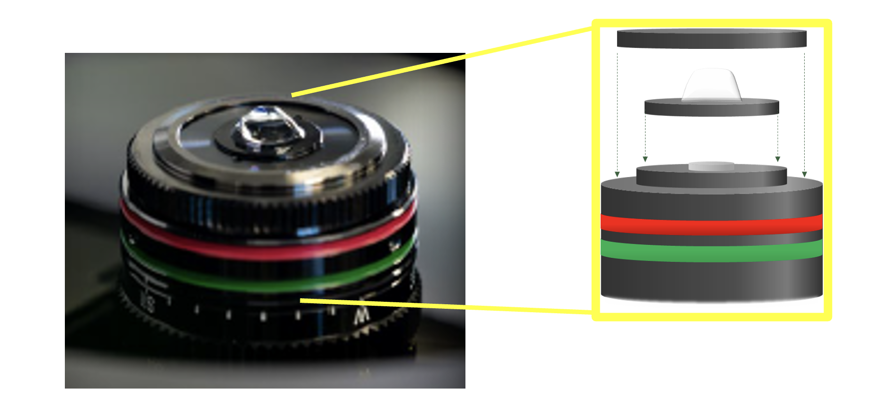
Evident’s LUPLAPO25XS objective lens (NA 0.85, WD 2 mm) with silicone gel pad technology (yellow).
Q: What are your future plans for research and experiments?
Dr. Yoneyama: As I mentioned in my current research, we are developing a device that can circulate blood through extracorporeal liver organoids to achieve a function similar to dialysis. This effort aims to develop a new approach that could one day help support liver function. The liver has a detoxification function, but in patients with poor liver function, waste products and toxins can easily accumulate in the blood. If liver organoids could take on that role, then they could one day help support patients awaiting transplants.
Organ transplants are still very difficult, and there has not yet been a single clinical application case of organoids in the field of liver regenerative medicine. I believe this research is significant for that reason.
Disclaimer: The opinions and statements expressed in this interview are those of the individual researcher and do not necessarily reflect the views or claims of Evident. The products and technologies mentioned are intended for research use only and are not designed for clinical or diagnostic applications.
Related Content
New Imaging Possibilities Using the World's First Multi-Immersion Silicone Gel Objective
Intelligent Shading Correction for High-Quality Stitched Images in Advanced Microscopy