Intelligent Shading Correction for High-Quality Stitched Images in Advanced Microscopy
Introduction
In recent years, advancements in microscope hardware and data handling have enabled the capture of large sample areas at high resolution. This has facilitated detailed observation and analysis, playing a crucial role in both life sciences and medical research. For instance, the IXplore™ IX85 platform by Evident offers an industry-leading field number (FN) of 26.5 mm for spinning disk confocal microscope systems, enabling the acquisition of wide-area images in a single shot.
However, capturing large samples at high resolution presents several challenges. Even with high pixel count cameras, achieving high optical resolution with low magnification objectives results in a low numerical aperture (NA). This leads to insufficient resolution and difficulty in observing fine details. To address this, high magnification objectives are used, data is captured while moving the sample in the XY direction using a motorized stage, and the images are stitched together. This method enables high-resolution observation of large sample areas.
Even with these advancements, this method faces the issue of shading. Evident has developed Intelligent Shading Correction, an algorithm for shading correction that does not require prior calibration. This white paper introduces this technology and explores how it supports high-quality stitched images in advanced microscopy.
The Challenges of Shading in Microscope Images
Shading typically manifests as a bright center and dark periphery in the microscope’s field of view. It may be caused by uneven illumination, lens characteristics, sample thickness, or refractive index differences. When shading occurs, image quality deteriorates during stitching, hindering observation and analysis.
Traditional shading correction involves capturing calibration samples and using the data for correction. However, this method has several drawbacks. Calibration requires preparing uniform fluorescent samples or specific patterned samples, which is time-consuming and labor-intensive. Additionally, differences between calibration samples and actual samples can affect correction accuracy. Biological samples often have heterogeneous optical properties and non-flat shapes, leading to varying shading even within the same sample type.
To overcome these issues, more efficient and accurate shading correction technology is needed.
Introducing Intelligent Shading Correction
Evident’s newly developed Intelligent Shading Correction function estimates and corrects shading using only the images acquired during stitching. This technology eliminates the need for prior calibration using samples, significantly reducing effort. Using sample images for correction also yields better results compared to calibration methods.
During image stitching, images are acquired with a slight overlap. The overlap region captures the same sample area, so the brightness values of the two images should be similar. However, shading causes differences in brightness values, which become more pronounced with greater shading. By analyzing these differences, the shading magnitude can be evaluated and estimated (Figure 1). Intelligent Shading Correction analyzes the brightness differences in the overlap regions of all image data, accurately estimating and modeling the shading state. Based on this model, it corrects the brightness values of each image, removing the shading effect (Figure 2).
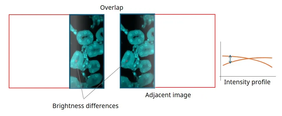
Figure 1. Brightness differences in overlap regions. The two corresponding intensity values are different in the adjacent images.
Figure 2. Example of Intelligent Shading Correction applied when stitching fluorescence widefield images (speed adjusted). Sample: mouse brain. Cyan (DAPI): nuclei. Green (Alex Fluor 488): MAP2. Magenta (Alexa Fluor 568): MBP. Sample courtesy of EnCor Biotechnology Inc.
This method not only corrects shading in typical fluorescence microscope images but also applies to differential interference contrast (DIC) images, which were previously difficult to correct (Figure 3).
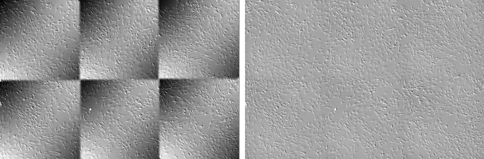
Figure 3. 2x3 stitched DIC image of cultured NIH 3T3 cells without shading correction (left) and with Intelligent Shading Correction (right). Sample courtesy of EnCor Biotechnology Inc.
The Intelligent Shading Correction method can also be applied to confocal laser scanning microscopes using point scanning (Figures 4 and 5).
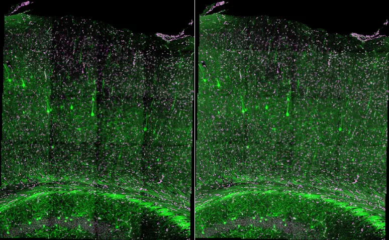
Figure 4. Stitched confocal image of a cleared mouse brain without shading correction (left) and with Intelligent Shading Correction (right). Magenta (DAPI): nuclei. Green (EGFP): neuro cells. Captured on the FLUOVIEW FV4000MPE. Sample courtesy of Sinjun Lab. Co., Taiwan.
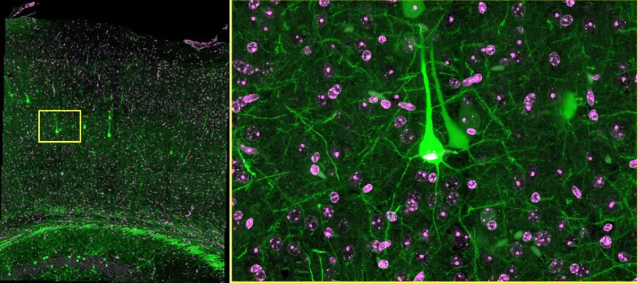
Figure 5. Z-projection image (right) acquired from the yellow rectangle area (left) in the seamlessly stitched image of Figure 4, right. This close-up image shows clear details of nuclei (magenta, DAPI) and neuro cells (green, EGFP) within a cleared mouse brain. TruSight processed to improve the contrast and sharpness of the image. Sample courtesy of Sinjun Lab. Co., Taiwan.
When correcting images based on the estimated shading state, the average brightness value of the image is maintained. This minimizes artifacts during correction, even when combining Z-stack and image stitching. Additionally, color balance is preserved during RGB channel correction, resulting in high-quality stitched images.
Summary
In summary, the advantages of Intelligent Shading Correction include:
- No calibration required: Eliminates the need for prior calibration, significantly reducing effort and time.
- High-precision correction: Uses actual sample images, achieving comparable or improved correction accuracy compared to calibration methods.
- Brightness and color balance maintenance: Corrects image brightness to the average brightness of the original image and maintains color balance during RGB channel correction.
- Artifact reduction: Minimizes artifacts during correction, even in Z-stack and time-lapse images.
- Adaptability: Applicable to a wide range of conditions and sample characteristics.
This technology helps prevent the degradation of quality due to shading, enabling efficient acquisition of high-quality stitched images. It also maintains color balance and brightness in color and Z-stack images, broadening its applications in life science.
Author
Yosuke Tani
Micro-Imaging Solutions R&D, Program Architect
Evident, Japan
Products Related to This Application
Sorry, this page is not
available in your country.