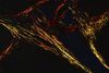
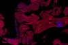
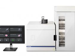
In den Teilgebieten Zytologie und Pathologie werden Proben von Zellen, Gewebe oder Organen entnommen und mit dem bloßen Auge oder einem Mikroskop untersucht, um krankheitsbedingte Abweichungen zu studieren. Unsere Lösungen eignen sich, dank unserer spezialisierten Mikroskope, Kameras und Bildgebungssoftwaresysteme besonders für pathologische Forschungsanwendungen. Unsere Serie von aufrechten Mikroskopen bietet eine hohe optische Leistung mit einer Reproduzierbarkeit von hoher Farbtreue. Dank ihres ergonomischen Designs ermöglichen sie eine komfortable Betrachtung der Aufnahmen während langen Arbeitszeiten. Unser robustes Objektträgersystem eignet sich für die Forschung und Pathologie, denn es ermöglicht die gleichzeitige Betrachtung des gesamten Objektträgers mit einem vergrößerten Bereich. Es verfügt zudem über eine innovative Synchronisierungsfunktion für die Analyse mehrerer virtueller Objektträger. Dieses innovative virtuelle Objektträgersystem für eine hochauflösende digitale Bildgebung ermöglicht zudem eine einfache Ansicht der gesamten Probe auf dem Objektträger. Es werden ebenso Lösungen zu Probenaufbewahrung, Datenbankerstellung, Weiterbildungen und Konsultationen für diesen Forschungsbereich angeboten.
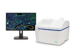
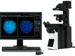
The APEXVIEW™ APX100 benchtop fluorescence microscope makes it fast and simple to acquire expert-quality microscope images. Built with our renowned optics, an intuitive user interface, a powerful AI, and a suite of smart features, the APX100 system combines ease of use with high-quality image data to fit your research needs.
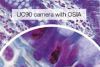
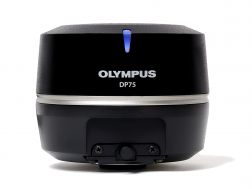
The high-performance DP75 digital microscope camera makes it easy to capture high-resolution brightfield or fluorescence images using a single color camera. It simplifies your microscopy imaging, so you can focus more on your work.
Integrated TruAI denoising maximizes the camera’s image quality in real time
Exceptional color reproduction, making your images as vivid as looking through the microscope oculars
Supports multiple staining combinations and wavelengths up to 1000 nm with a switchable infrared (IR) cut filter
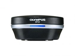
Enabling fast, easy capture of high-quality images that can be clearly observed on a large screen, the DP23 microscope digital camera eases routine life science and clinical research, conferencing, or teaching. Integrate it seamlessly into your microscopy workflow and easily share or stream images.
Share images using the DP23-AOU network solution
Clearly observe live images on a large screen
Fast, high-quality imaging for conferences and teaching
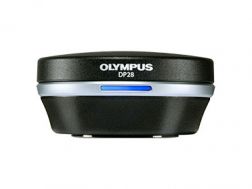
Providing color accuracy and 4K resolution, the DP28 digital microscope camera’s powerful features and wide field of view capture images that enhance tasks such as conferencing, teaching, and clinical research. Integrate it seamlessly into your microscopy workflow for improved work efficiency and image quality.
Providing intuitive operations and a seamless workflow, cellSens software’s user interface is customizable so you control the layout. Offered in a range of packages, cellSens software provides a variety of features optimized for your specific imaging needs. Its Graphic Experiment Manager and Well Navigator features facilitate 5D image acquisition. Achieve improved resolution through TruSight™ deconvolution and share your images using Conference Mode.
Improve experiment efficiency with TruAI™ deep-learning segmentation analysis, providing label-free nuclei detection and cell counting
Modular imaging software platform
Intuitive application-driven user interface
Broad feature set, ranging from simple snapshot to advanced multidimensional real-time experiments