HPF Counting Remains a Core Tool in Clinical Microscopy
Introduction
Clinicians and pathologists continue to rely on high-power field (HPF) counting for its speed, simplicity, and diagnostic utility. This method enables fast, standardized assessment of cells and structures under high magnification, directly supporting decisions in fields such as nephrology, pathology, and microbiology.
With minimal equipment required and immediate results, HPF counting remains integral to medical guidelines and daily lab practice—even as digital tools evolve.
Table 1. Correction Methods by Field Number
Minimizing Counting ErrorsMost diagnostic thresholds are based on a standard microscope field number (FN) of 22, which defines an HPF area of approximately 0.237 mm² at 400X magnification.
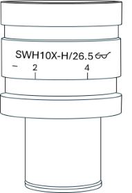
| Figure 1. The field number is indicated on the ocular, such as FN26.5 in this example. |
| Field number (FN) | HPF area (mm²) | Correction factor (to match FN22) | HPFs to count (to equal 10 FN22 HPFs) |
|---|---|---|---|
| FN18 | 0.159 | × 1.49 | 15 HPFs |
| FN20 | 0.196 | × 1.21 | 12 HPFs |
| FN22 (Standard) | 0.237 | × 1.00 (no adjustment) | 10 HPFs |
| FN26.5 | 0.344 × 0.69 | × 0.69 | 7 HPFs |
Optimizing Field Number SelectionUsing a larger FN offers practical advantages. With FN26.5, only 7 HPFs are needed to examine the same area as 10 HPFs with FN22. If the result is already diagnostically significant at this point, stop counting. If the result is borderline, continue to 10 HPFs and apply the interpolating correction factor. Since you will have evaluated 31% more slide area than with FN22, you have a statistical advantage when confirming your diagnosis. |
|
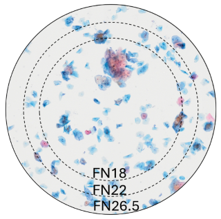
Figure 2. Thin layer of cells. Field of view at 20X magnification for FN18, FN22, and FN26.5. |
Conclusion
Despite the growing presence of digital imaging and automated analysis in clinical microscopy, HPF counting remains a cornerstone technique due to its reliability, accessibility, and diagnostic relevance. Understanding the impact of a microscope’s field number on an HPF area is essential to maintain accuracy, especially when deviating from the standard FN22.
By applying correction factors or adjusting the number of fields counted, clinicians can help ensure consistent and meaningful results. Larger FNs offer efficiency and statistical advantages, while smaller FNs require careful compensation to avoid sampling bias. Ultimately, mastering these adjustments reinforces HPF counting as a robust and adaptable tool in modern diagnostic workflows.
Products Related to This Application
Sorry, this page is not
available in your country.