We are thrilled to announce the winners of our 5th annual Image of the Year contest, recognizing the world’s best in scientific microscopic imaging. This year’s contest included our first-ever video category. This year’s winners were selected from entries from 29 countries. We thank everyone who entered, and we look forward to your participation again next year. |
|
 | The Global WinnerBeauty of Cosmic ProportionsCross-section of a Cosmic Orange aster flower with pollen grains maturing inside anthers. Captured by Igor Siwanowicz of the United States. |
|---|
Materials Science WinnerConnecting All Living ThingsHuman hair (vertical) knotted onto horsehair (horizontal). Captured by Gerd Günther of Germany. | 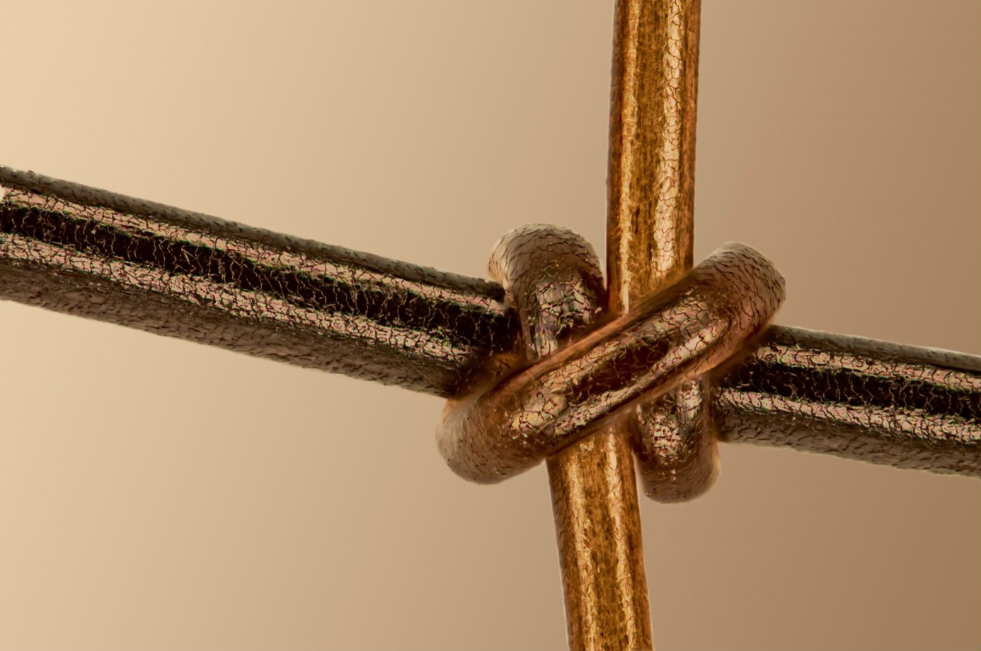 |
|---|
Video WinnerA Vast & Boundless UniverseAmino acid crystallization process. Captured by Zhigang Zheng of China. |
Regional Award Winners
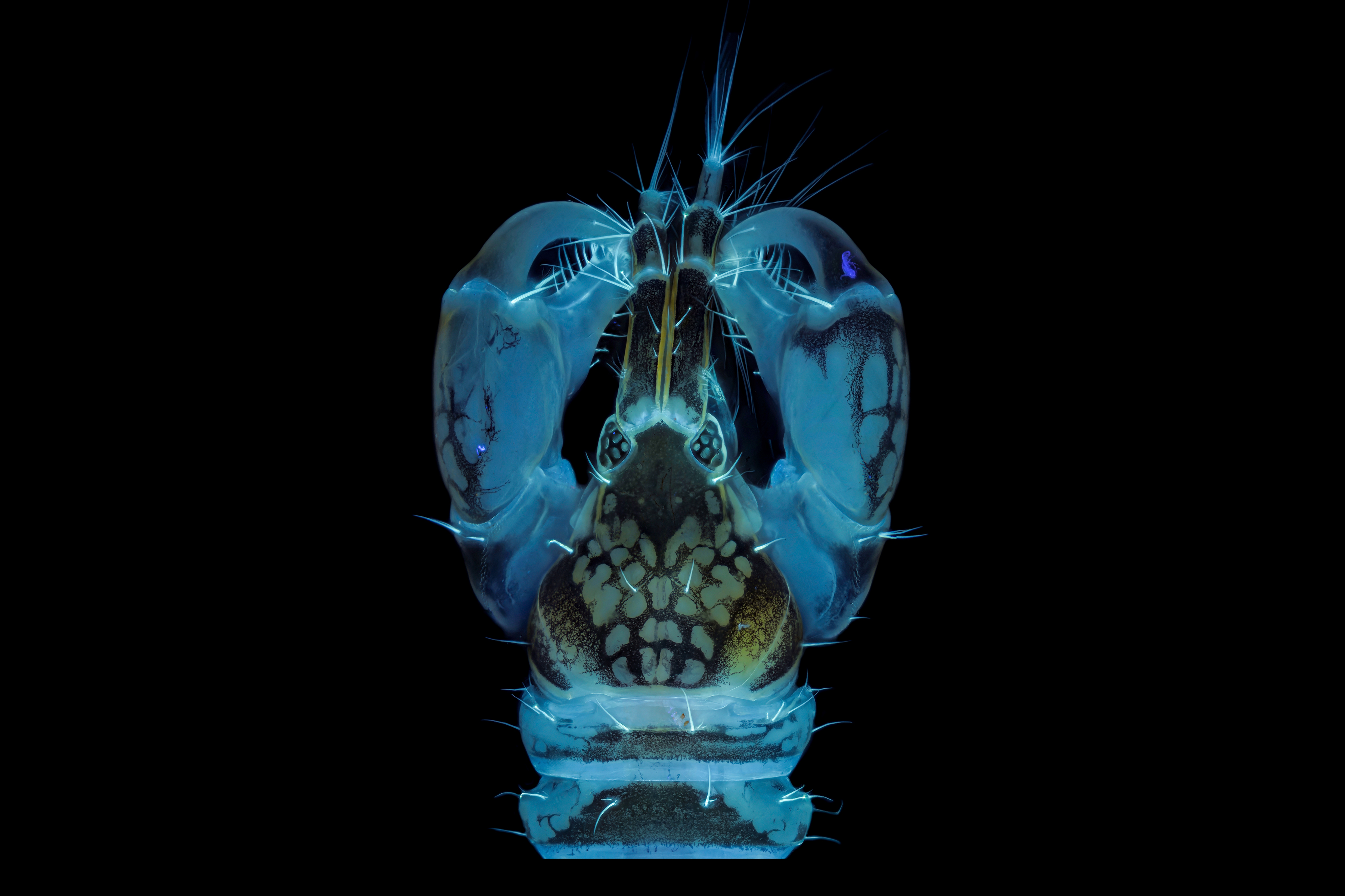
Americas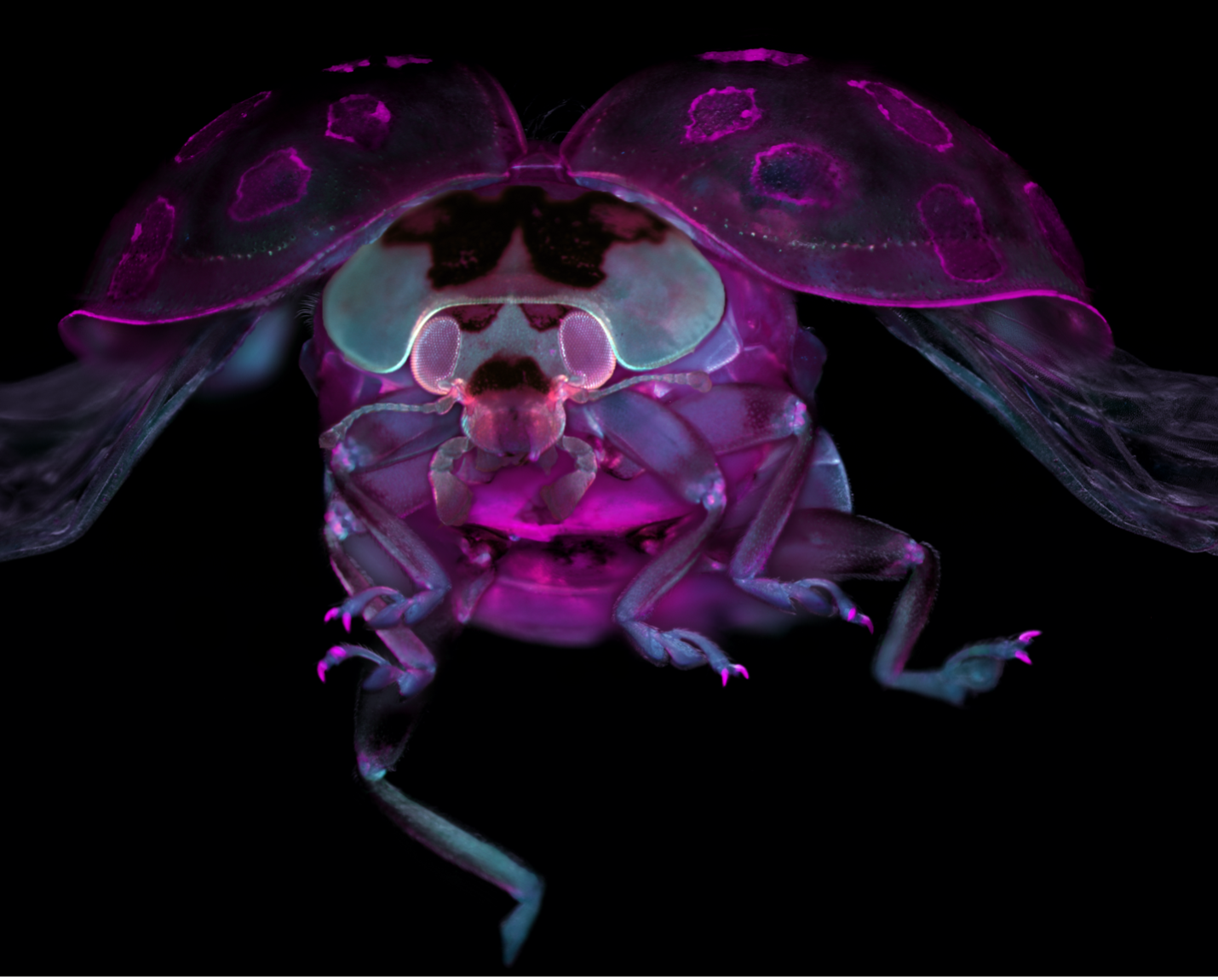 The Revelation of New Perspective Tissue-cleared ladybug. Captured by Marko Pende (USA). | Europe, the Middle East, and Africa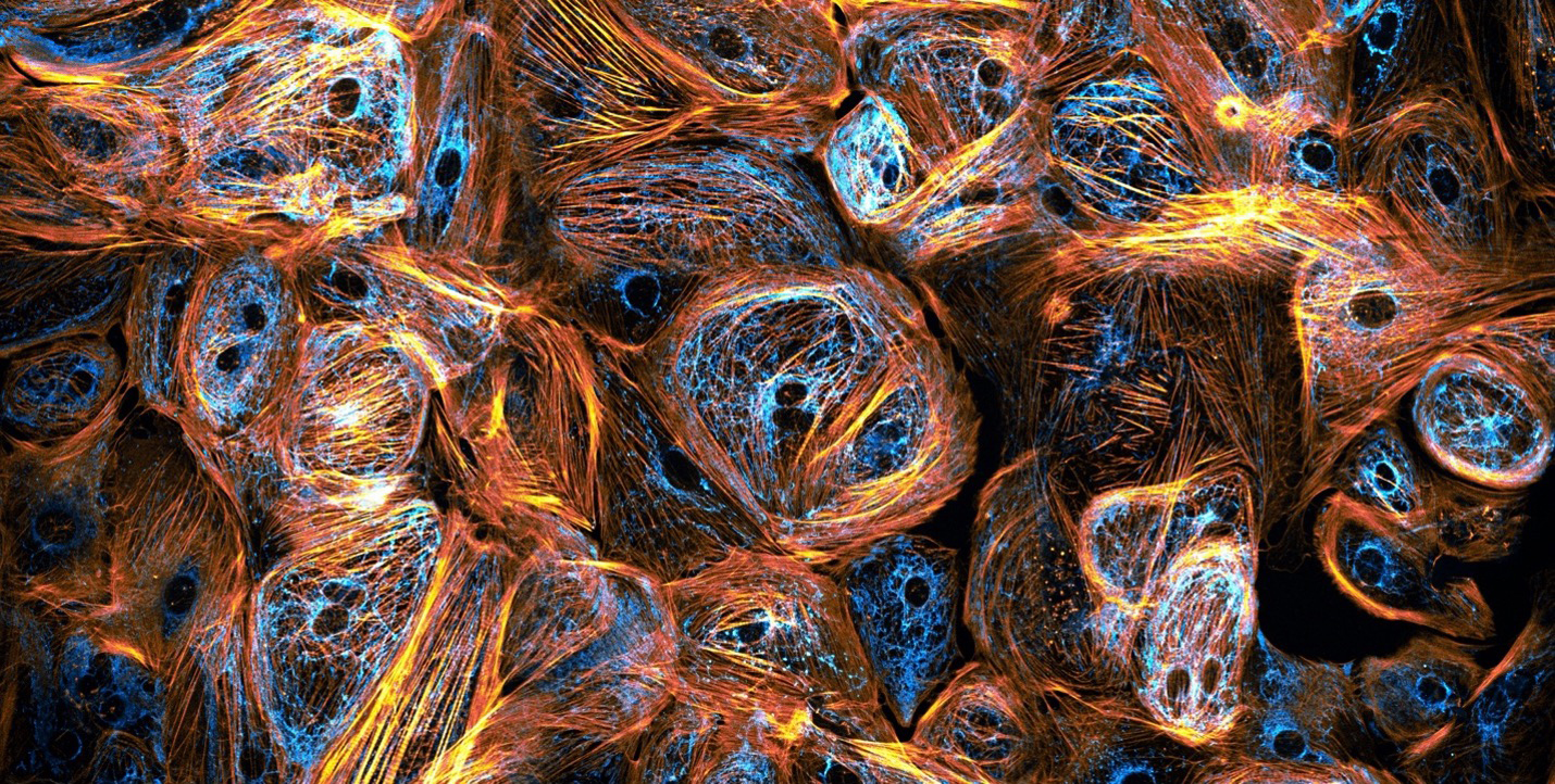 Nature as the Ultimate Artist Heart cells derived from induced pluripotent stem cells. Captured by Till Stephan (Germany). | Asia-Pacific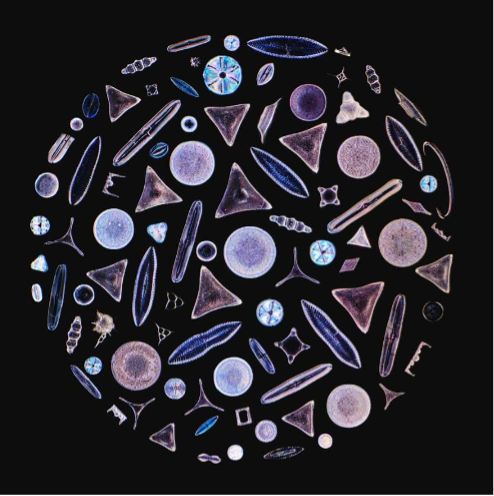 Nature’s Glass Gems Diatom arrangement. Captured by Daniel Han (Australia). |
Honorable Mentions
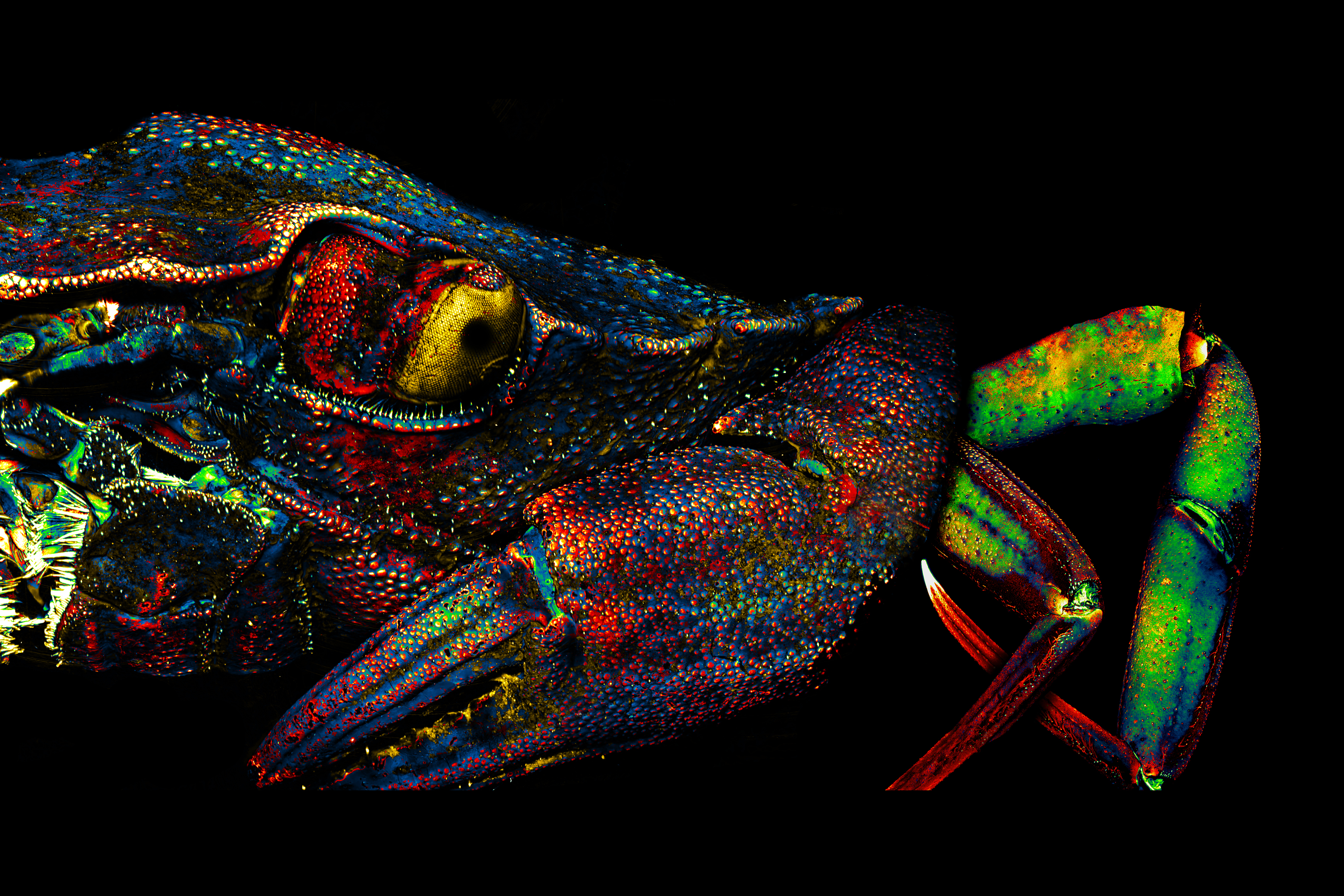 Green crab, labeled with DAPI and imaged autofluorescence, reflected light. | 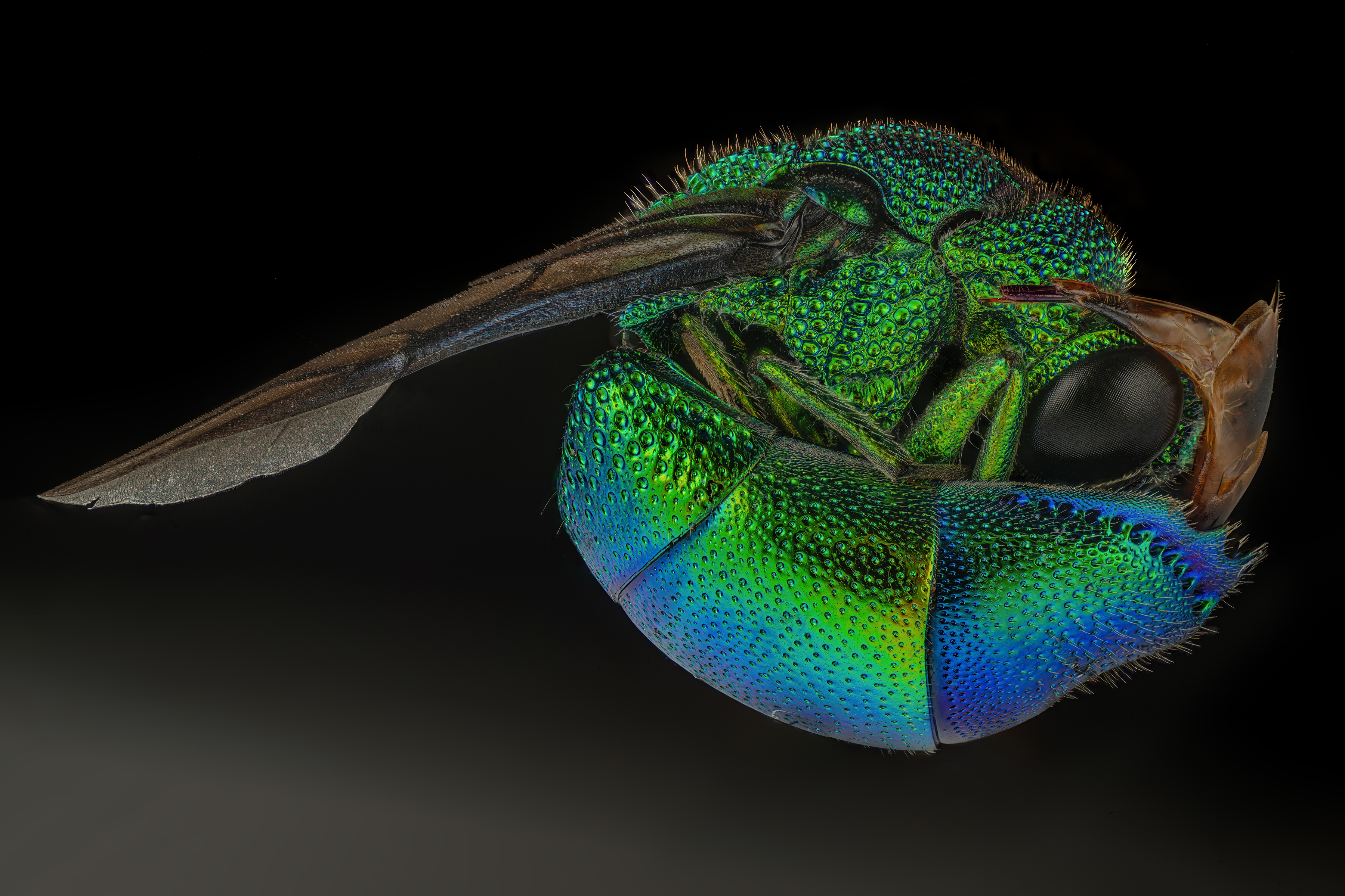 A cuckoo wasp. Cuckoo wasps are captivating insects, showcasing a dazzling array of iridescent colors. |  Juvenile sea star skeleton. Sea star (Patiria miniata) juvenile stained with calcein and DAPI. |
 Butterfly image consists of more than 200 scale patterns taken from different butterfly wings. | 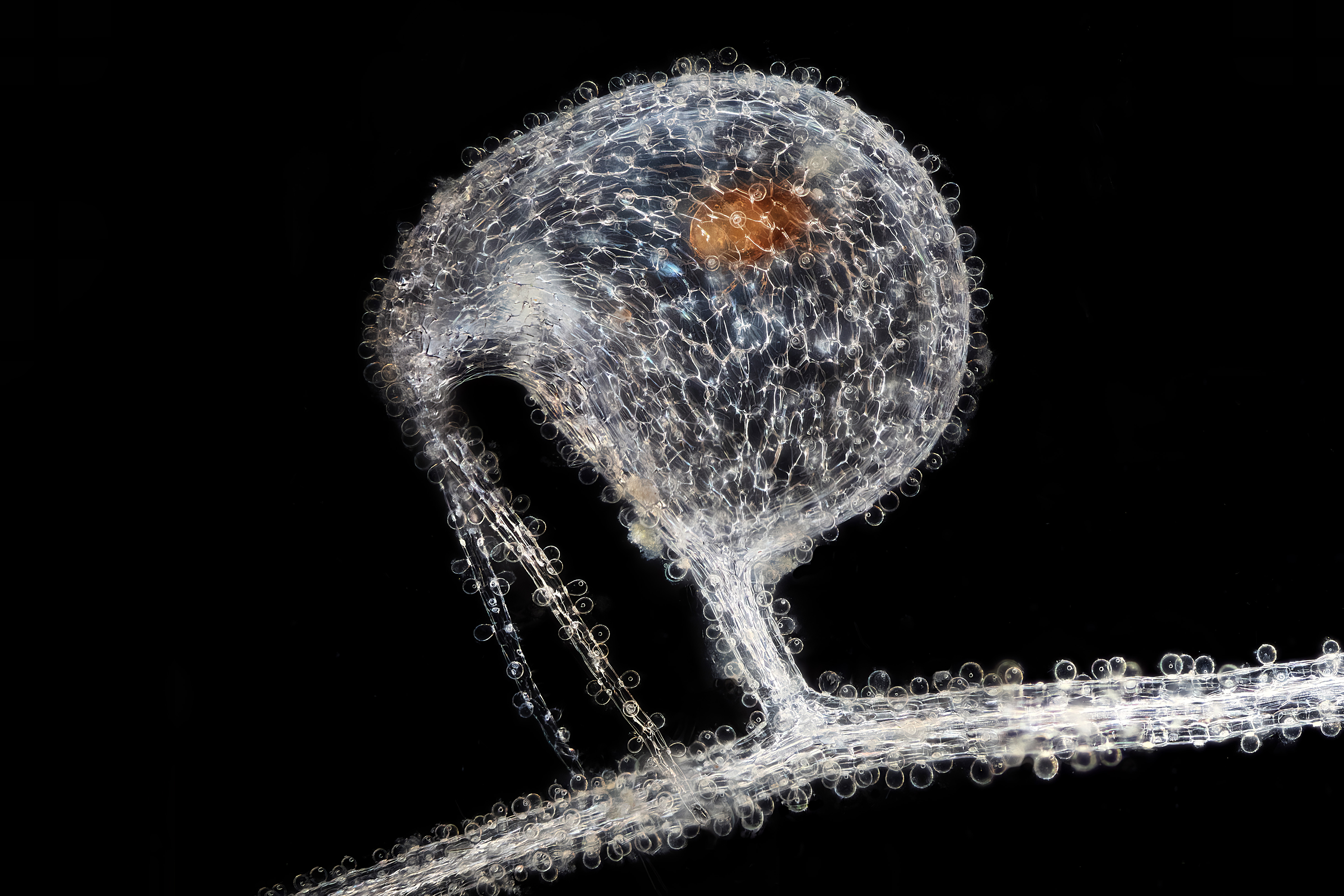 Trap of subaquatic carnivorous bladderwort Utricularia humboldtii with trapped water mite Hydrachnidia. |  Collage of different fish parasites observed in dark field in Argentine Patagonia. |
 A warty leaf beetle (Poropleura) that looks like a prehistoric tyrannosaurus. |  Fluorescent images of normal rod-shaped filamentous cells of the common soil bacterium Bacillus subtilis and a spherical cell representative that has lost its cell wall. | 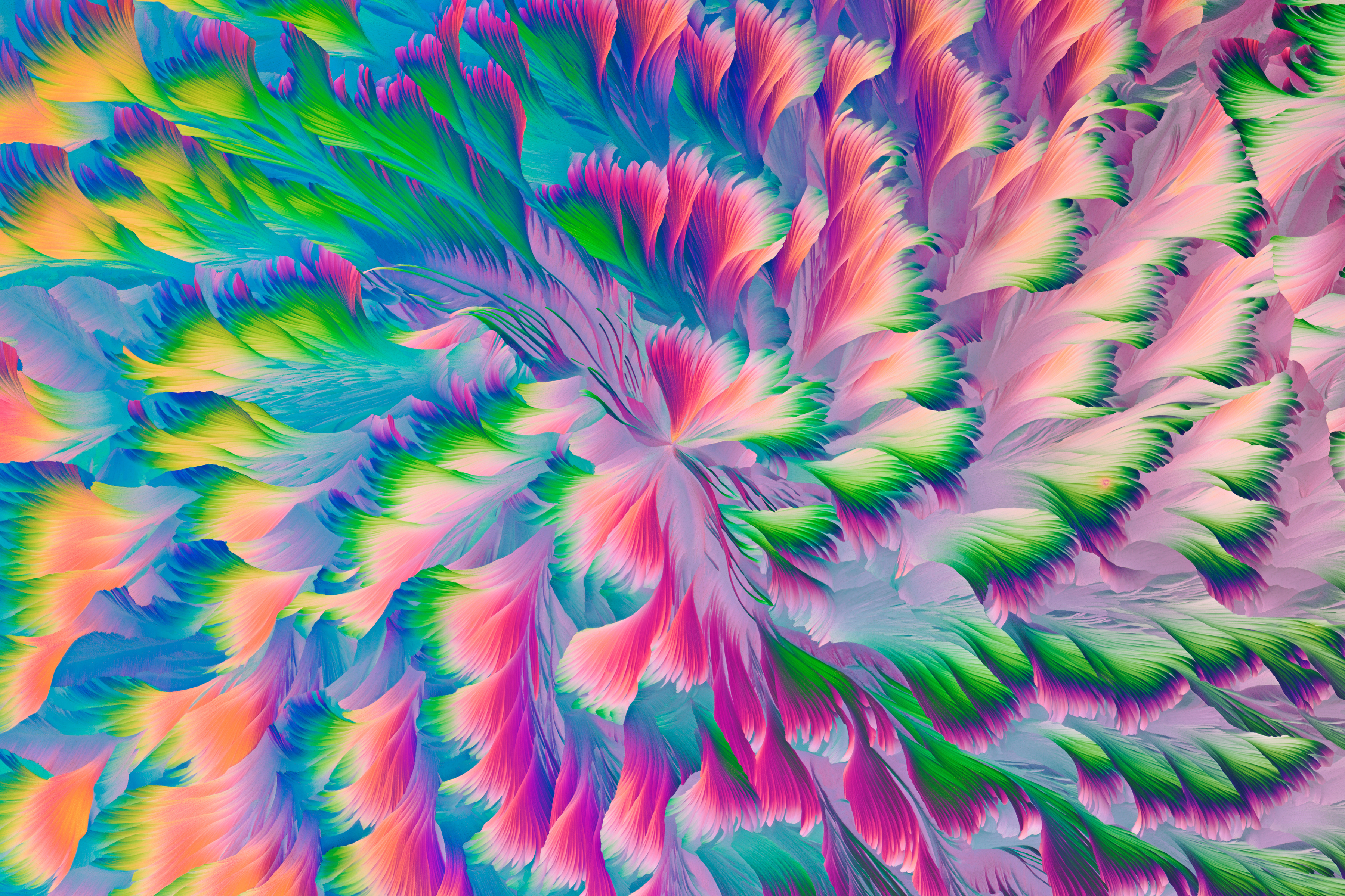 Crystalline pattern, like the big cotton padded jacket pattern handed down during the Chinese Spring Festival. |
 Tanaidacea crustacean swims in the water column during night and is attracted by light. Chitin emit light when excited using UV light. | 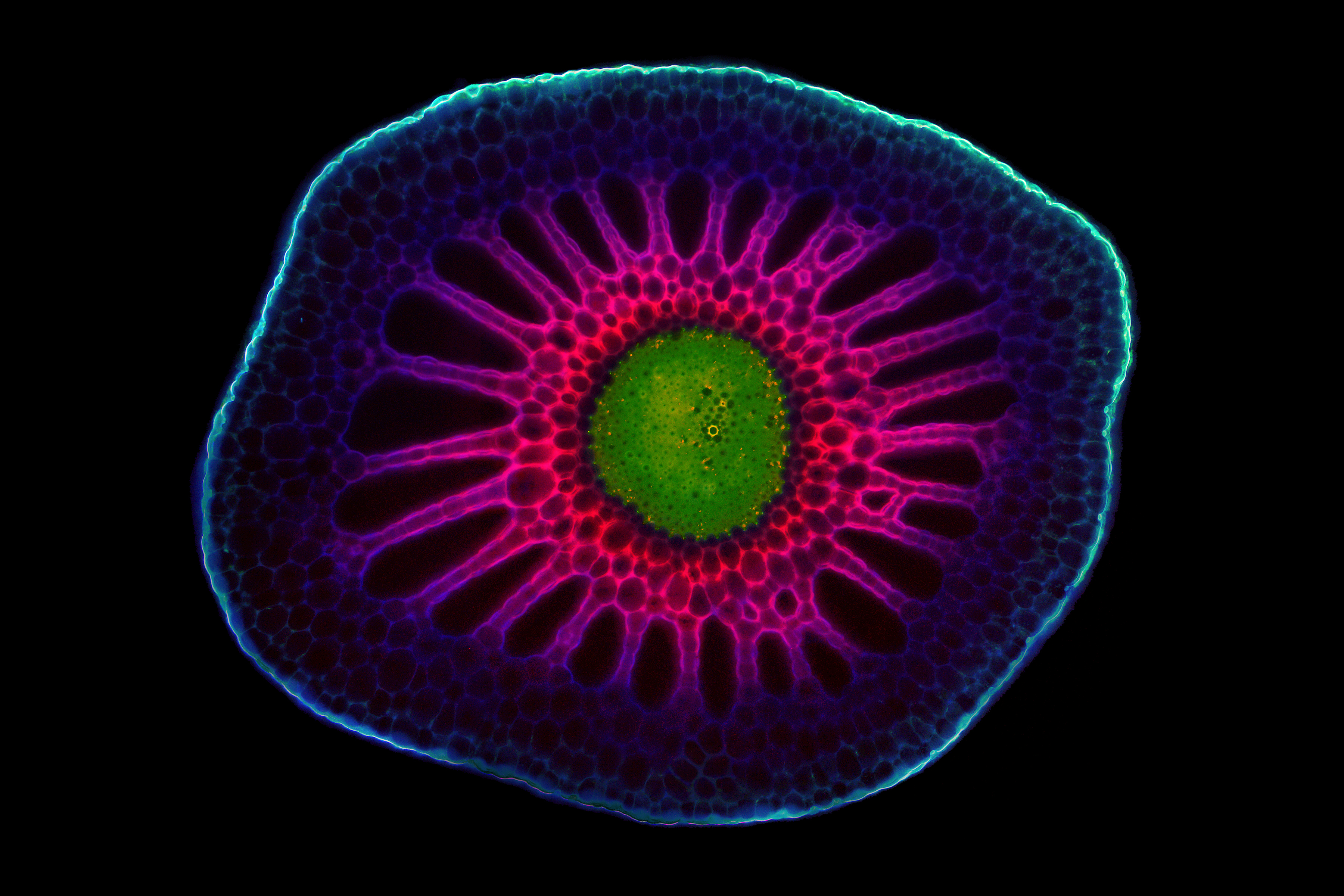 A look into the depths. Cross sections through the apical stem segments of the aquatic species Utricularia macrorhiza. |  The submarine. The image is a depict of a zebrafish head covered by epithelial cells expressing the palm-mTurquoise fluorescent protein and mCherry nuclear protein. |
Jurors
 | Geoff Williams, Manager of the Leduc Bioimaging Facility at Brown UniversityGeoff Williams is in his 14th year as manager of the Leduc Bioimaging Facility at Brown University. The opportunity to combine visual arts, science, technology, and mastery of a skill clicked with his discovery of microscopy (electron and light) as an undergraduate at Connecticut College. Geoff transitioned from a graduate program at Michigan State University to running the imaging facility at Central Michigan University before arriving at Brown. Over the past 20-plus years, he has been honing his craft as both an electron and light microscopist, paying more attention to the aesthetics of each image collected than is typically required of a purely scientific investigation. Geoff’s work, under the name Nanoscape, provides a tactile and striking view of samples we may or may not encounter in our day-to-day lives. |
 | Harini Sreenivasappa, Manager of the Cell Imaging Center at Drexel UniversityHarini Sreenivasappa is the manager of Drexel University’s light microscopy core facility, the Cell Imaging Center. She was introduced to microscopy during graduate school at Texas A&M University (TAMU), where she studied the role of microenvironment stimuli on cellular sensing and adapting as it takes place in blood vessel wall remodeling in cardiovascular disease. This led to a PhD in biomedical engineering. She has more than 10 years’ experience working with various microscopy techniques, such as atomic force microscopy (AFM), spinning disk confocal, and total internal reflection fluorescence (TIRF) microscopy. With ASCB’s COMPASS Outreach grant, she created and curated a Traveling Micrographs exhibit showcasing micrographs by TAMU researchers that was free and open to the public. The goal of the series of exhibits was to share research at TAMU with the local community and stimulate interest in imaging science. |
 | Rachid Rezgui, Research Instrumentation Scientist, Microscopy, New York University Abu DhabiRachid Rezgui is a microscopist and an active research scientist. Rachid studied physics at the Leibniz University of Hanover in Germany, then completed his PhD in biophysics at the Ecole Polytechnique in France studying DNA-protein interactions at the single molecule level. He joined the microscopy core facility at New York University Abu Dhabi in 2014, and has since worked with all types of microscopes, including two-photon, super-resolution, confocal, fluorescence lifetime, and widefield. He is involved in all aspects of optical imaging, such as sample preparation, training, acquisition, and post-processing, as well as core facility management. |
 | Nicolas Schilling, Microscopy Application Specialist, Center for Microscopy and Image Analysis, University of ZurichNicolas Schilling is a microscopy application specialist at the University of Zurich's light and electron microscopy core facility. His journey in optical imaging started during his biomedicine graduate studies at the University of Zurich. After earning his master's degree, he engaged in the university's technology platform program, where he worked on a novel sample preparation method that integrates high-pressure freezing cryofixation with super-resolution light microscopy, enhancing structural preservation. In 2019, he joined the university's microscopy core facility team and has since worked with all types of microscopes, including super-resolution, confocal, widefield, and electron microscopes. His responsibilities are diverse, covering sample preparation, training researchers, data acquisition, and post-processing. |
 | Songhai Shi, Professor and Doctoral Supervisor at the School of Life Sciences, Tsinghua UniversityDr. Songhai Shi is a renowned professor and doctoral supervisor at the School of Life Sciences, Tsinghua University in Beijing, China. He has long been dedicated to studying the development, formation, and functioning mechanisms of the brain using methods such as neurobiology, genetics, cell, and developmental biology. |
 | Dr. Wen-Tai Chiu, Professor and Department Chair, Department of Biomedical Engineering, National Cheng Kung UniversityDr. Wen-Tai Chiu specializes in optogenetics, calcium signaling, live-cell molecular imaging, and cancer research. He actively serves as a professor at his alma mater, the National Cheng Kung University. His recent projects include investigating the regulation of calcium ions (Ca2+) on focal adhesion dynamics, carcinogenesis, and chemoresistance, as well as exploring advanced bioimaging. His lab has also combined engineering technology to construct an optogenetic platform, which can accurately create different Ca2+ oscillations in time and space scales different from traditional chemical stimulation. As director of the Bioimaging Core Facility at National Cheng Kung University, which is affiliated with the National Core Facility for Biopharmaceuticals of the National Science and Technology Council, Dr. Chiu advocates for the collaborative use of high-end microscopic instruments by internal and external academic and industrial units. He believes that the researchers can learn from each other and practice the ideals of collaboration through the shared platform. |
Past Winners
|
The global winning image was taken by Laurent Formery (USA). Nervous system of a juvenile sea star (Patiria miniata) about 1 cm wide. Labeled with an antibody against acetylated tubulin after optical clearing, and captured using a color-coded Z-projection. |

Sorry, this page is not
available in your country.
Sorry, this page is not
available in your country.