
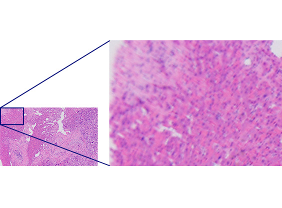
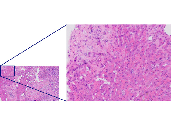
  | Uniform Quality from the Center to the EdgeImproved flatness produces images with uniform high quality from the center all the way to the edge. The high numerical aperture (NA) enables the objectives to gather more light for brighter, higher resolution images. Pairing X Line objectives with a BX53 upright microscope greatly improves the overall quality of images. |
|---|
Efficient Image StitchingStitching enables you to produce images of whole tissue sections, but if the image flatness is poor, the stitched image can be blurry and present a "patchwork" artifact. X Line objectives provide uniformly flat images from the center to the edge, enabling you to acquire uniform high definition stitched images. You can also create a large stitched image using a fewer number of images than before, improving efficiency and saving you time. | 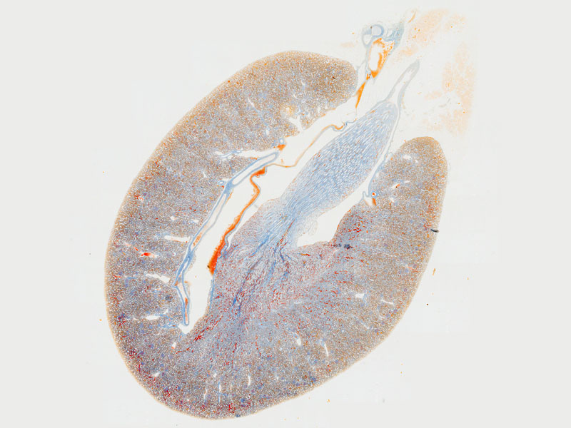 |
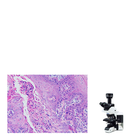
 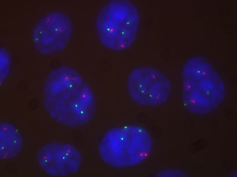 | Improved Images for Clinical Research ApplicationsImproved flatness, numerical aperture, and chromatic aberration combine to deliver clear, high-resolution images with excellent color reproduction. The objectives' superior chromatic aberration management delivers better color accuracy across the entire spectrum. The elimination of violet color aberration creates clear whites and vivid pinks, improving contrast and sharpness. Chromatic aberration correction in a wide wavelength range (400–1000 nm) enables you to acquire high resolution multicolor images during fluorescence observation, such as FISH. |
|---|
Related Products
BX53 Upright Microscope
| BX63 Upright Microscope
|
Sorry, this page is not
available in your country.
Sorry, this page is not
available in your country.