Not Available in Your Country
Sorry, this page is not
available in your country.
- Overview
- Absolute Quantitation
- Speed and Resolution
- Unmatched Simplicity
- Reliability and Flexibility
- Specifications
- Resources
Overview
Related Videos | Simply Powerful Imaging: Faster, Smarter, ClearerThe FLUOVIEW FV5000 is a platform built for every dimension of discovery. From crisp, photon-level quantitation at the surface to deep, multiphoton imaging in thick, living samples, the FV5000 captures biology at every scale. SilVIR™ detectors deliver exceptional sensitivity, a wide dynamic range, and photon-level precision, while 2K resonant and 8K galvo scanners freeze motion in real time for exceptional clarity. Smart automation simplifies setup and workflow and ensures consistent, reproducible results. Combining advanced detection, intelligent design, and intuitive operation, the FV5000 makes powerful imaging more accessible than ever, helping researchers capture more detail, more reliably, in less time. |
Precision Imaging You Can TrustBorn from more than 100 years of optical excellence, the FV5000 is setting a new standard in life science imaging for neuroscience, cell biology, drug discovery, cancer research, and developmental biology—delivering clear answers to challenging biological questions. The FV5000 is a next-generation platform designed to capture sharper, fully quantifiable data faster and easier than ever before:
Try the FV5000 yourself—it will be the last laser scanning microscope you demonstrate. |
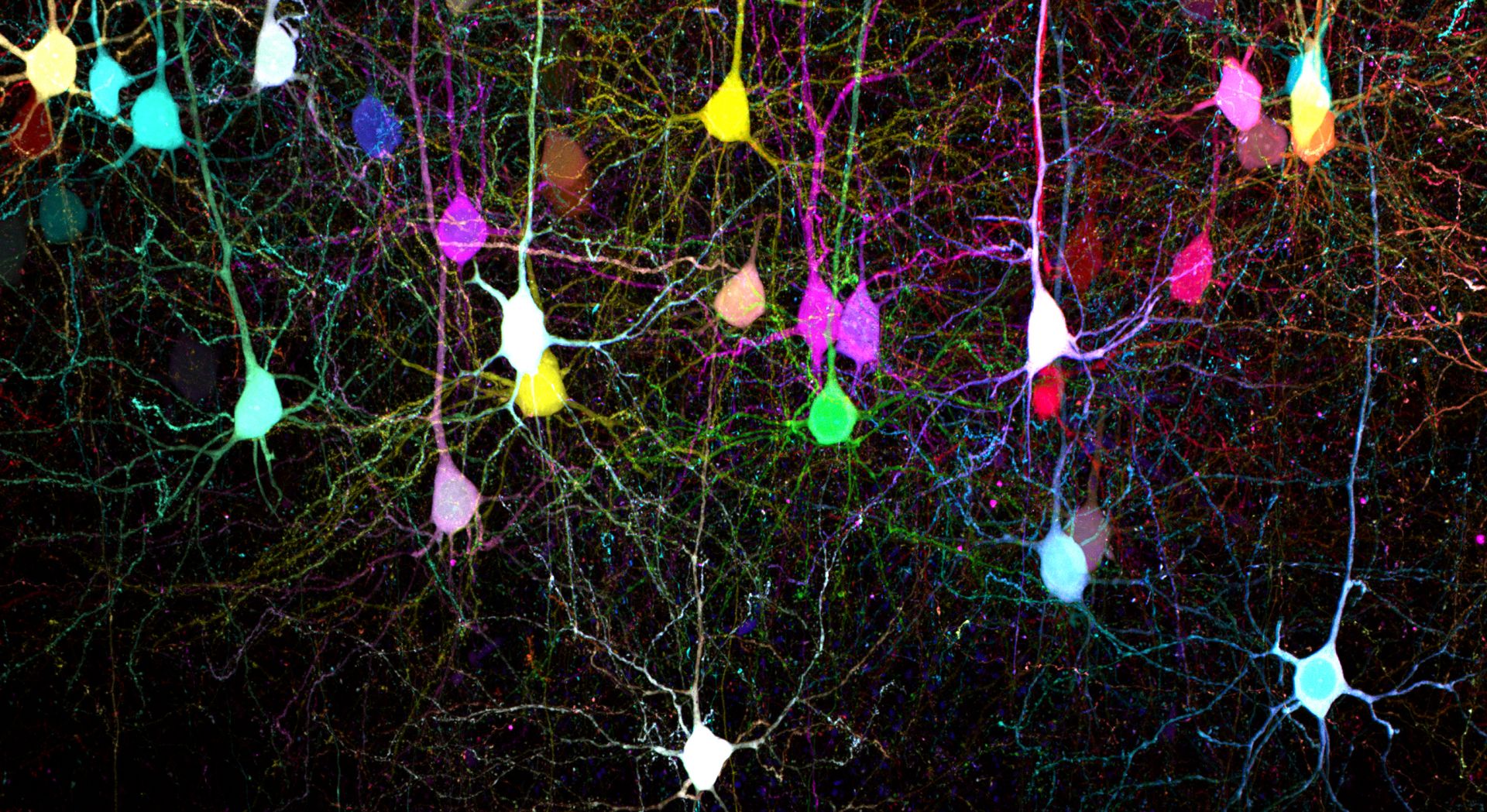
Mouse brain slice expressing 7-color Tetbow cleared with SeeDB2 (in utero electroporation). Sample courtesy of: Drs. Satoshi Fujimoto and Takeshi Imai, Graduate School of Medical Sciences, Kyushu University. |
Absolute Quantitation
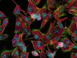
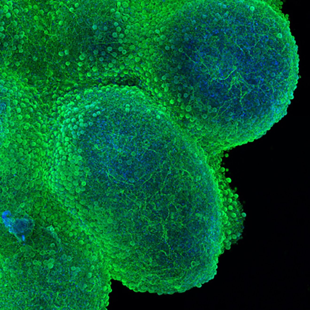
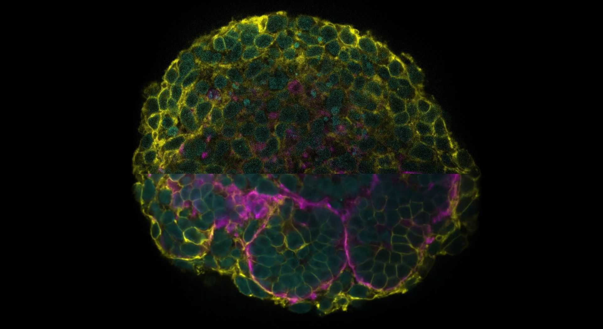
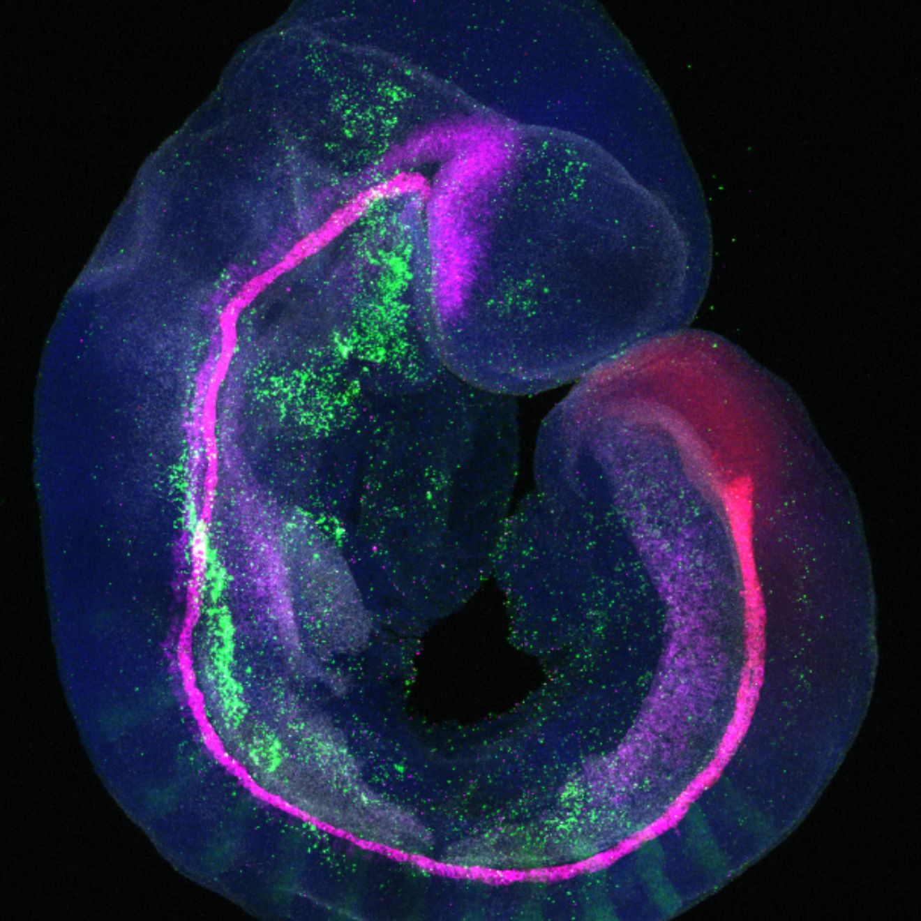
MAP2 (green) and Hoechst (blue) in a cortical organoid at DIV 45 with cell line KOLF2.1J. Sample courtesy of: Declan J. Brennan, Nygaard Lab at UBC. | Absolute Quantitation—Every Pixel, Every RunThe new benchmark in advanced microscopy, our SilVIR™ detectors deliver photon-level quantitation with exceptional sensitivity and ultra-high signal-to-noise across the industry’s widest dynamic range. Trust the industry’s first built-in laser power monitor to ensure consistent, reproducible illumination from samples captured today and in the future. Finally, combine beautiful images with beautifully quantifiable results. |
| “The dynamic range of the detectors has allowed us to image a number of different labels that we were not able to previously accomplish without making compromises over what gets over- or under-exposed.” Jonathan Epp, PhD |

SilVIR™ Next-Generation Detector TechnologyBuilt on Evident’s patented silicon photomultiplier design, SilVIR technology captures every photon with exceptional sensitivity and the industry’s widest dynamic range. Low-noise electronics and a built-in laser power monitor maintain illumination stability and ensure reproducible, quantitative results from faint signals to deep-tissue imaging. |
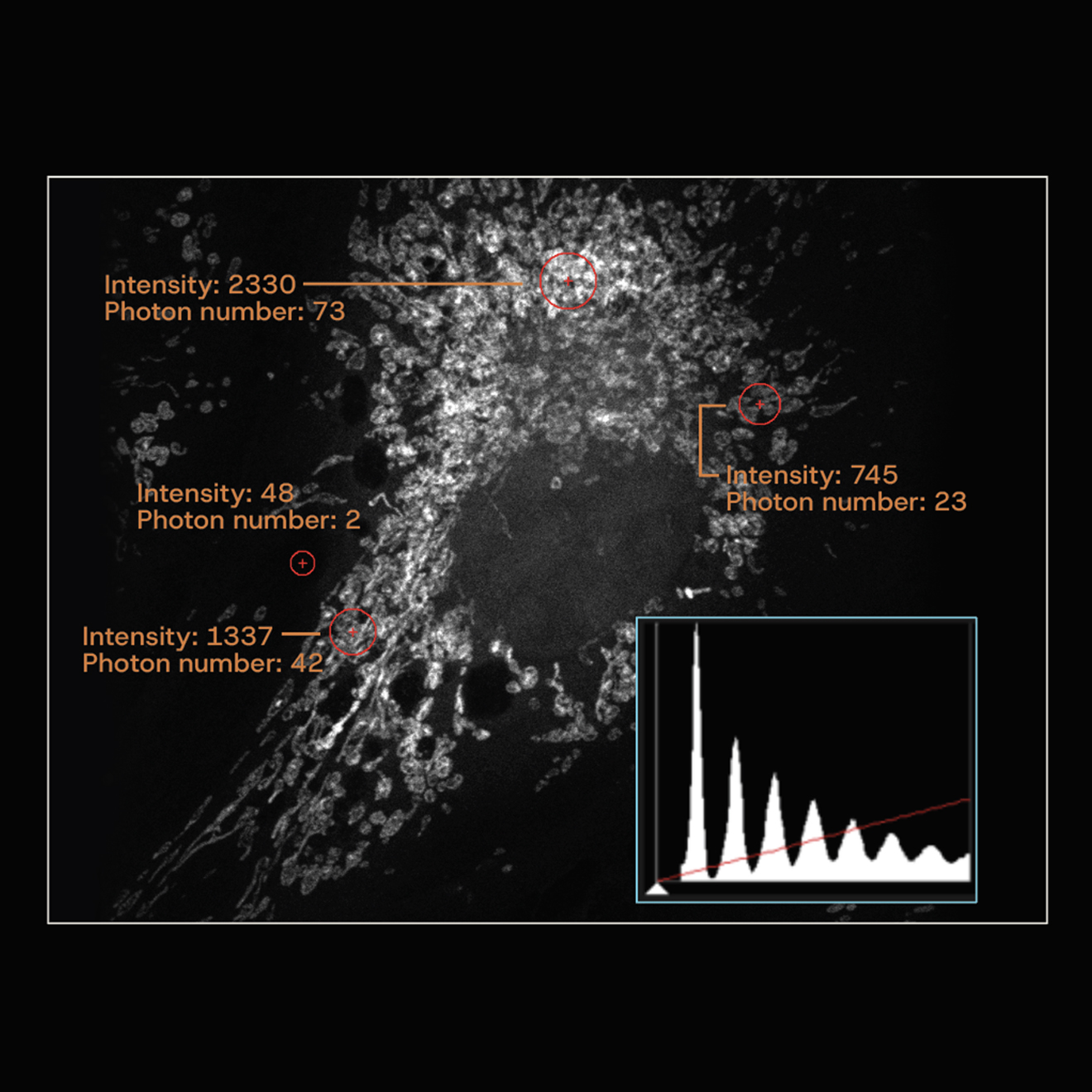
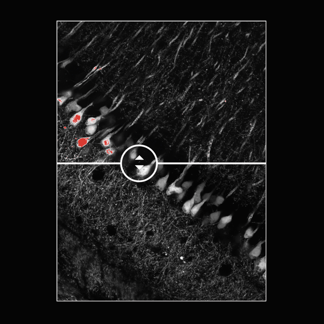
Expect exceptional imaging from every level of signal. Clear high and ultra-low signals from the same sample with no compromise in quality. Histograms show discrete photon counts with quantifiable intensity and minimum background. |
Top: Image saturation seen with a GaAsP-PMT detector (in red). | Leave Saturated Images in the PastWith its expansive dynamic range, the SilVIR detector prevents signal clipping and minimizes time spent adjusting settings. Each capture delivers valid, unsaturated data ready for deconvolution, stitching, or spectral unmixing. From single-photon events to intense fluorescence, SilVIR records the full signal spectrum in one acquisition. High dynamic range preserves faint details while preventing saturation of bright regions, reducing the need for reacquisition and ensuring consistent, quantitative image analysis. |
Speed and Resolution
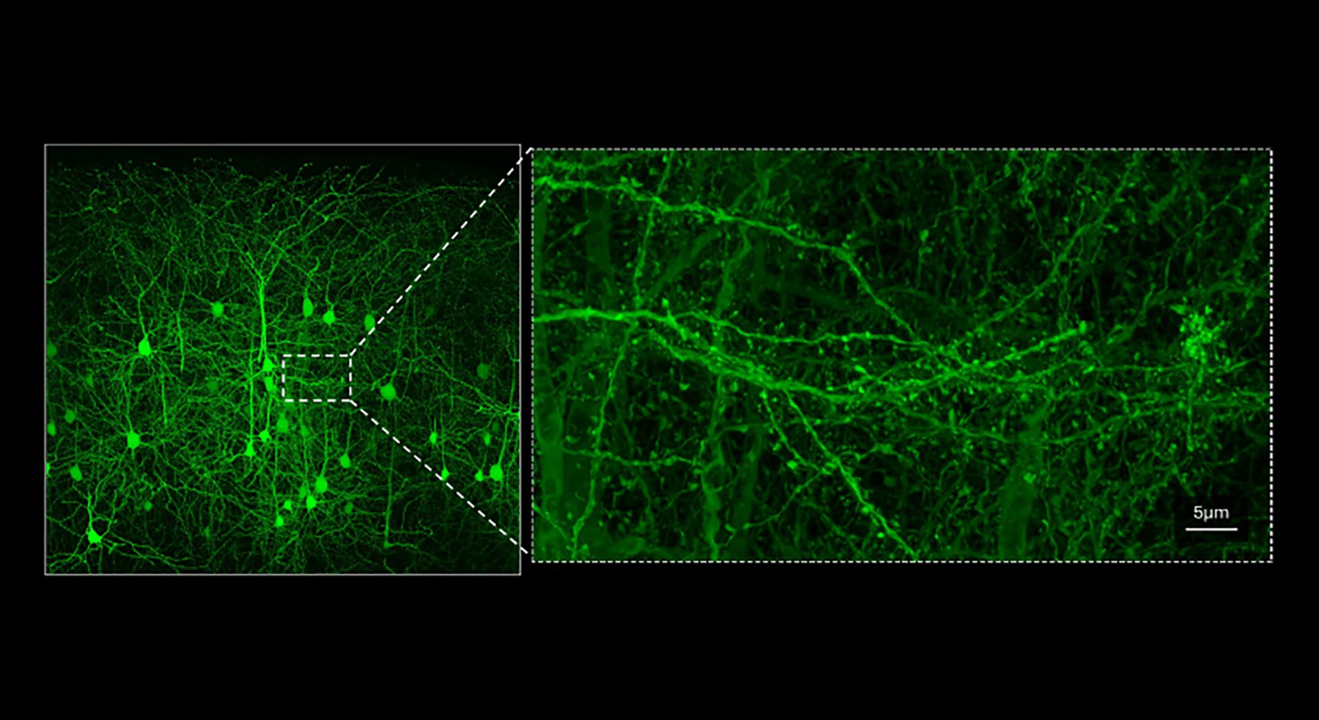
Two Scanners, One Workflow: High-Density Sampling and High-Speed Imaging. No Compromises.Whether you need high-speed series or pixel-dense maps, the FV5000 adapts. Our resonant scanner captures fast cellular dynamics at full clarity across a 20 mm FOV with little to no averaging, giving you high-quality, high-SNR raw images, in up to 438 FPS. Switch to 8K × 8K galvo scanning to image large areas at high spatial resolution with ultra-fast pixel dwell times as short as 0.2 µs. Achieve up to 120 nm XY resolution across six spectral channels with high NA objectives and FV-OSR software—no additional hardware required. The result: fewer compromises and faster time to data. |
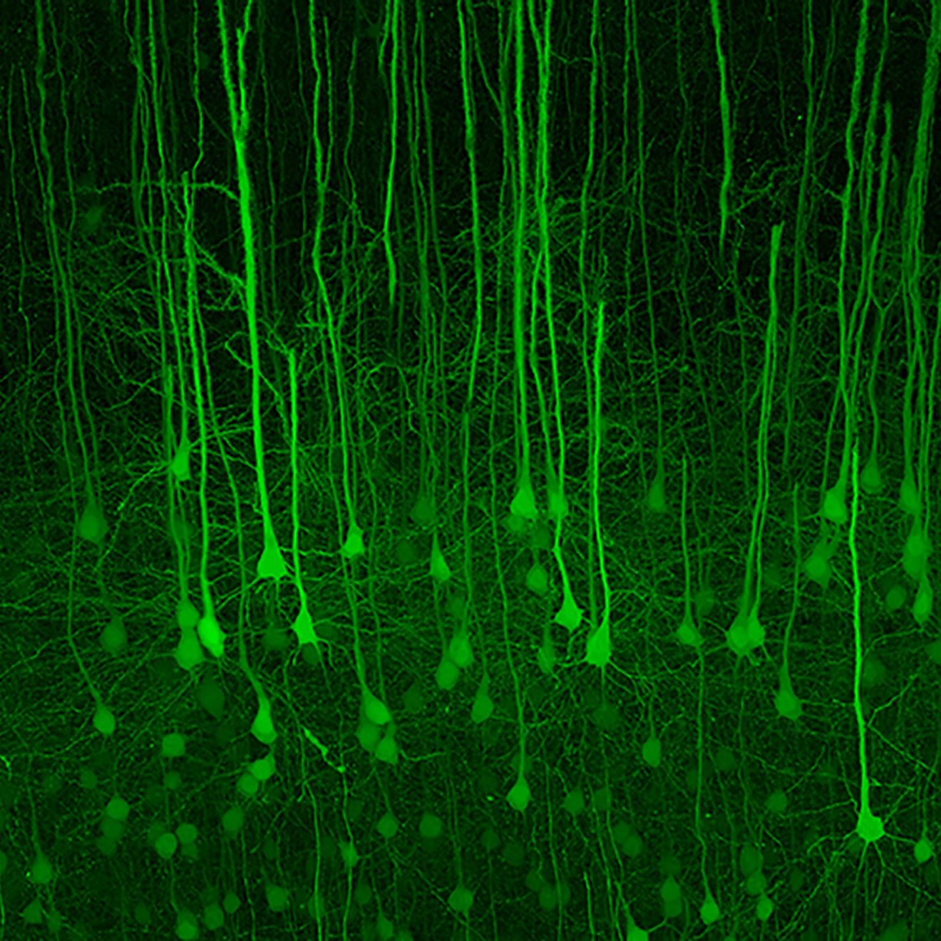
Stitched mouse brain slice cleared with SeeDB2. EYFP is expressed in cortical layer 5 pyramidal neurons in Thy1-YFP-H transgenic mice, acquired with a LUPLAPO25XO objective lens and resonant scanner. Sample courtesy of: Drs. Satoshi Fujimoto and Takeshi Imai, Graduate School of Medical Sciences, Kyushu University. |
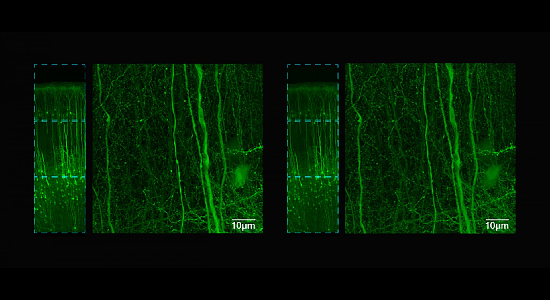
Acquire up to nine times faster than galvo with the same stunning clarity. Left: 43.5 minutes with 2K galvo Z-stack (galvo scan with no accumulation). Right: 4.6 minutes with 2K resonant Z-stack (resonant scan with 4x accumulation). Mouse brain cleared with SeeDB2. EYFP is expressed in cortical layer 5 pyramidal neurons in Thy1-YFP-H. Sample courtesy of: Drs. Satoshi Fujimoto and Takeshi Imai, Graduate School of Medical Sciences, Kyushu University. |
Effortless Super-Resolution ImagingPowerfully Accessible Subcellular AnswersAchieve super-resolution imaging on the FV5000 with no additional hardware. By pairing high NA objectives, such as our A Line™ HR series, with FV-OSR software, you can resolve subcellular structures down to 120 nm in XY with ease. FV-OSR automatically adjusts the confocal aperture to capture and enhance high-frequency signal components, producing crisp, detailed images in real time. Combined with the sensitivity of the SilVIR detector, the FV5000 delivers simultaneous super resolution across up to six spectral channels. Take your imaging—and your discoveries—further than ever before. |
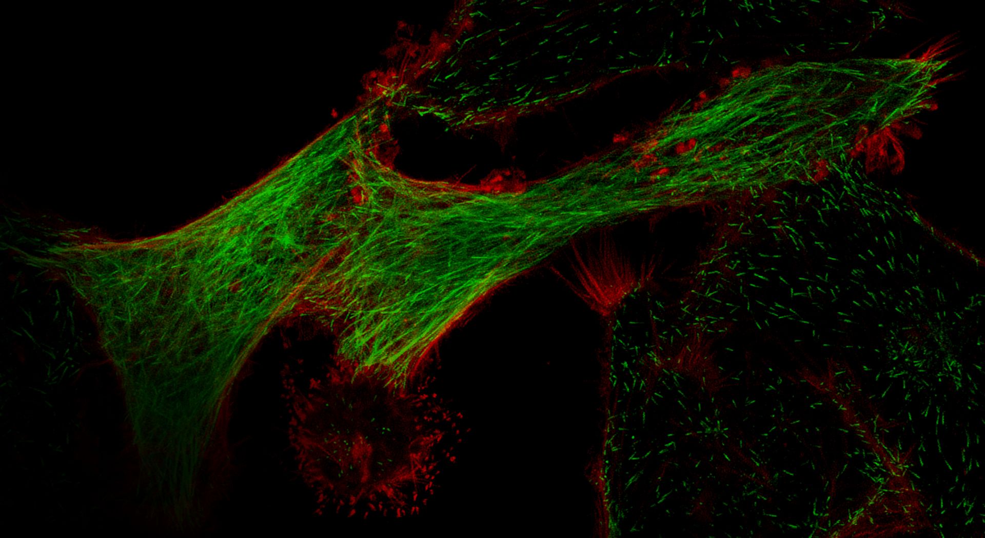
Cultured HeLa cells expressing Lifeact-mScarlet-I and EB3-3xmNeonGreen. Acquired with super-resolution mode on the FV5000. Sample courtesy of: Haruka Mii, Prof. Kazuhiro Aoki, Graduate School of Biostudies, Kyoto University. To learn more about the life of Henrietta Lacks and her contribution to modern medicine, visit henriettalacksfoundation.org. |
Unmatched dynamic range combined with effortless Nyquist, big picture, and fine details. Galvo 8192 × 8192, zoom 0.9x, 0.8 a.u., LUPLAPO25XO objective lens, Ypet: 514 / 530-570, Z: 60-150 µm, Z step: 0.82 µm, MIP. Mouse brain slice cleared with SeeDB2. YPet is expressed in layer 2/3 pyramidal neurons (in utero electroporation). Sample courtesy of: Drs. Satoshi Fujimoto and Takeshi Imai, Graduate School of Medical Sciences, Kyushu University. |
Capture Fast ResponsesThanks to the high SNR of the SilVIR detector, only minimal image averaging or accumulation is required to achieve high-quality results. |
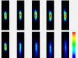
Related VideosLive imaging of an acute olfactory bulb slice expressing GCaMP6f cleared with SeeDB-Live. Image acquired with a LUPLAPO25XS objective lens at 70 µm depth from the surface. Sample courtesy of: Drs. Shigenori Inagaki, Takeshi Imai, Graduate School of Medical Sciences, Kyushu University. | Related VideosLive imaging of an acute olfactory bulb slice expressing GCaMP6f cleared with SeeDB-Live. Image acquired with a LUPLAPO25XS objective lens at 120 µm depth from the surface. Sample courtesy of: Drs. Shigenori Inagaki, Takeshi Imai, Graduate School of Medical Sciences, Kyushu University. | Related VideosLive imaging of an acute olfactory bulb slice expressing GCaMP6f cleared with SeeDB-Live. Image acquired with a LUPLAPO25XS objective lens at 200 µm depth from the surface. Sample courtesy of: Drs. Shigenori Inagaki, Takeshi Imai, Graduate School of Medical Sciences, Kyushu University. |
Biology in Motion, from Cells to EmbryosSee biology in motion, in detail. From FRAP to ablation, SilVIR quantitative detection and fast scanning allow you to capture weak signals with minimal averaging, delivering high SNR at real-time frame rates. In living samples, precise stimulation enables repeatable, targeted perturbations, so you can track repair, migration, and signaling over time. A wide linear dynamic range helps prevent saturation, keeping time-lapse data analysis-ready. |
Time-lapse sequence in a living zebrafish embryo showing the repair response after localized multiphoton ablation of microtubules (green). Sample courtesy of: Soraya Villaseca, PhD, Department of Physiology, Development and Neuroscience, University of Cambridge. |
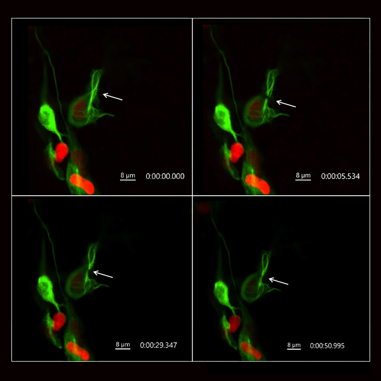
| “The quality of imaging, I’ve never seen something similar before." Soraya Villaseca, PhD |

Unmatched Simplicity
End-to-End Simplicity with FLUOVIEW Smart™ SoftwareInspired by real researchers tackling the challenges of science, the FV5000 is transforming confocal imaging, making it smarter and faster.
Note: FLUOVIEW Smart is available with FV5000 inverted configurations. FLUOVIEW Smart is not compatible with gantry, upright, or MPE configurations. | 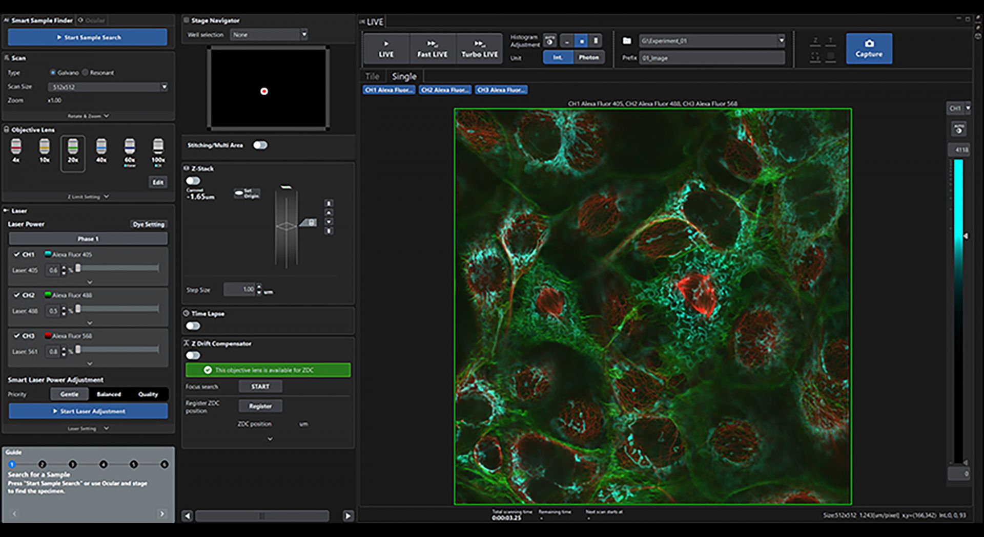 |
 TruResolution technology automates correction collar adjustment. It fine-tunes objective settings to minimize spherical aberrations from cover glass and sample heterogeneity over depth, with broad objective compatibility. | Smart Simplicity in ActionAutomated Correction Collar AdjustmentThe FV5000’s integrated TruResolution™ technology simplifies one of microscopy’s most tedious alignment tasks: objective collar adjustment. In a single click, the system automatically locates the optimal collar position for your sample, eliminating manual trial and error. For thick specimens, TruResolution dynamically fine-tunes the collar during XYZ scans to maintain consistent image sharpness throughout the entire volume. Compatible with many standard objectives, it delivers uniform clarity across diverse samples and imaging conditions. |
Intelligent Shading CorrectionFor edge-to-edge clarity and precision, automatically create seamless, high-quality stitched images with Intelligent Shading Correction. | |
 Left: Without auto correction collar. Right: With auto correction collar. Automated correction collar adjustment delivers sharper, more detailed images. | 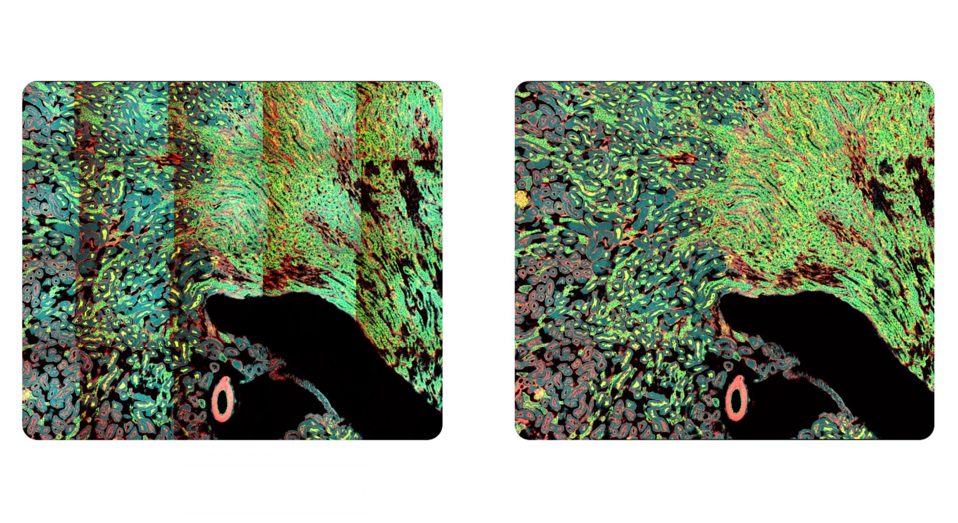 Left: Without Intelligent Shading Correction. Right: With Intelligent Shading Correction. Intelligent Shading Correction automatically compensates for uneven illumination across the field of view to generate stitched images without manual adjustment, improving efficiency and consistency in large-area imaging. |
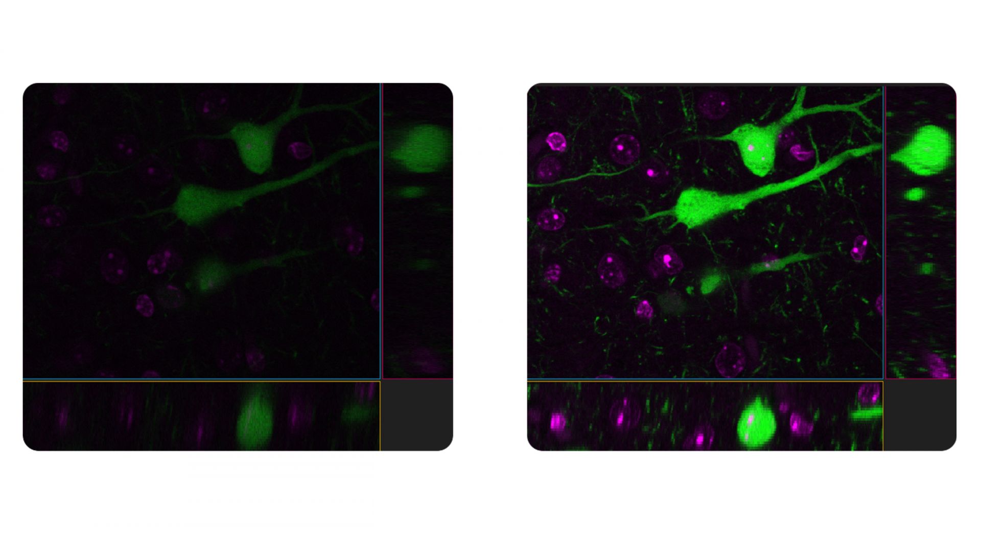
 Above line: Raw image. Below line: TruAI image. Human iPSC-derived kidney organoids with membrane-GFP. GFP signal immuno-amplified using anti-GFP primary and Alexa Fluor 488 secondary; laminin-111/211 labeled with Alexa Fluor 568; nuclei stained with DAPI. Captured with single-wavelength fiber-pigtailed IR lasers at 920 nm and 1064 nm for simultaneous 3CH multiphoton imaging at 2K resonant imaging. Sample courtesy of: Dr. Robert Turnbull and Prof. Katja Röper, Department of Physiology, Development and Neuroscience, University of Cambridge. | Software Tools as Dynamic as Your ScienceMinimize Noise, Maximize Data The FV5000’s software ecosystem includes advanced AI tools that enhance image quality, accelerate analysis, and streamline complex workflows, all without sacrificing scientific rigor. TruAI noise reduction further improves the FV5000’s already high signal-to-noise ratio by using neural networks trained on SilVIR detector noise patterns. Whether applied in real time or post-processing, TruAI restores clarity in resonant images and preserves temporal resolution while reducing photodamage. To speed downstream analysis, pretrained AI models can automatically segment image data, minimizing manual workload and ensuring faster, more consistent results across experiments. |
Image Segmentation Powered by Deep LearningGo Beyond Conventional ThresholdsTraditional intensity-based thresholding can be slow, inconsistent, and highly sensitive to sample conditions. TruAI image segmentation uses deep learning to recognize subtle patterns and faint signals that conventional methods miss, enabling accurate, reproducible segmentation of weakly labeled structures and complex tissues. Clearly distinguish cells—and quickly find answers. | 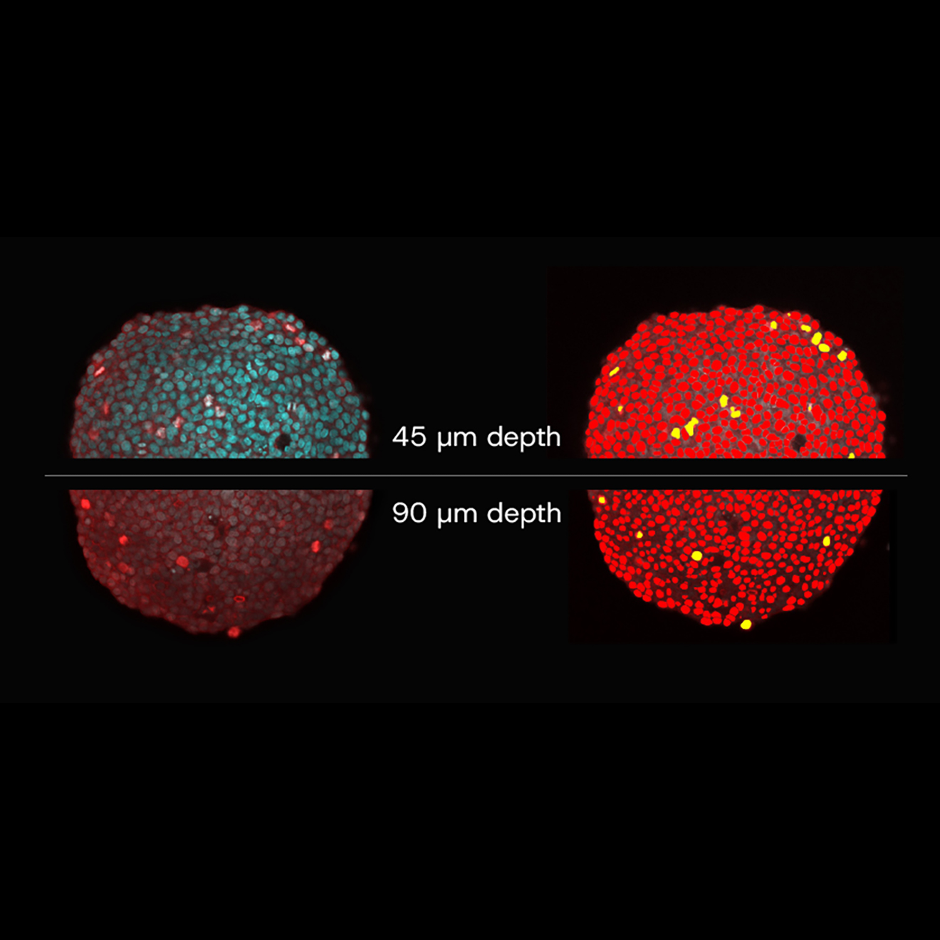 Spheroid imaging and analysis with TruAI. Left: Without TruAI. Right: With TruAI. TruAI segments and classifies cells (images on the right), even at high-penetration depths when the nuclei DAPI signal becomes weaker due to scattering. |
Reliability and Flexibility
Engineered for Long-Term Precision, Built to LastThe FV5000 is engineered for long-term precision and adaptability, delivering the reliability and flexibility your science demands. Configure your system for today’s needs and expand effortlessly as your research evolves. Add detectors, cameras, or lasers as workflows advance, or upgrade to multiphoton imaging with the MPE module, enabling single- and multiphoton acquisition as well as second and third harmonic generation. Smart hardware and software continuously monitor and optimize imaging performance, ensuring consistent, reproducible results. And with Evident’s global service and support network, every system is built to deliver lasting confidence and uptime. Built for ReproducibilityThe FV5000 maintains measurement precision through active system monitoring. The Laser Power Monitor (LPM) ensures consistent laser output across sessions, allowing different users to acquire images under identical conditions, even days or weeks apart. This stability supports the reproducibility required for quantitative and longitudinal studies. To further safeguard performance, the Microscope Performance Monitor (MPM) automatically evaluates system sensitivity and imaging consistency. It detects deviations early, helping researchers maintain confidence in their results and ensuring every dataset reflects true experimental conditions. | 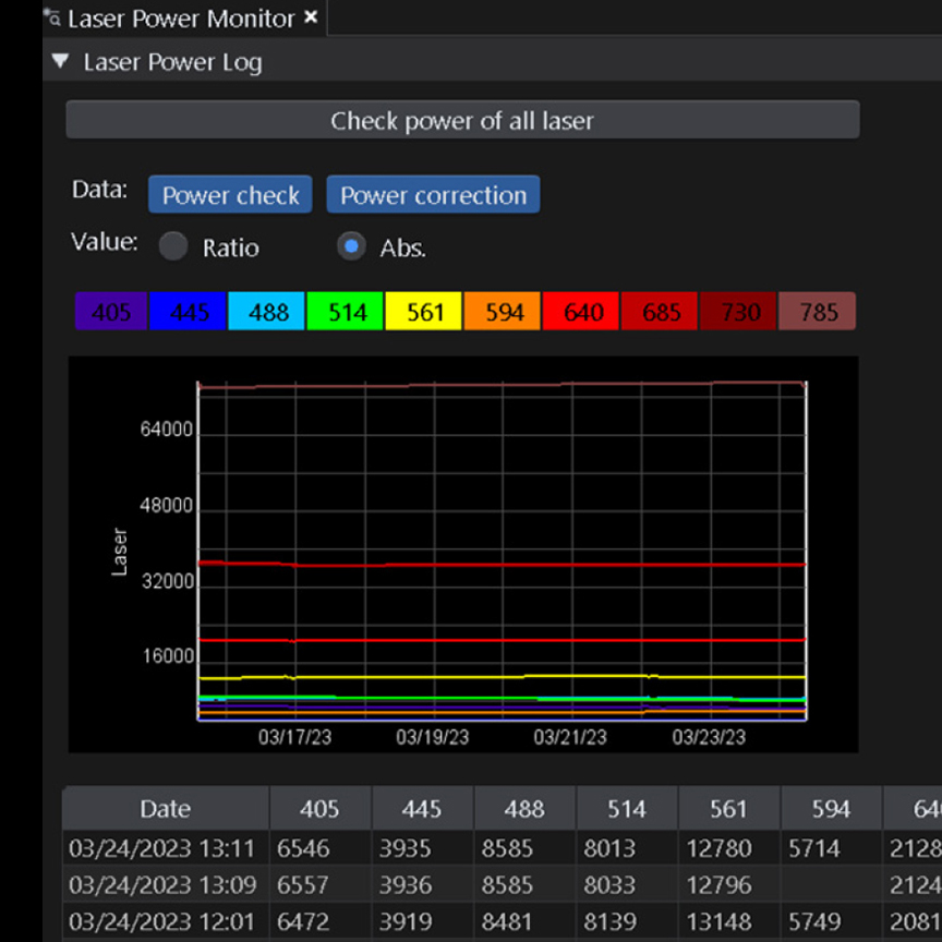 |
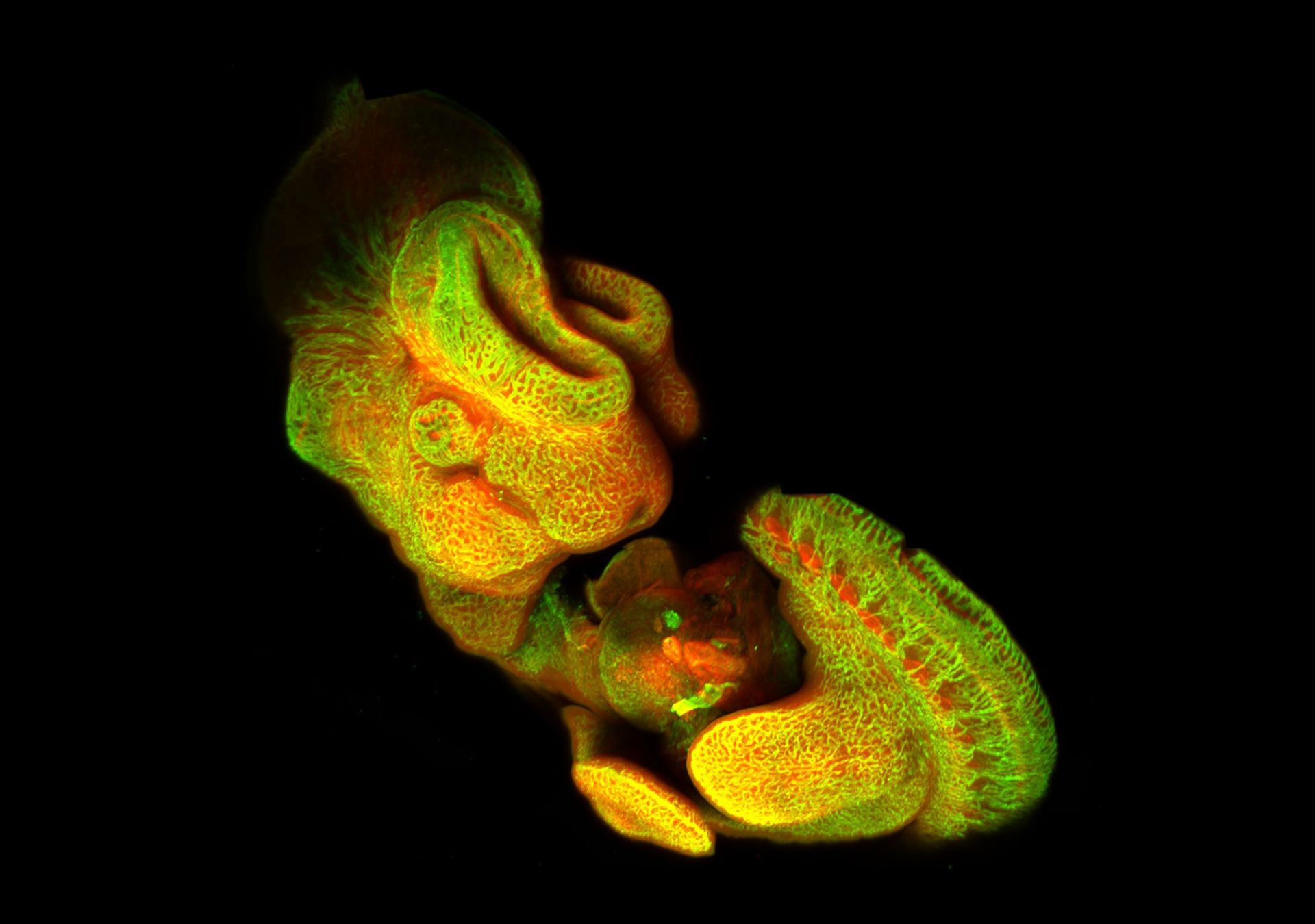
 High-resolution five-channel confocal image of an embryo captured using DAPI and Alexa Fluor™ 488, 568, 647, and 750—revealing detailed structures across multiple fluorescence spectra. | More Colors and More InformationCapture more colors and extract more information from every image using the FV5000’s enhanced multiplexing capabilities. Updated TruSpectral™ technology, in combination with the high-sensitivity SilVIR detector, enables simultaneous acquisition of up to six channels, configurable with a selection of broadband and red-shifted detectors to accommodate a broader range of fluorochromes. This configuration supports flexible experimental design, helping to reduce autofluorescence and photodamage during live-cell imaging. Modular laser combiners allow the integration of up to 10 laser lines, spanning wavelengths from 405 to 785 nm, to operate in parallel. |
ConfigurationsDesigned for Every ApplicationThe FV5000 platform supports a full range of configuration, from IX85 inverted systems for high-speed live-cell imaging and upright frames for general imaging or electrophysiology, to gantry setups for large or irregular specimens. For deeper imaging, the MPE (multiphoton excitation) configuration enables small-animal and thick-tissue studies, including large-frame and 3D organoid-optimized designs. Confocal and multiphoton modes can also be combined in a single system, giving researchers unmatched versatility within one platform. Up to six SilVIR detector channels can be configured for confocal imaging and an additional six for multiphoton detection, offering up to 12 simultaneous channels with photon-level sensitivity across modalities. |
CONFOCAL |
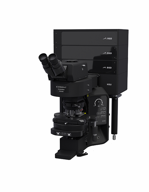 Upright SystemFor glass slide sample imaging. | 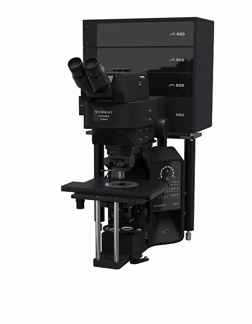 Upright SystemFor electrophysiology. | 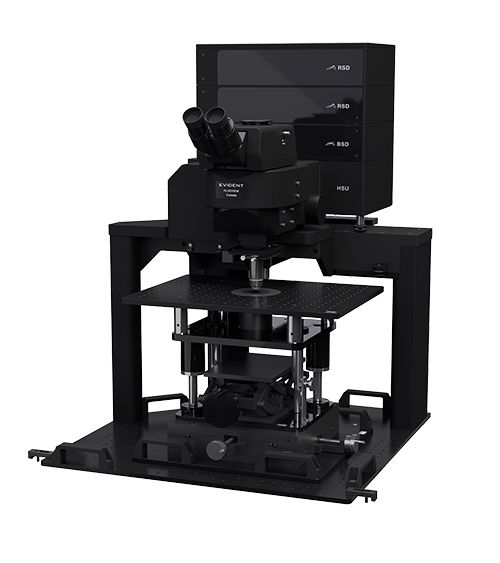 Gantry SystemFor in vivo observation that requires maximum space. | 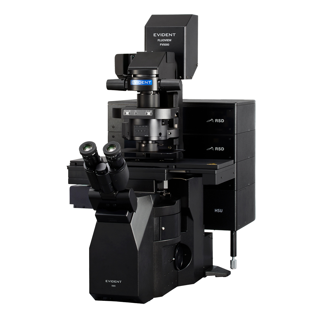 Inverted SystemFor observing tissue cultures, 3D cultures, and cell cultures (spheroids). |
MULTIPHOTON |
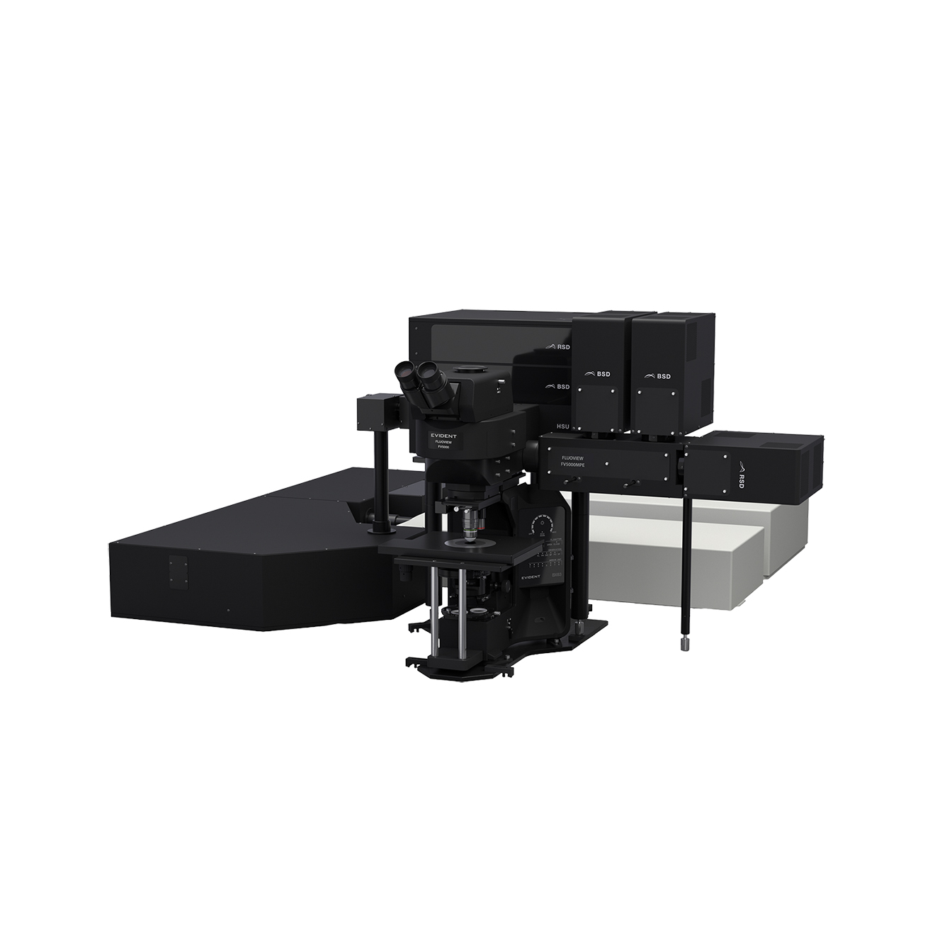 Upright SystemA large focus stroke accommodates a range of specimens, from tissue slices to live mice and other small animals. | 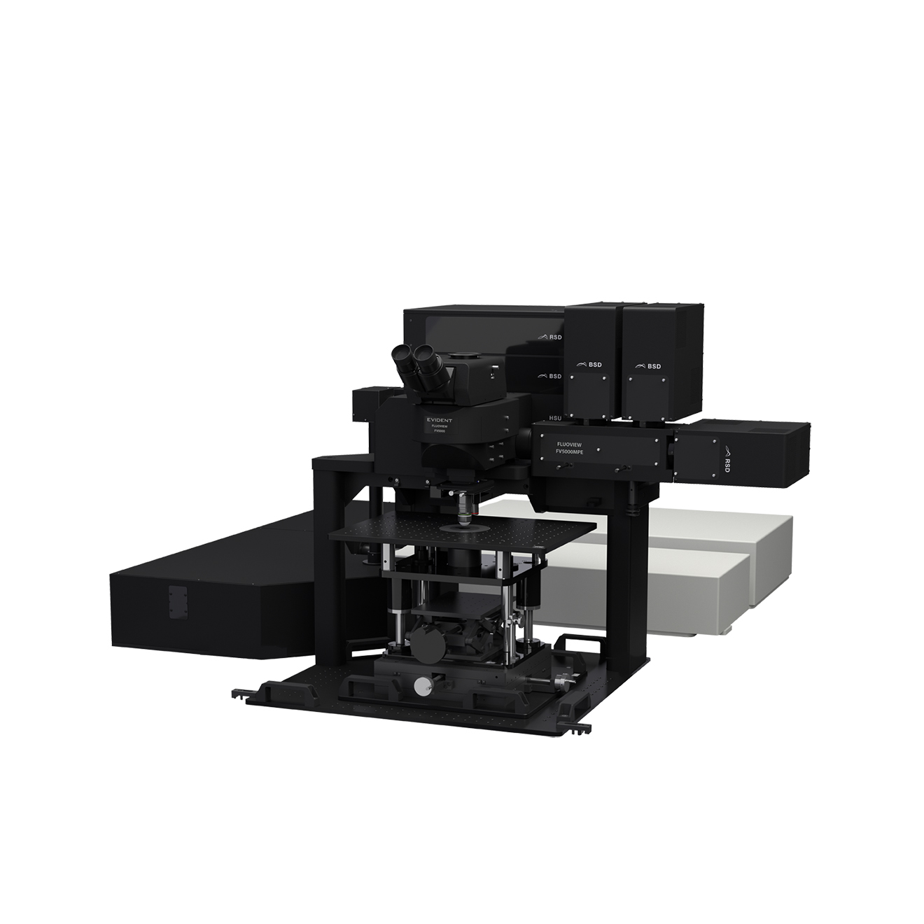 Gantry SystemThe frame maintains a large working space below the objective, making it easier to position experiment equipment. | 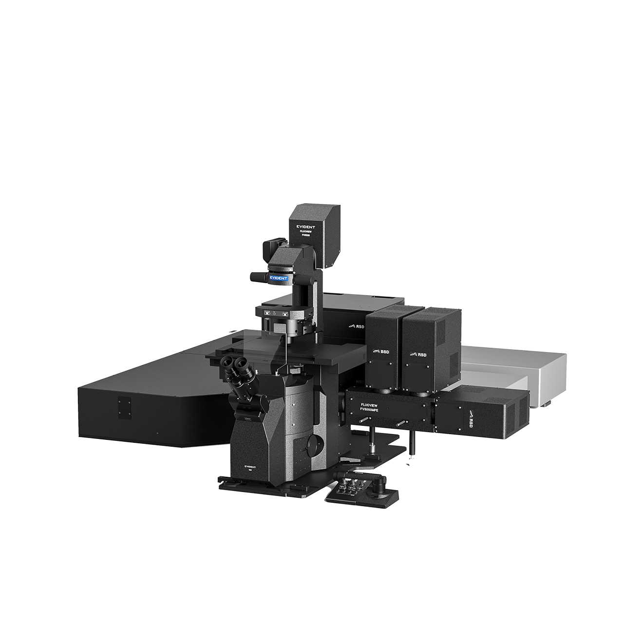 Inverted SystemThe frame supports observation of 3D cultures and multicellular clusters that are difficult to image using an upright frame. |
Learn more about the FV5000MPE for multiphoton applications. | ||
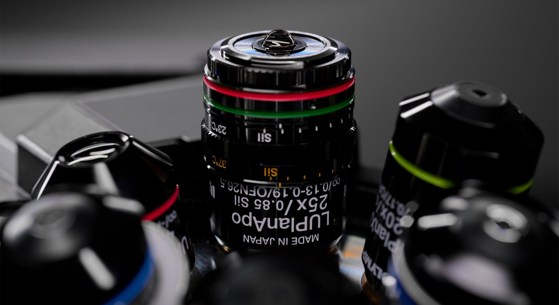
World-Class Imaging ObjectivesIn addition to our award-winning X Line™ objectives, Evident offers an expansive range of A Line™ objectives that can meet any research need, pushing your confocal system even further. Use our Objective Finder to locate the ideal objective for your application. |
| “The gel objective is my favorite thing ever. Image quality and acquisition speed, and also sensitivity, were all very impressive.” Emma Steijvers, MSc, MPhil |
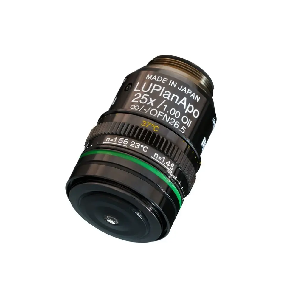
New Oil Immersion ObjectiveOur new long WD oil immersion objective lens allows you to see details deep in cleared samples.
| 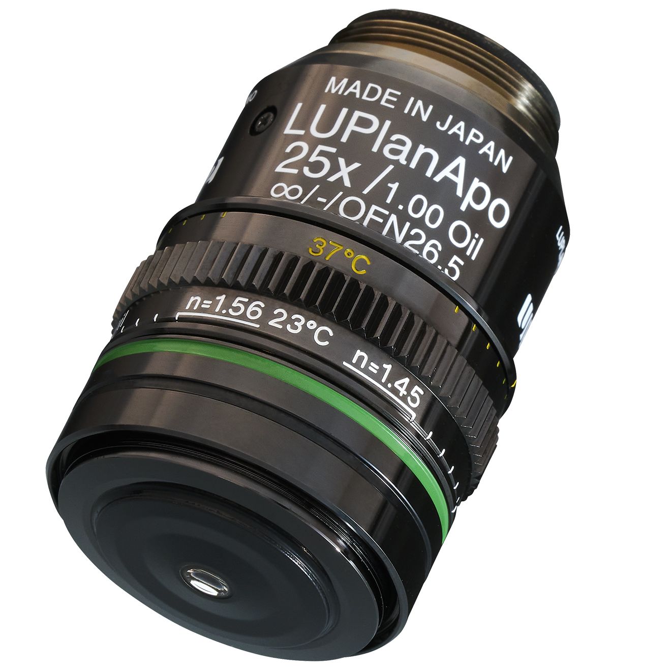 |
 Whole mouse embryo cleared with Ethyl cinnamate, labeled with Alexa Fluor 405, 488, and 568. Captured in confocal mode (170 tiles in XY, 700 microns depth) with a 25X oil immersion lens (1 mm WD). Sample courtesy of: Dr. Emma Siragher, Hanna Group, Department of Physiology, Development and Neuroscience, University of Cambridge. |
 | Driving the Future of High-End Imaging ResearchThe FV5000 combines precision engineering with intelligent automation to deliver reliable, reproducible results for every user—from core facilities to individual researchers.
|
Support and Service You Can Count OnWhen it comes to protecting your investment and the integrity of your research, your needs come first. We stand behind our products with a commitment to prompt service and technical support to help you achieve your goals. Available in three convenient tiers—Maintenance, Protection, and Performance Plus—our FV5000 Service Plans* include priority support to help minimize downtime, regular scheduled maintenance to keep your equipment in peak condition, predictable repair costs to eliminate unplanned expenses, and direct, efficient solutions when you need them most. *Regional variations in service offerings may apply. | 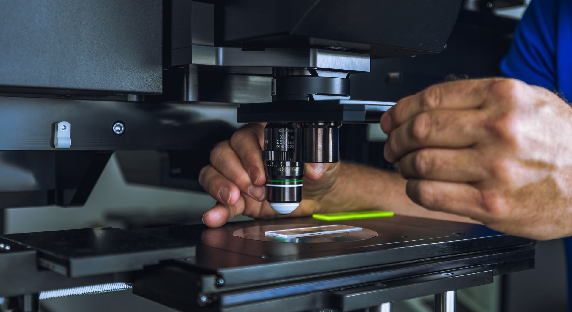 |
| Maintenance | Protection | Performance Plus | |
|---|---|---|---|
| Priority remote support | ✓ | ✓ | ✓ |
| Preventative maintenance | ✓ | ✓ | ✓ |
|
Repair coverage
(parts, labor, travel) | 10% discount | ✓ | ✓ |
| Fast on-site response | - | - | ✓ |
Specifications
| SPECIFICATIONS | FV5000 | FV5000-RS | |
|---|---|---|---|
| Scanner | Galvanometer Scanner | 64 × 64 – 8192 × 8192 pixels, 0.2 μs/pixel – 1000 μs/pixel | |
| Resonant Scanner | 512 × 512 pixels, 1024 × 1024 pixels, 2048 × 2048 pixels | ||
| Field Number | 20 (for both scanner types) | ||
| Spectral Confocal Detector | Detector | SilVIR detector (cooled SiPM, broadband type/red-shifted type) | |
| Maximum Channels | Six channels | ||
| Spectral Method | VPH, detectable wavelength range: 400 nm-900 nm | ||
| Laser | VIS Laser | 405 nm, 445 nm, 488 nm, 514 nm, 561 nm, 594 nm, 640 nm | |
| NIR Laser | 685 nm, 730 nm, 785 nm | ||
| Laser Power Monitor | Built in | ||
| Image | High dynamic range photon counting (1G cps, 16-bit) | ||