Not Available in Your Country
Sorry, this page is not
available in your country.
Overview
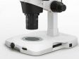
 | SZX16 Stereo MicroscopeDesigned for advanced research, materials science, QC inspection, and failure analysis, the SZX16 stereo microscope features an ultra-high resolution (0.3 NA), an ultra-wide zoom ratio (16.4:1), and versatile imaging with darkfield, fluorescence, and polarization. With these broad capabilities, you can image whole samples, such as organisms or components, down to fine microscopic details, such as individual cell structures or micro-defects. Equip your lab with the optical clarity needed to detect, document, and identify even the smallest details. |
|---|
Meeting the Challenges of Life Science and Materials Science ImagingDesigned for advanced research, materials science, QC inspection, and failure analysis, the SZX16 stereo microscope features:
With these broad capabilities, you can perform demanding applications—from imaging whole samples, such as organisms or components, down to fine microscopic structures, such as individual cell structures or micro-defects. Equip your lab with the optical clarity needed to detect, document, and identify even the smallest details. |
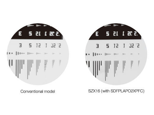
Advanced Stereo Microscope with a Wide Zoom RatioOutstanding Optical PerformanceThe SZX16's optics, which include observation tubes, a zoom body, and objectives, are fully apochromatic to reduce chromatic blur over the entire magnification range. The super depth of focus (SDF) objectives can deliver clear and high-contrast images of virtually any sample—from micron-scale defects to cell component structures. The maximum NA of 0.3 produces a resolution of 900 line pairs per mm, enabling clear visualization of fine microscopic cell and cell component structures. Outstanding resolution and peerless magnification make work more efficient, more precise, and reveal much more information from specimens. This higher resolution also means the stereo microscope can distinguish smaller, more closely spaced features in components, which is essential for high-precision failure analysis and QC inspection. Spot micro-cracks or fractures, surface scratches or contamination, or manufacturing detects invisible at lower resolutions. Wide 16.4:1 Zoom RatioThe SZX16 boasts an ultra-wide zoom ratio of 16.4:1, enabling 7x-115x magnification with a 1X objective, and up to 230x magnification with a 2X objective. This wide zoom range makes it suitable for a wide range of research analysis applications, with low-magnification macro views for dissection and specimen manipulation, and sharp high-magnification views for close observation of microstructures. In failure analysis and QC inspection work, this ultra-wide zoom range is ideal for inspecting everything from large components or assemblies down to micron-scale defects like cracks, scratches, or contaminant particles. |
|
|---|
|
|---|
Double Objectives for 3.5x to 230x ZoomWith the parfocal objectives and two-position revolving nosepiece, switching between objectives is quick and easy, requiring little refocusing. With the 0.5X and 2.0X objectives, the uninterrupted magnification range is 3.5x - 230x. This represents an effective zoom ratio of 65.7:1. |
|
|---|

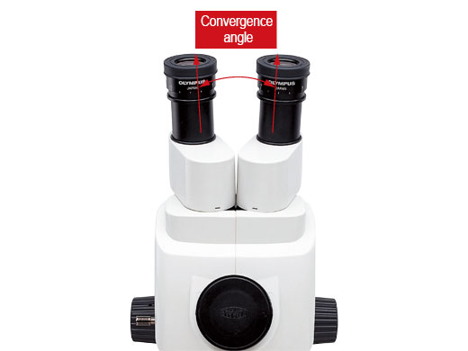
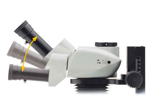
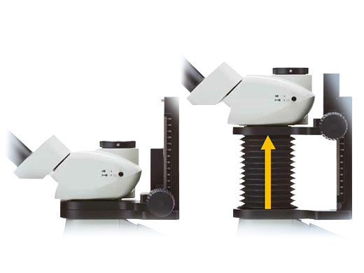
Ergonomic Observation Tube and Extendable Eyepoint AdjusterThe SZX2 series offers observation tubes with an optimized convergence angle, enabling users to observe from a natural position that reduces eye fatigue during extended use.
Together, these ergonomic solutions help reduce physical strain and fatigue, supporting more comfortable microscope operation. |
Convergence Angle
| Tilting Observation Tube
| Extendable Eyepoint Adjuster
|
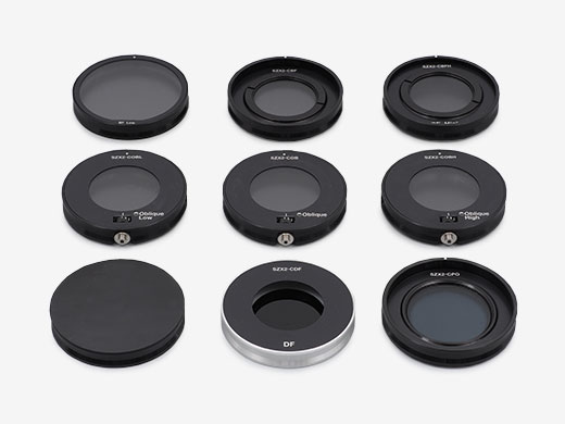
Versatile Contrast and Observation MethodsSZX2-ILLTQ / SZX2-ILLTSWith a slim 41.5 mm profile—about half the thickness of previous halogen lamp transmitted light bases—our LED transmitted light illumination bases offer a lower height for a more comfortable eyepoint and easier access to base-mounted samples during observation and operation.
An additional benefit of LED illumination is a cooler base surface, making it more suitable for long-duration manipulation of live specimens. Compared to a conventional 30 W halogen light source, LED illumination consumes less power and offers a significantly longer service life—over 60,000 hours—helping to reduce operating costs. |  |
|---|
|
|
|

Need assistance? |
Fluorescence Observation
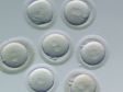
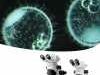
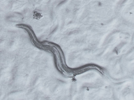
SZX16-RFA Illumination System for Advanced Fluorescence ObservationThe SZX16 works as a fluorescence microscope for observing everything from whole organisms to nuclear detail, thanks to its precisely engineered optics and high NA. The advanced glass materials with low autofluorescence and new surface coatings greatly increase the transmission of light, improving image clarity. These features result in an excellent signal-to-noise ratio (SNR) and high fluorescence signal intensity. Full control of the excitation light is facilitated by the 5-position filter wheel, which enables flexible illumination for low-magnification, high-resolution imaging. The near-vertical reflected light illuminator’s excitation light paths are independent from the observation paths, allowing for substantially improved excitation light efficiency. These features provide far brighter fluorescence observation than conventional stereo microscopes at all magnifications. Transmitted light observation for verification of the sample outline is possible even under reflected light fluorescence observation. |
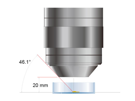
High NA Provides Bright Fluorescent ObservationA near-vertical reflected light illumination system produces illumination that is almost coaxial to the observation path and enables substantially improved excitation light efficiency. These features provide an average of two to three times better fluorescent observation than conventional stereo microscopes at all magnifications. In addition to using reflected light, transmitted light can be also be used for sample confirmation. |
Five-Position Turret with Nine-Filter SelectionThe fluorescence illumination system for the SZX16 has five-position turrets with a five-filter selection for different samples. Nine fluorescent filter units capture the details of bright and high-contrast fluorescent images. |
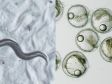
 SZX16 Filter Wheel |  Fluorescence Imaging Samples |
Need assistance? |
Digital Imaging and Documentation
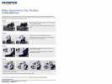
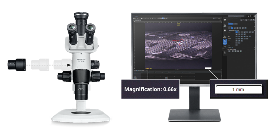
Digital Imaging and DocumentationEquip the SZX16 with a trinocular tube and a digital camera from our DP series for high-resolution imaging and documentation. DP series cameras offer high sensitivity for a wide range of applications, including high-sensitivity fluorescence imaging. They can be easily controlled using cellSens™ software for life science research or PRECiV™ software for materials science and inspection. |
All-in-Focus Imaging Made SimpleReconstructing an all-in-focus image is simple in our software. Manually acquire images from the Live EFI (extended focus imaging) function to obtain fully focused views of thick, rough, or uneven samples.
|
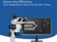
Flexible Inspection Workflows in PRECiV SoftwarePRECiV software offers a flexible workflow solution for QC inspection, materials analysis, and failure analysis. Pair it with the SZX16 and a digital camera to seamlessly acquire images, process them, and make 2D measurements. Advanced imaging and analysis functions include panoramic views, extended focus images, and particle analysis. Zoom into Efficiency
To speed up QC inspection or failure analysis work, combine the SZX16 with our SZX2-ZMS zoom magnification sensor. It automatically records the zoom magnification in your PRECiV software, enabling the correct 2D geometric measurements without the need to manually enter the magnification. From imaging to reporting, all dimensions are under control. Now you can zoom in and measure the details faster, adding confidence and efficiency to your inspections. |
Need assistance? |
Specifications
| Observation Method > Fluorescence (Blue/Green Excitations) | ✓ | |
|---|---|---|
| Observation Method > Fluorescence (Ultraviolet Excitations) | ✓ | |
| Observation Method > Simple Polarized Light | ✓ | |
| Observation Method > Brightfield | ✓ | |
| Observation Method > Darkfield | ✓ | |
| Observation Method > Oblique | ✓ | |
| Zoom > Zoom Ratio | 16.4 | |
| Zoom > Magnification Indication | 0.7, 0.8, 1, 1.25, 1.6, 2, 2.5, 3.2, 4, 5, 6.3, 8, 10, 11.5 | |
| Zoom > AS |
| |
| Zoom > Features | Zoom variable magnification system with a parallel optical axis | |
| Zoom > Zoom Magnification Sensor | Optional SZX2-ZMS records the zoom magnification in PRECiV v. 2.2.1 or later | |
| Optics > Galilean Optical System | ✓ | |
| Illuminator > Fluorescence Illuminator > Hg Lamp |
| |
| Illuminator > Fluorescence Illuminator > Xenon Lamp |
| |
| Illuminator > Fluorescence Illuminator > Light Guide Illumination |
| |
| Focus > Focusing Mechanism > Coarse/Fine Focus | ✓ | |
| Focus > Load Capacity |
| |
| Focus > Coarse Handle Stroke |
| |
| Focus > Coarse Handle Stroke per Rotation |
| |
| Focus > Fine Handle Stroke per Rotation |
| |
| Revolving Nosepiece > Manual > Standard Type | ✓ | |
| Observation Tubes > Widefield (FN22) > Trinocular | ✓ | |
| Observation Tubes > Widefield (FN22) > Tilting Trinocular | ✓ | |
| Observation Tubes > Widefield (FN22) > Ergonomic Long Tilting Trinocular | ✓ (Intermediate magnification is 1.25X) | |
| Observation Tubes > Tube Inclination Angle |
| |
| Observation Tubes > Trinocular Tube Light Path Selection (Camera: Observation) |
| |
| Observation Tubes > Interpupillary Distance Adjustment |
| |
| Extendable Eyepoint Adjuster | Height adjustment range: 30–150 mm (with a scale attached) | |
| Stands > Standard Stand | ✓ | |
| Stands > Optional Bases and Stands > Quad-Position LED Transmitted Light Illumination Base | ✓ | |
| Stands > Optional Bases and Stands > Single-Position LED Transmitted Light Illumination Base | ✓ | |
| Stands > Optional Bases and Stands > Large Stand | ✓ | |
| Stands > Optional Bases and Stands > Universal Stand | ✓ | |
| Dimensions (W × D × H) | 268 mm × 386 mm × 413 mm (10.55 in. × 15.20 in. × 16.26 in.) in the standard set configuration | |
| Operating Environment > Indoor Use > Ambient Temperature | 0 °C to 40 °C (32 °F to 104 °F) | |
| Operating Environment > Indoor Use > Maximum Relative Humidity | 30–90% | |
| Eyepiece |
| |
| Objectives | SDFPLFL0.3X, WD 141 mm, NA 0.045 | |
| Fluorescence Illuminators | Near vertical fluorescence illuminator |