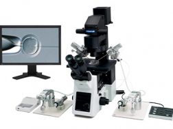
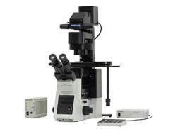
La inyección intracitoplasmática de espermatozoides (ICSI, por sus siglas en inglés) es una técnica de reproducción in vitro, en donde un solo espermatozoide es inyectado en el citoplasma de un oocito/ovocito usando una micropipeta. Nuestro sistema de microscopio invertido ayuda a mejorar la calidad de la ICSI, en donde la velocidad y precisión son aspectos esenciales. Gracias a su sistema óptico, desarrollado exclusivamente para la inyección intracitoplasmática de espermatozoides, el microscopio otorga una visualización clara del huso mitótico en un oocito/ovocito de metafase II a través de los oculares. Observar el huso mitótico ayuda a aumentar la precisión de la ICSI, ya que es posible confirmar la fase de maduración del ovocito y la posición de la inyección con la micropipeta, evitando así daños en el huso mitótico. La unidad motorizada también contribuye a acelerar el proceso de la ICSI a través de una operación más eficiente que ayuda a aliviar el estrés ante el cual se expone al ovocito. Unidades opcionales para la observación por contraste de interferencia diferencial (DIC) permiten a los investigadores usar la inyección intracitoplasmática de espermatozoides morfológicamente seleccionados (IMSI) para confirmar la forma, el tamaño y la cantidad de vacuolas presentes en la cabeza del espermatozoide.
The IX3-ICSI system is designed to facilitate the use of intracytoplasmic sperm injection (ICSI) as part of the in vitro fertilization (IFV) process. Olympus’ relief contrast method enables 3D oocyte observation in plastic dishes to check the zona pellucida condition. The IX3 semimotorized system has an ICSI-dedicated hand switch to instantly change the observation method and magnification with the simple touch of a button. Use the polarization method to observe the spindle and its
position to confirm oocyte maturity and avoid damage during injection and the differential interference contrast (DIC) method to perform intracytoplasmic morphologically selected sperm injection (IMSI).
Optimized for a smoother ICSI workflow
Easy oocyte condition checks through spindle visualization
Streamlined ICSI steps through motorized operation
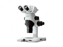
Image macro views of whole organisms to micro views of individual cell structures using the SZX16 research stereomicroscope. Its wide zoom ratio (16.4:1) enables magnifications of 7x–115x with a 1X objective and up to 230x with a 2X objective. Clearly observe fine details owing to the apochromatic optics, reducing chromatic blur for the entire magnification range, and its 0.3 numerical aperture (NA), providing a high 900 line pairs per mm resolution.
Advanced model offering darkfield, brightfield polarization, oblique, and advanced fluorescence observation
Ergonomically designed and provides ample space for manual and automated manipulation and injection tools
2 energy-efficient, long-life LED transmitted light illumination base options
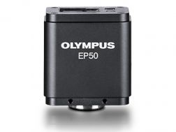
Promoting interactive learning, our wireless-enabled EP50 microscope digital camera converts a microscope into a wireless imaging system. Equipped with full stand-alone configuration capabilities, you can control the EP50 camera with a mobile device or PC and stream images to a monitor or projector via WLAN and HDMI.
Wireless digital imaging
Simultaneous direct output of WLAN and HDMI
Full stand-alone configuration available
Flexible camera control options for use with mobile devices, PCs, or direct streaming to a monitor/projector with the stand-alone setup
Enabling fast, easy capture of high-quality images that can be clearly observed on a large screen, the DP23 microscope digital camera eases routine life science and clinical research, conferencing, or teaching. Integrate it seamlessly into your microscopy workflow and easily share or stream images.
Share images using the DP23-AOU network solution
Clearly observe live images on a large screen
Fast, high-quality imaging for conferences and teaching