Evident LogoOlympus Logo

Regenerative Medicine

ライフサイエンスリサーチアプリケーション

Applications: Regenerative Medicine

再生医療は、幹細胞治療や組織工学などの最先端技術を活用して身体を修復するもので、怪我から変性疾患まで、さまざまな病状に対する革新的な解決策を提示しています。この分野では、顕微鏡が研究ワークフローの各段階で極めて重要な役割を果たします。当社の顕微鏡ソリューションは、幹細胞の培養、人工組織のモニタリング、綿密な細胞解析などの分野で、お客様の研究のお役に立てるよう設計されています。当社のインキュベーションモニタリングシステムで、細胞培養中の標準化と効率化を促進します。オールインワンデジタルイメージングシステムで、簡単に専門家品質の明視野および蛍光画像を取得できます。高速かつ精密な細胞や組織の動態解析には、当社のレーザー走査型顕微鏡およびスピニングディスク型共焦点超解像システムをご利用ください。

お客様のニーズに適した顕微鏡ワークフロー構築についてのご相談は、最寄りの当社代理店までお問い合わせください

再生医療研究向けソリューション―お客様に最適なシステムを構築

Successfully added visible items to your Bookmarks list

View Bookmarks

Microscope System/Frame

FV5000Show Description
FV5000
  • Extraordinary clarity, speed, and reliability driven by groundbreaking innovations 
  • SilVIR™ detectors deliver photon-level quantitation, exceptional sensitivity, and ultra-high signal-to-noise 
  • Unmatched dynamic range captures the full signal spectrum and prevents saturation 
  • High-speed 2K resonant scanning and high-density 8K galvo scanning in one platform  
  • FLUOVIEW Smart™ software simplifies operation with intuitive controls and AI-powered automation  
  • TruResolution™ auto correction collar optimizes focus for over 20 objectives 
  • Modular design supports up to 10 laser lines and future multiphoton upgrades 
  • Laser Power Monitor (LPM) ensures stable illumination and reproducible results over time 

FV5000

Confocal Laser Scanning Microscope
ChooseSelected  
APX100Show Description
APX100
  • Easy-to-use, all-in-one microscope system
  • Publication-quality images in a few clicks
  • Fast, efficient data management

APX100

Digital Imaging System
ChooseSelected  
FV4000Show Description
FV4000

  • Game-changing dynamic range for imaging from the macro scale to subcellular structures
  • Multiplex up to six channels simultaneously with TruSpectral technology
  • Redesigned high-speed, high-resolution scanners for fixed and live cell imaging
  • Improved depth and photosensitivity with pioneering NIR capabilities and renowned optics
  • Peace of mind with the reliable, repeatable SilVIR detector
  • Industry leading* ten laser lines with a broader spectral range from 405 nm to 785 nm

*As of October 2023.

FV4000

Confocal Laser Scanning Microscope
ChooseSelected  
IXplore IX83 ProShow Description
IXplore IX83 Pro

The IXplore Pro automated system acquires panoramic, multichannel images without a complex workflow. Easing experiment setup, the Graphical Experimental Manager offers fully automated multidimensional observation capabilities. The microscope’s high-precision ultrasonic stage, automated focusing, and bright, uniform fluorescence illumination facilitate image stitching applications.  

  • Automated multidimensional observation (X, Y, Z, T, wavelength, and positions)
  • Boost your statistics with multiwell plate screening
  • Acquire fluorescence panoramic images of large samples, such as brain slices
  • Create 3D optical sections and enhance resolution with TruSight™ deconvolution

IXplore IX83 Pro

Automated Microscope System
ChooseSelected  
IXplore IX83 SpinShow Description
IXplore IX83 Spin

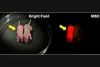
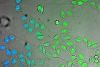
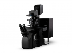
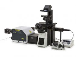
The IXplore Spin system features a spinning disk confocal unit that enables fast 3D image acquisition, a large field of view, and prolonged cell viability in time-lapse experiments. Researchers can use it to perform rapid 3D confocal imaging with high resolution and contrast at greater depths for imaging into thicker samples. The spinning disk also helps to cut down on photobleaching and phototoxicity of samples upon excitation.

  • Real-time controller (U-RTCE) helps optimize the device’s speed and precision during automated acquisition
  • TruFocus™ Z-drift compensation system maintains focus for each frame
  • Precise 3D imaging with improved light collection using X Line™ objectives
  • Upgrade to the IXplore SpinSR super resolution system as your research progresses

IXplore IX83 Spin

Confocal Imaging Microscope System
ChooseSelected  

Cell Culture Equipment

CM30Show Description
CM30

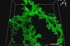
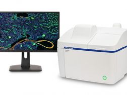
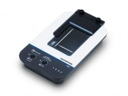
Remotely monitor, analyze, and share your cell cultures’ health, cell count, and confluency using the reliable quantitative data provided by the automated CM30 incubation monitoring system. The system enables label-free observation, reduces the risk of damage to your cultures, and standardizes your culture workflow.

  • Automatically collects quantitative data on the health and confluency of your cultures
  • Monitor, analyze, and share your cultures' progress remotely from a PC or tablet
  • Equipped with oblique epi-illumination for label-free observation

CM30

Incubation Monitoring System
ChooseSelected  
CKX53Show Description
CKX53

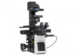
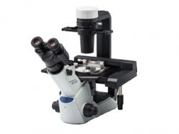
The compact, ergonomic CKX53 inverted microscope’s simple phase contrast system enables simple, high-contrast cell culture observation, accommodating a variety of containers. Its inversion contrast (IVC) technique offers clear pseudo-3D observation, easing workflows including live cell imaging, cell sampling and handling, and fluorescence.

  • Simple phase contrast with no centering needed
  • Inversion contrast (IVC) technique provides clear three-dimensional views
  • Fluorescence with a 3-position slider option

CKX53

A Compact, Ergonomic Cell Culture Solution
ChooseSelected  

このページはお住まいの地域ではご覧いただくことはできません。

Sorry, this page is not available in your country