Evident LogoOlympus Logo

Ask the Experts

Olympus Organoid Conference: Think Deep, See Deeper | 3-Day Virtual Event | September 7-9, 2021

Olympus Organoid Conference- Think Deep, See Deeper

From 7 – 9 September, learn from Olympus microscope users and technology partners on how they can support your scientific research. Cell Biologists, Microscopists and Image analysis experts will share their experiences and answer your questions.

Come Join Us! Register for free today!


Agenda

Time (GMT +8) Day 1
7 September 2021
Day 2
8 September 2021
Day 3
9 September 2021

1.30pm - 1.40pm

Opening Address by Mr. Tommy Martel
Division Manager
Scientific Solutions Business Division
Olympus Corporation of Asia Pacific Limited


Chairperson: Dr. Graham Wright
Acting Director
Research Support Centre (RSC) A*STAR, Singapore

Welcome Address by Mr. Kunihiro Idei
Managing Director
Scientific Solution Business Group, Olympus Sales and Service Co. LTD.


Chairperson: Dr. Anil Kumar PR (PhD)
Scientist In-charge
Division of Tissue Culture, Sree Chitra Tirunal Institute for Medical Sciences and Technology

Welcome Address by Dr. Kim Everuss
Sales Manager
SSBU Life Sciences
Olympus Australia


Chairperson: Dr. Richard De Mets
Research Fellow
Mechanobiology Institute, National University of Singapore

1.40pm - 2.25pm

Session 1
Culture and Quantitative 3D Imaging of Organoids: Challenges and Solutions

Presenter:
Dr. Anne Beghin

Assistant Professor, Research
Mechanobiology Institute, National University of Singapore
 

More information

Session 1
3D Microscopy: Understanding The Give and Take on Instrument Performance to Enable Informed Decisions

Presenter:
Dr. Graham Wright

Acting Director
Research Support Centre (RSC)
A*STAR, Singapore
 

More information

Session 1
3D Segmentation for Fluorescence Images: From Qualitative to Quantitative

Presenter:
Dr. Yu Weimiao

Head of Computational & Molecular Pathology Lab (CMPL)
Agency of Science, Technology and Research
 

More information

2.25pm - 2.55pm

Session 2
Converting from 2D to 3D: Bio-Techne Solutions for Your 3D Culture

Presenter:
Dr. Xiaotong Cui

Field Application Specialist
Bio-Techne
 

More information

Session 2
Advances in 3D Optical Imaging Technologies: An Overview

Presenter:
Mr. Srivats Hariharan

Manager
Application & Marketing
Olympus Singapore
 

More information

Session 2
An In Vitro System for Evaluating Anticancer Drugs Using Patient-Derived Tumor Organoids

Presenter:
Dr. Motoki Takagi

Professor
Medical-Industrial Translational Research Center
Fukushima Medical University
 

More information

2.55pm - 3.40pm

Session 3
Utilising Tumoroids to Explore Anti-Tumour Immunity in Rectal Cancer

Presenter:
Dr. Kasmira Wilson

Researcher
Peter MacCallum Cancer Foundation
 

More information

Session 3
Investigating Spheroid Architecture Using FV3000

Presenter:
Ms. Gency Gunasingh

Research Assistant
The University of Queensland Diamantina Institute
 

More information

Session 3
Study the Function of Stromal Cells Through Intestinal Organoid Co-Culture Technology 

Presenter:
Dr. Ningbo Wu

Associate Professor 
Shanghai Institute of Immunology, Shanghai Jiao Tong University School of Medicine
 

More information

3.40pm - 4.25pm

Session 4
Tissue Optical Clearing Imaging: From in Vitro to in Vivo

Presenter:
Dr. Dan Zhu

Professor
Wuhan National Laboratory for Optoelectronics
Huazhong University of Science and Technology, Wuhan, China
 

More information

Session 4
Prostate Cell Lineage Hierarchy and Plasticity

Presenter:
Dr. Dong Gao

Principal Investigator
Shanghai Institute of Biochemistry and Cell Biology, Chinese Academy of Sciences
 

More information

Session 4
NoviSight Demonstration: 3D Image Analysis and Statistical Software for Organoids and Spheroids

Presenter:
Mr. Hiroya Ishihara

Applications Scientist
Olympus Technologies Asia, Olympus Corporation
 

More information

4.25pm - 4.30pm

Closing Address

Closing Address

Closing Address


Speakers

Dr. Anne Beghin

Assistant Professor, Research
Mechanobiology Institute, National University of Singapore

Dr. Xiaotong Cui

Field Application Specialist
Bio-Techne

Dr. Anne Beghin

Biography

Dr. Anne Beghin is a multidisciplinary scientist with fifteen years of extensive research experience across academia and industry. She obtained her PhD in oncology in 2007 at the University Claude Bernard in Lyon (France). She then moved to optical microscopy at the Université de Lyon, where she established the microscopy platform and developed live cell imaging solutions and image analysis services for 4 years.

In 2011, she was recruited by a biotechnology company based in Bordeaux, where she spent 3 years in charge of a tissue analysis service: from biologic samples (whole tissue sections and Tissue Micro Arrays) to image acquisition and analysis with database establishment. She has been part of the Interdisciplinary Institute of NeuroScience IINS for 3 years where she successfully developed a new platform linking the High Content Screening (HCS) approach with super resolution microscopy such as Single Molecule Light Microscopy (HCS-SMLM), a collaboration with pharmaceutical company, Sanofi. Subsequently, she moved to the MechanoBiology Institute (MBI) in Singapore to study organoids using advanced imaging and HCS. This work has resulted in a patent and publications are on-going.

Abstract

Culture and Quantitative 3D Imaging of Organoids: Challenges and Solutions

Turning organoids into impactful translational models includes being able to culture them and assess those that develop robustly with physiologically relevant architecture. However, quantitative comparisons and statistical analysis at high content, which are mandatory to describe the complexity of such multicellular 3D objects are not possible owing to the lack of high-throughput 3D imaging methods. We have thus engineered a versatile High Content Screening (HCS) device to streamline all the steps of organoid culture to exploit its potential in morphogenesis understanding. Our approach comprises a new generation of versatile scaffolding cell culture multiwell chips with embedded optical components (= lighting JeWells) that enables rapid 3D imaging.

The device is fully compatible with classical imaging techniques such as brightfield, widefield, light sheet or confocal microscopy. The defined positioning of organoids allows correlations between all these different techniques without any loss of organoids. The high surface density of JeWells meets HCS standards: we can generate more than hundred organoids in a surface equivalent to a single well of a 384 wellplate. Our platform allows one to follow the morphogenesis of live organoids by non-toxic fluorescent live imaging based on light sheet microscopy over weeks (validated on hESC, hIPSC and primary cells). Moreover, the large number of 3D images can be used to train convolutional neural networks to precisely detect and quantify subcellular and multicellular features, such as mitotic and apoptotic events, multicellular structures (rosettes), and classify whole organoid morphologies. The combination of high-resolution 3D microscopy techniques with HCS and machine learning approaches allow us to quantitatively describe the morphogenesis of hundred living organoids correlated with phenotypic characterization to decipher mechanisms involved in human developmental biology, tissue pathology, and to enrich drug discovery pipelines.

Xiaotong Cui

Biography

Dr. Xiaotong Cui received his B.S. and M.Sc. from the University of Leicester, UK. He went on to receive aPh.D. from the Institute of Life Science, Kyoto University in 2018, where he worked under the direction of Prof. Osamu Takeuchi. From 2018-2020, he was a program-specific researcher in Shenzhen Digital Life Institute and ASHBi, Japan. Dr. Xiaotong has a solid background in molecular cell biology, immunology and he participated in one special program for Key Basic Research of Ministry of Science and Technology, China. Now, he serves as a Field Application Specialist in Bio-Techne China.

Abstract

Converting from 2D to 3D: Bio-Techne Solutions for Your 3D Culture

Organoid and three-dimensional (3D) cell culture are emerging as pivotal systems for understanding human organ development, modeling disease, screening for drug efficacy or toxicity, and investigating personalized medicine. Usually they are derived from primary tissue, embryonic stem cells (ESCs), or induced pluripotent stem cells (iPSCs), which are capable of self-renewal and differentiation.

This session will provide an overview of the methods to establish an organoid cell culture in vitro and how organoids can be used as a physiologically relevant method for disease modeling, drug screening and biobank establishment. We will highlight our available and fully optimized applications for organoid cell culture that enable researchers to focus efforts on addressing their scientific questions quickly and precisely.

Dr. Kasmira Wilson

Researcher
Peter MacCallum Cancer Foundation

Dr. Dan Zhu

Professor
Wuhan National Laboratory for Optoelectronics 
Huazhong University of Science and Technology, Wuhan, China

Dr. Kasmira Wilson

Biography

Dr. Kasmira Wilson is a general surgeon and CSSANZ trainee, having previously completed a BSc(Hons) and MBBS. She is currently undertaking a PhD through the University of Melbourne at Peter MacCallum cancer centre which focuses on translational research in rectal cancer. Her research utilises tumouroid models to explore anti-tumour immunity in rectal cancer in a novel functional cytotoxic assay.

Abstract

Utilising Tumoroids to Explore Anti-Tumour Immunity in Rectal Cancer

Dr. Kasmira Wilson is a general surgeon and CSSANZ trainee, having previously completed a BSc(Hons) and MBBS. She is currently undertaking a PhD through the University of Melbourne at Peter MacCallum cancer centre which focuses on translational research in rectal cancer. Her research utilises tumouroid models to explore anti-tumour immunity in rectal cancer in a novel functional cytotoxic assay.

Globally colorectal cancer is a significant public health burden. It is the third most commonly diagnosed cancer and fourth leading cause of cancer related deaths in the world. A subset of patients diagnosed with rectal cancer require neoadjuvant chemoradiotherapy (NACRT) prior to surgery. However, there is a spectrum of response to this therapy with only 10-20% of patients achieving a complete pathological response. In addition, 20-40% of patients will demonstrate no response to this treatment. There is currently no method that predicts how a patient will respond to NACRT accurately. In order to investigate the mechanisms underpinning how patients respond to therapy, patient derived tumouroids have been utilised. These personalised in vitro three-dimensional tumour models recapitulate the in vivo tumour of origin genotypically. The Ramsay laboratory (Peter MacCallum, Melbourne) has successfully co-cultured patient-matched rectal cancer tumouroids with tumour infiltrating lymphocytes (TILS) in a novel in vitro assay, preliminary data generated suggests this assay has the ability to predict the response of a patient to treatment with NACRT prior to instigation of neo-adjuvant therapy. This assay provides a pre-clinical platform that encapsulates the hosts immune response toward their tumour. However, manual analysis of the data generated from this assay is time consuming and limits the clinical utility of this platform. Machine-based learning to develop artificial neural networks capable of analysing data produced from the killing assay has been developed to automate analysis. Automated analysis utilising artificial neural networks is a feasible approach to expedite the processing of data generated from the cytotoxic killing assays and will improve the clinical utility of this platform to direct personalised patient therapy.

Dr. Zhu Dan

Biography

Dr. Dan Zhu is a professor of Huazhong University of Science & Technology, and Vice-Director of Wuhan National Laboratory for Optoelectronics. Her research interests mainly focus on tissue optical clearing imaging and applications. She is the pioneer in the field of in vivo tissue optical clearing, and also developed fast, label-compatible in vitro optical clearing methods. She has authored more than 150 papers including Science Advances, Nature Communications, et al. She is also Fellow of SPIE, and Secretary General & Vice President of Biomedical Photonics Committee of Chinese Optical Society. She serves for journals as editorial member or guest editor, including Biomedical Optics Express, Journal of Biomedical Optics, Scientific Reports, Journal of Innovative Optical Health Sciences, Frontier of Optoelectronics et al.

Abstract

Tissue Optical Clearing Imaging: From in Vitro to in Vivo

Biomedical optical Imaging, as a powerful tool has been applied for observing biomedical tissue structural and functional information with high resolution and contrast unattainable by any other method. However, the high scattering of turbid biological tissues limits the penetration of light, leading to strongly decreased imaging resolution and contrast as light propagates deeper into the tissue. Fortunately, novel tissue optical clearing technique provide a way for reducing the scattering of tissue and improving the optical imaging quality. This presentation will introduce our progress from in vitro and in vivo of tissue optical clearing imaging, including developing in vitro optical clearing methods, such as FDISCO and MACS. Meanwhile, we will also demonstrate in vivo skull/skin optical clearing window for imaging structural and functional of cutaneous / cortical vascular and cells, also manipulating cortical vasculature.

Dr. Graham Wright

Acting Director
Research Support Centre (RSC)
A*STAR, Singapore

Mr. Srivats Hariharan

Manager
Application & Marketing
Olympus Singapore

Graham Wright

Biography

Dr. Graham Wright holds an interdisciplinary PhD in cell biology and physics from The University of Edinburgh and an MBA from the Warwick Business School at The University of Warwick. He is the Acting Director of A*STAR’s Research Support Centre (RSC) and the Director of the A*STAR Microscopy Platform (AMP). RSC offers everything from on-demand access to sophisticated scientific instruments and services, located within varied Technology Platforms, to a research consumables webstore, playing a crucial role in powering biomedical innovation in Singapore.

Dr. Graham has extensive experience, and a strong publication record, in applying advanced light microscopy to a wide range of biomedical research projects. Outside the laboratory, he is committed to science outreach and has featured as a judge on MediaCorp’s National Science Challenge TV show, presented a TEDx talk, had microscopy images displayed on the big screen in Times Square, New York and exhibited work at the National Museum of Singapore.

Abstract

3D Microscopy: Understanding The Give and Take on Instrument Performance to Enable Informed Decisions

Biologists have a significant toolbox at their disposal when it comes to microscopically imaging 3D samples, such as organoids. From widefield microscopy to confocal, superresolution, multiphoton and lightsheet, each have their own set of pros and cons that must be carefully considered before making an informed choice on the most suitable to address your biological question. Often a correlative approach is required, applying several techniques to address the question from different perspectives. It is also crucial to consider the method of sample preparation and optimise each of the potential steps which can include fixation, permeabilisation, labelling and mounting. Further, the images generated by all techniques can be enhanced with post-processing techniques, such as deconvolution, which can enable or help to improve subsequent image analysis and interpretation.

This presentation aims to introduce each of the microscopy techniques that may be applicable for imaging 3D samples and highlight their relative performance attributes in terms of sample viability, speed, resolution, contrast and depth penetration – highlighting that each technique and instrument will come with trade-offs between these parameters1.

1 Jonkman, Brown, Wright, Anderson & North (2020) Tutorial: guidance for quantitative confocal microscopy. Nature Protocols 15:1585–1611

Srivats Hariharan

Biography

Mr. Srivats Hariharan is an Applications & Marketing Manager in Olympus life science team in the Asia Pacific region. He holds a bachelor’s degree in Mechanical Engineering from Nanyang Technological University, Singapore and has experience working in biomedical research labs and A*STAR Microscopy Core Facility where he supported researchers on confocal and live cell imaging technologies and help setup single molecule super-resolution and light sheet microscopes. He joined the life-science team of Olympus Singapore in 2011 as Product Manager and in-charge of supporting research customers and business partners in South-East Asia and Taiwan.

Abstract

Advances in 3D Optical Imaging Technologies: An Overview

With rapid development in fluorescent proteins, synthetic fluorochromes, and digital imaging, advanced 3D imaging technologies are now available to investigators to provide critical insights into the fundamental nature of cellular and tissue functions. 3D and 4D imaging systems have become very common tools among biologists. However, there are several technical challenges and limitations in performing successful 3D and 4D imaging. Olympus has developed a wide range of 3D imaging microscopes to overcome these challenges and to satisfy the requirements of researchers across different disciplines.
In this talk, we will be taking you through the different imaging modalities that Olympus offers including widefield based fluorescence microscope like the Olympus IXplore and how in combination with image processing tools like Olympus TruSight can render usable 3D data sets. 
However, more sophisticated 3D imaging systems are required for imaging samples such as organoids and spheroids. We will therefore illustrate the importance of laser-based systems that have been effectively used for deeper imaging into samples, like the Olympus FV3000 laser scanning confocal microscope, IXplore SPIN spinning disk confocal microscope and finally the FVMPE-RS multiphoton-excitation microscope. Along with these, we will stress the importance of high-quality objective lenses optimised for 3D imaging.

Ms. Gency Gunasingh

Research Assistant
The University of Queensland Diamantina Institute

Dr. Dong Gao

Principal Investigator
Shanghai Institute of Biochemistry and Cell Biology, Chinese Academy of Sciences

Gency Gunasingh

Biography

Ms. Gency Gunasingh completed her Master of biotechnology degree from University of Queensland in 2012 and did her Masters project under Dr. Andrew Prowse and Prof. Peter Gray at Australian Institute for Bioengineering and Nanotechnology. Her primary area of research was large scale generation of cardiac progenitors from embryonic stem cells in 3D. She then worked under Prof. Brian Gabrielli at UQ Diamantina institute on developing 3D tumour spheres in melanoma for in vitro drug testing. She currently works for Prof. Nikolas Haass at UQDI on understanding tumour heterogeneity and tumour architecture in melanoma spheroids.

Abstract

Investigating Spheroid Architecture Using FV3000

Phenotypic tumour heterogeneity arising due to differentially cycling cell populations has been implicated in increased therapy resistance. This phenomenon cannot be assessed in adherent cell culture, where microenvironmental conditions are homogeneous. Thus, we utilise melanoma spheroids to model the 3D tumour microenvironment including the extracellular matrix (ECM) and study spheroid structure, necrotic region, individual cell arrangement within and gene expression patterns. We achieve this by exploiting the fluorescence ubiquitination cell cycle indicator (FUCCI) system to monitor cell cycle stages as a surrogate marker for phenotypic tumour heterogeneity, tissue clearing and confocal microscopy using FV3000.

Dr. Dong Gao

Biography

Dr. Dong Gao is the Principal Investigator of Shanghai Institute of Biochemistry and Cell Biology, Chinese Academy of Sciences. His research interests mainly focus on t prostate adult stem cell and the establishment of patients-derived cancer organoid biobank. He has authored more than 40 papers including Cell, Nature Genetics, Cell Stem Cell et al.

Abstract

Prostate Cell Lineage Hierarchy and Plasticity

Prostate cancer is one of the most common cancers worldwide and also the second leading cause of cancer-related death in males in Western countries. Although the majority of human primary prostate cancers have a luminal phenotype, both basal cells and luminal cells can serve as cellular origins of prostate cancer in model systems. However, the stem cell-like plasticity of defined prostate epithelial cells and the cellular origin of prostate cancer under physiological conditions have not been identified. Recently, prostate basal and luminal cell populations were both shown to be self-sustaining, and both cell types could initiate prostate cancer. However, the oncogenic transformation of basal cells requires basal to luminal cell transition. In addition, luminal cells were shown to have greater tendency to be the cells of origin for prostate cancer in some contexts. 

These studies highlight the need to characterize the prostate cell lineage hierarchy and their behaviors in various contexts. We are studying the prostate cell lineage hierarchy using single-cell RNA sequencing, cell lineage tracing and organoid culture technologies. We have identified a novel prostate luminal progenitor cell population (termed as Luminal-C) which located at the prostate the distal prostate invagination tips (termed as Dist-Luminal-C) and proximal prostate region (termed as Prox-Luminal-C). Furthermore, we are interested in prostate cell lineage plasticity at the different stages of prostate tumor initiation, progression, and therapy resistance. We have identified defined ERG as a master transcription factor to regulate prostate luminal lineage through orchestrating chromatin interactions.

Dr. Yu Weimiao

Head of Computational & Molecular Pathology Lab (CMPL)
Agency of Science, Technology and Research

Dr. Motoki Takagi

Professor
Medical-Industrial Translational Research Center
Fukushima Medical University

Dr. Yu Weimiao

Biography

Dr. Yu Weimiao obtained his Ph.D. from the National University of Singapore (NUS) in 2007, majoring in image processing and machine vision. He joined the Agency of Science, Technology and Research (A*STAR) in 2007. He is currently heading Computational Digital Pathology Lab (CMPL) in BII to deepen and extend the R&D with clinical and industrial partners. He is also the joint PI in IMCB leading the Computational & Molecular Pathology Lab (CMPL). His research interests are Computational Biomedical Image Analysis and Quantitative Imaging Informatics. He applied 3D image analysis solution to segment and tracking the cells in 3D to understand the developmental problem and collective cell migration mechanisms. His work was published in Nature Communication, Current biology, Nature Cell Biology, etc. To enhance the applications in clinical diagnosis/prognosis, he co-founded a biotech company, known as A!maginostic Pte. Ltd. He established a world-class joint platform for the immunodiagnosis at the tissue level. The platform allows the researchers, clinicians, and pharma to profile the patient immune signature for diagnosis, prognosis, and drug response study.

Abstract

3D Segmentation for Fluorescence Images: From Qualitative to Quantitative

Cells are 3D functional elements in biology science and they are actively moving to perform their functions. Collective cell migration is appreciated as an important model for the understanding of the mechanism governing the cell movement in Vivo and in Vitro. It is a highly kinetic process involved in immune response, wound healing, tissue development and cancer metastasis. Recent decades have seen the fast development of various optical imaging techniques with excellent spatial-temporal resolution, dimensionality and scale. The generation of novel probes have also allowed us to acquire the movies of migrating cells with specific proteins/molecules. However, we lack of advanced solution to analyse such high-content and highly dynamic images/videos.

 In this talk, we will review some important components for 3D image analysis using two cell migration problems:  I). cancer B cell migrating in given Chemokine gradient; and II). border cell cluster migrating in Drosophila egg chamber. The extracted quantitative information provided us new insight on how a cluster of cells coordinates and collectively moves to the target.

Dr. Motoki Takagi

Biography

Dr. Motoki Takagi received PhD from Graduate School of Agriculture and Life Sciences, the University of Tokyo in 2001. Then he continued his research as a postdoctoral fellow at Institute of Molecular and Cellular Biosciences, the University of Tokyo. He was engaged in the research of nucleic acid drugs at Genencare Research Institute, Co. Ltd. since 2002. Since 2006, he conducted drug discovery research at Biological Systems Control Team, Biomedicinal Information Research Center, Japan Biological Informatics Consortium. Since 2012, he has been researching chemical biology as an associate professor at Fukushima Medical University and became a professor in 2014.

Abstract

An In Vitro System for Evaluating Anticancer Drugs Using Patient-Derived Tumor Organoids

Patient-derived tumor organoids (PDOs) represent a promising preclinical cancer model that better replicates disease, compared with traditional cell culture models. We have established a novel series of patient-derived tumor organoids (PDOs) from various types of tumor tissues from the Fukushima Translational Research Project, which are designated as Fukushima (F)-PDOs. F-PDOs could be cultured for >6 months and formed cell clusters with similar morphologies to their source tumors. Comparative histological and comprehensive gene-expression analyses also demonstrated that the characteristics of PDOs were similar to those of their source tumors, even following long-term expansion in culture. In addition, suitable high-throughput assay systems were constructed for each F-PDO in 96- and 384-well plate formats.

This presentation represents the characteristics of F-PDOs and an in vitro evaluation of different classes of anticancer drugs, including chemotherapeutic, molecular targeted, antibody and immunotherapeutic drugs, using F-PDOs. We firstly evaluated chemotherapeutic drugs such as paclitaxel and carboplatinand, and molecular targeted drugs such as epidermal growth factor receptor (EGFR) inhibitors using a suitable high-throughput assay system. In addition, an evaluation system for the antibody-dependent cellular cytotoxic activity of anti-EGFR antibody and the cytotoxic activity of activated lymphocytes, such as cytotoxic T lymphocytes and natural killer cells, was constructed. Moreover, an evaluation system was developed for the immune checkpoint inhibitors, nivolumab and pembrolizumab, using F-PDOs. Our results demonstrate that the in vitro assay systems using F-PDOs were suitable for evaluating different classes of anticancer drugs under conditions that better reflect pathological conditions. In addition, our results indicated that F-PDOs have structural characteristics enabling the construction of an evaluation system for anticancer drugs, based on structural changes of F-PDO found with a 3D cell-analysis system, demonstrating that 3D cell analysis is a powerful tool for analyzing the characteristics of PDOs.

Dr. Ningbo Wu

Associate Professor
Shanghai Institute of Immunology, Shanghai Jiao Tong University School of Medicine

Mr. Hiroya Ishihara

Applications Scientist
Olympus Technologies Asia, Olympus Corporation

Dr. Ningbo Wu

Biography

Dr. Ningbo Wu is an Associate Professor at Shanghai Institute of immunology, Shanghai Jiaotong University School of Medicine. His research interests mainly focus on the function of intestinal stromal cells in intestinal homeostasis and related work was published in Nature and Science CHINA Life Sciences, et al.

Abstract

Study the Function of Stromal Cells Through Intestinal Organoid Co-Culture Technology 

For a long period of time, intestinal mesenchymal stromal cells have been considered as a relatively simple and homogeneous group of cells. With the help of single cell transcriptomics studies, it has now been clear that these cells are quite complex and heterogeneous. However, the detailed cellular and molecular mechanisms that regulate the function of these cells remains poorly understood. Therefore, the ability to perturb and evaluate the function of these stromal cells is critical to the understanding of intestinal stem cell niche and the etiology of the inflammatory bowel diseases and colitis associated colorectal cancer.

This presentation will introduce our work recently published in Nature, which identified a new subset of intestinal stromal cell by combining single-cell sequencing technology and intestinal organoid co-culture technology.

Hiroya Ishihara

Biography

Mr. Hiroya Ishihara is an Application Scientist at Olympus. He was studying the epigenetic factors involved in plant regeneration using omics and microscopy in Tokyo University of Science. Confocal and two-photon microscopy were his trusted partner at that time. Therefore, he joined Olympus to make life science more exciting with microscopes. Currently, he is working on a wide range of projects from basic research to product/sales strategy.

Abstract

NoviSight Demonstration: 3D Image Analysis and Statistical Software for Organoids and Spheroids

Three-dimensional cell culture models such as patient-derived organoids (PDO) and spheroids have increased in popularity because they can provide a 3D microenvironment that more closely reproduces in vivo conditions compared to 2D monolayer culture. Phenotypic and functional heterogeneity arise among cancer cells within the same tumor because of genetic change, environmental differences and reversible changes in cell properties. Therefore, evaluation of cell-specific responses is important for accurate prediction of drug efficacy and kinetics in vivo.

Microscopic imaging technique such as confocal microscopy is promising to monitor the cell-specific responses at higher spatial resolution. Here, we introduce a novel 3D cell analysis software “NoviSight”. “NoviSight” recognizes 3D objects based on fluorescence intensity and analyzes them based on various information such as their sizes, the cell number, viability, and subcellular features contained in the recognized objects. In this webinar, we will introduce a case study of the analysis of patient-derived cancer organoids and spheroids using NoviSight. Then, we will demonstrate NoviSight software.

Three-dimensional high-throughput cell analysis platform represents the first step toward the development of tools for understanding the pharmacological mechanisms of drugs or drug targets in the preclinical tissue models. We hope that our platform will provide invaluable information for the study of basic research, drug efficacy, dosage, pharmacology and ADMET before clinical phases of drug development.


Related Products

Light Sheet Microscope

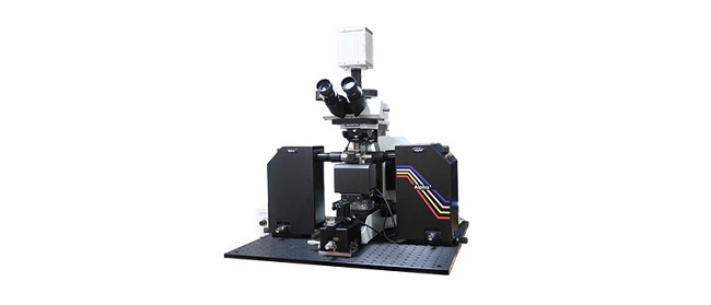
Alpha3

The Alpha3 advanced fluorescence light sheet microscope provides researchers with a flexible, high-performing light sheet solution. Using smart optical focus sweeping, it acquires uniform, artifact-free imaging across the entire field of view. Easily upgrade the Alpha3 system to meet your specific needs by selecting from options such as Smart 3D scanning, XY tiling, or temperature controls, based on your goals.

  • Multidirectional light sheet with real-time focus sweeping for rapid, artifact-free 3D imaging
  • Compatible with a large selection of high-quality Olympus optics
  • Accessories support a variety of specimens and are resistant to organic solvents or aqueous buffers
  • User-friendly QtSPIM software and workstation enables seamless 3D image acquisition at maximum speed
Incubation Monitoring System

CM20

Remotely monitor, analyze, and share your cell cultures’ health, cell count, and confluency using the reliable quantitative data provided by the automated CM20 incubation monitoring system. The system enables label-free observation, reduces the risk of damage to your cultures, and standardizes your culture workflow.

  • Automatically collects quantitative data on the health and confluency of your cultures
  • Monitor, analyze, and share your cultures' progress remotely from a PC or tablet
  • Equipped with oblique epi-illumination for label-free observation
Confocal Laser Scanning Microscope

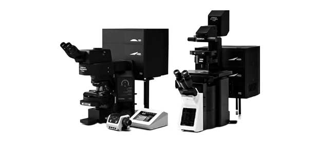
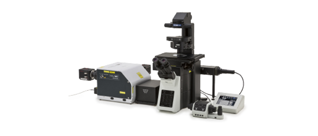
FV3000

  • Available with either galvanometer-only (FV3000) or hybrid galvanometer/resonant (FV3000RS) scanner configurations
  • Highly efficient and accurate TruSpectral detection on all channels
  • Optimized for live cell imaging with high sensitivity and low phototoxicity
  • Inverted and upright frame options to suit a variety of applications and sample types
Multiphoton Laser Scanning Microscope

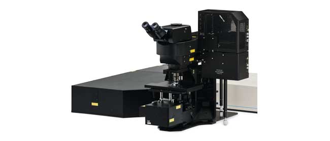
FVMPE-RS

  • Maximized resolution and contrast in deep imaging with TruResolution objectives
  • High-speed imaging with resonant scanner
  • Extended IR multiphoton excitation up to 1300 nm
  • Triple scanner option for multiphoton and visible light laser stimulation
  • Highly efficient transmission with 1600 nm optical coatings on multiphoton-dedicated objectives and scanning unit
  • 4-axis auto-alignment of IR laser beams
Confocal Imaging of Rapid Cell Dynamics

IXplore Spin (Inactive)

  • Rapid and high-resolution confocal imaging with a spinning disk system
  • 3D confocal time-lapse imaging of live cells with less phototoxicity and bleaching
  • Precise 3D imaging with improved light collection using silicone oil immersion objectives
  • Upgrade to the IXplore SpinSR super resolution system depending on your research progress and/or budget
3D Cell Analysis Software

NoviSight

NoviSight 3D cell analysis software provides statistical data for spheroids and 3D objects in microplate-based experiments. Use it to quantify cell activity in 3D, easily capture rare cell events, obtain accurate cell counts, and improve detection sensitivity. NoviSight software works with a range of imaging techniques, including point-scan confocal imaging, two-photon imaging, spinning disk confocal imaging, and super resolution live cell imaging.

  • Fast 3D image recognition from whole structures to subcellular features
  • Accurate statistical analysis
  • Equipped with a variety of ready-to-use default assays or easily design your own
Olympus Organoid Conference: Think Deep, See Deeper | 3-Day Virtual Event | September 7-9, 2021Dec 03 2024
Sorry, this page is not available in your country
Ask the Expert Sign-up

By clicking subscribe you are agreeing to our privacy policy which can be found here.

Sorry, this page is not
available in your country.

Sorry, this page is not available in your country