Not Available in Your Country
Sorry, this page is not
available in your country.
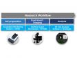
Overview
 | Insightful Analysis, Intelligent AnswersNoviSight 3D cell analysis software advances your discovery by providing statistical data for spheroids and other 3D objects in microplate-based experiments. The software enables you to quantify cell activity in three dimensions and more easily capture rare cell events, obtain accurate cell counts, and improve detection sensitivity. With a convenient user interface, NoviSight software offers the tools you need for recognition, analysis, and statistics. |
|---|
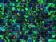
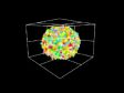
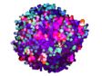
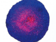
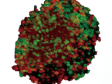
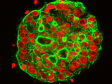
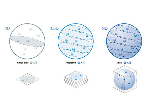
Accurate 3D Cell Analysis ResultsNoviSight software’s True 3D technology makes it easier to check the morphology of your samples. Measure a range of spheroid or cell nuclei parameters, including volume and sphericity, and measure and analyze physiologically relevant 3D cell models to speed up your research work.
|
|
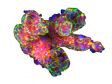
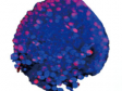
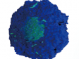
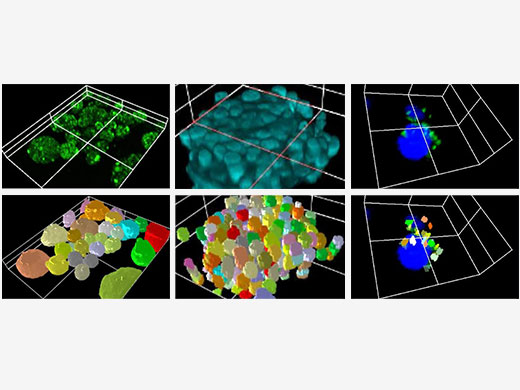
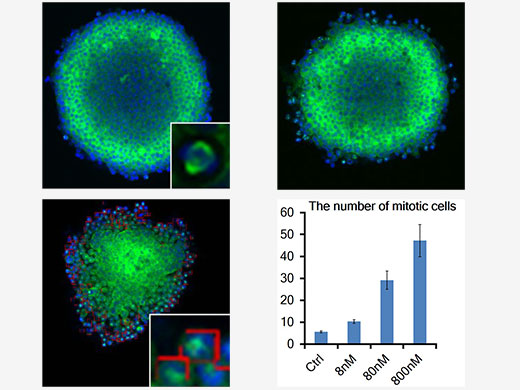
| Fast, Precise Object DetectionThe software can analyze objects of interest to provide morphology and spatiotemporal parameters in 3D space. Detect objects from whole structures to subcellular features and evaluate:
|
|---|
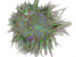
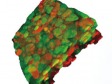
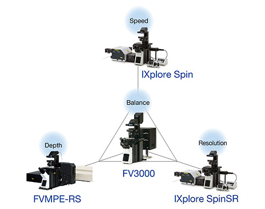
Works with Many Imaging TechniquesThe software is compatible with all Olympus confocal imaging systems. No matter which system you use, the software will quantify and analyze the objects you capture.
|
|
|---|
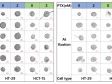
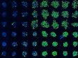
Ready-to-Use 3D Cell-Based AssaysReduce Your Assay Development TimeThe software comes with a variety of ready-to-use 3D cell assays. Simply choose the assay that suits your experiment and begin analyzing. You can also automate sequential processes for multiple data sets and assays. Easily Design Assays for Advanced 3D Cell AnalysisIf a suitable assay isn’t readily available, you can design your own. Easily create assays for multiwell, multichannel, and time-lapse experiments. |
|---|
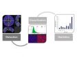
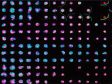
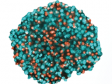
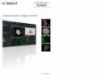
Fast Interactive 3D Cell AnalysisData and Images Are Always ConnectedNoviSight software’s accurate cell detection enables you to graph objects on a scatter plot or histogram. All the data are interactive—display your results in an image gallery, heat map, or table. Clicking a point in any of these display options shows the sample’s corresponding image. |
|---|
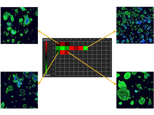
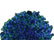
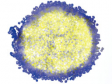
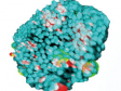
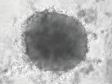
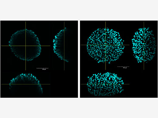
Image and Analyze Inside SpheroidsTreat your spheroids with a clearing reagent, such as Scale-S, and use NoviSight software to analyze information in your 3D sample from deep below the surface. |
|---|
Need assistance? |
Not for clinical diagnostic use. |
Specifications
Specifications |
| Image format | OIR format, VSI format |
|---|---|
| Applicable containers | Microplate :6, 12, 24, 48, 96, 384 wells |
| Save format | Dedicated (oxaf), FCS, and CSV |
| Convertible image format | TIFF |
| Image view | 2D view (single/three sides/MIP) |
| 3D view (isosurface/MIP/alpha blend) | |
| Graph view | Histogram, scattergram |
| Analysis/statistics | Various morphological measurements, table view, gating, gallery |
| Options | Recognition, measurement, statistics |
Equipment required for installation and operation |
| PC | HP Z4G4 or successor model |
|---|---|
| CPU | Intel Xeon CPU W-2123 (3.6 GHz) |
| Memory | 64 GB or more |
| HDD space | 2 TB or more |
| Graphics board | 1920 × 1080 monitor resolution with a 32-bit-video card |
| Drive | DVD-ROM drive |
| OS | Microsoft Windows 10 Pro for workstations, 64-bit English |
| .Net framework | .NET framework 4.7.0 or later (with Windows 10, as this is pre-installed, it is not necessary to install it with Installer) |
| Web browser | Internet Explorer 11 |
| Monitor | 22 inch (55.9 cm) |
| Network cable | Cable applicable to 10GBASE-T |