Advanced microscopy is more than a window into cells—it’s a way to uncover the hidden decisions that shape life at the cellular level. We spoke with Dr. Kazuhiro Aoki of the Graduate School of Biostudies at Kyoto University, whose work explores the mechanisms of cell fate decision: how cells process information and choose their path forward.
After using the IXplore™ IX85 motorized inverted microscope platform, Dr. Aoki shared his impressions on how the system supports his current research, future goals, and deeper understanding of cellular behavior.
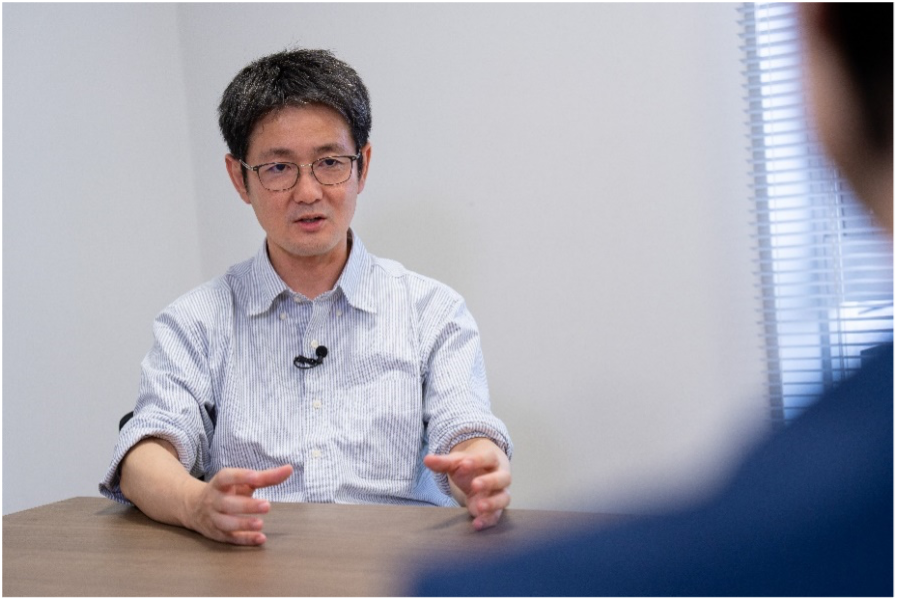
About Dr. Kazuhiro Aoki
Dr. Kazuhiro Aoki obtained his PhD from the Graduate School of Medicine at Osaka University. After engaging in research and education at the National Institutes of Natural Sciences, he is now a professor at the Graduate School of Biostudies at Kyoto University. Dr. Aoki's research lies in the interdisciplinary field of cell biology and systems biology. He is particularly focused on visualizing and quantifying information processing and signal transduction dynamics at the single-cell level with fluorescence imaging, optogenetics, and biosensor technologies.
Q: What research are you currently focusing on?
Dr. Aoki: I am currently focusing on the mechanisms of cell fate decision: how cells receive various stimuli from their external environment, such as temperature and nutrients, process that information within the cells, and decide cell fate, such as proliferation, differentiation, or death.
Even when cells look the same, each one has its own individuality and heterogeneity. Conventional research methods typically involve collecting about a million cells, lysing them, and studying the average value. This method, however, makes it impossible to grasp the individuality of each cell. To capture these differences caused by cellular individuality, we use a microscope to observe each cell one by one.
The strong desire to "see the individuality of cells" is my starting point. The methods I use are difficult and time-consuming, but that desire is what motivates me to continue my research.
Q: What led you to focus your research on the mechanisms of cell fate decision?
Dr. Aoki: Originally, I was interested in cancer signal transduction and was trying to reconstruct the cancer signaling pathways happening inside cells on a computer. By combining experiments and computer simulations, we made predictions and verified them. However, when we tried to observe the cellular phenotype—that is, how cells actually behave—we found that a single computer simulation model could not explain many aspects.
Even under the same conditions, some cells would divide while others wouldn't, and some would move while others remained static. Even for cancer-infiltrating cells, the infiltration often occurs not in individual cells but as a collective movement. I became increasingly interested in questions like: "How do cells behave as a collective?" and "Who is the leader, and who is the follower?" My strong desire to use microscope imaging to observe and research these questions led to my interest in the mechanisms of cell fate decision.
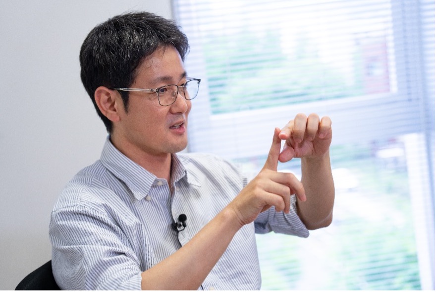
Dr. Kazuhiro Aoki, Professor at the Graduate School of Biostudies at Kyoto University.
Q: What do you value most when conducting experiments with a microscope?
Dr. Aoki: Cellular individuality and heterogeneity cannot be understood without directly looking at them with a microscope. That's why I place great importance on meticulously observing each and every cell under the microscope.
Since this can often become a vast and painstaking task, the ease of use of the microscope is also extremely important. While performance is of course crucial, I also value an intuitive user interface, such as a software layout that fits our specific style.
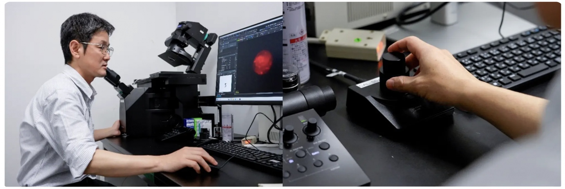
Dr. Kazuhiro Aoki uses the IXplore IX85 inverted microscope to explore the mechanisms of cell fate decision.
Q: What aspects of the new IXplore™ IX85 inverted microscope were effective in your experiments?
Dr. Aoki: The IXplore IX85 improved our throughput because of its wide field of view, which allows us to observe more cells at once. Previously, we could only image the center of the field of view, so the efficiency and amount of information were incomparable to now. The ability to acquire high-resolution image data over a wide field of view at once is extremely effective for microscopy-based research.
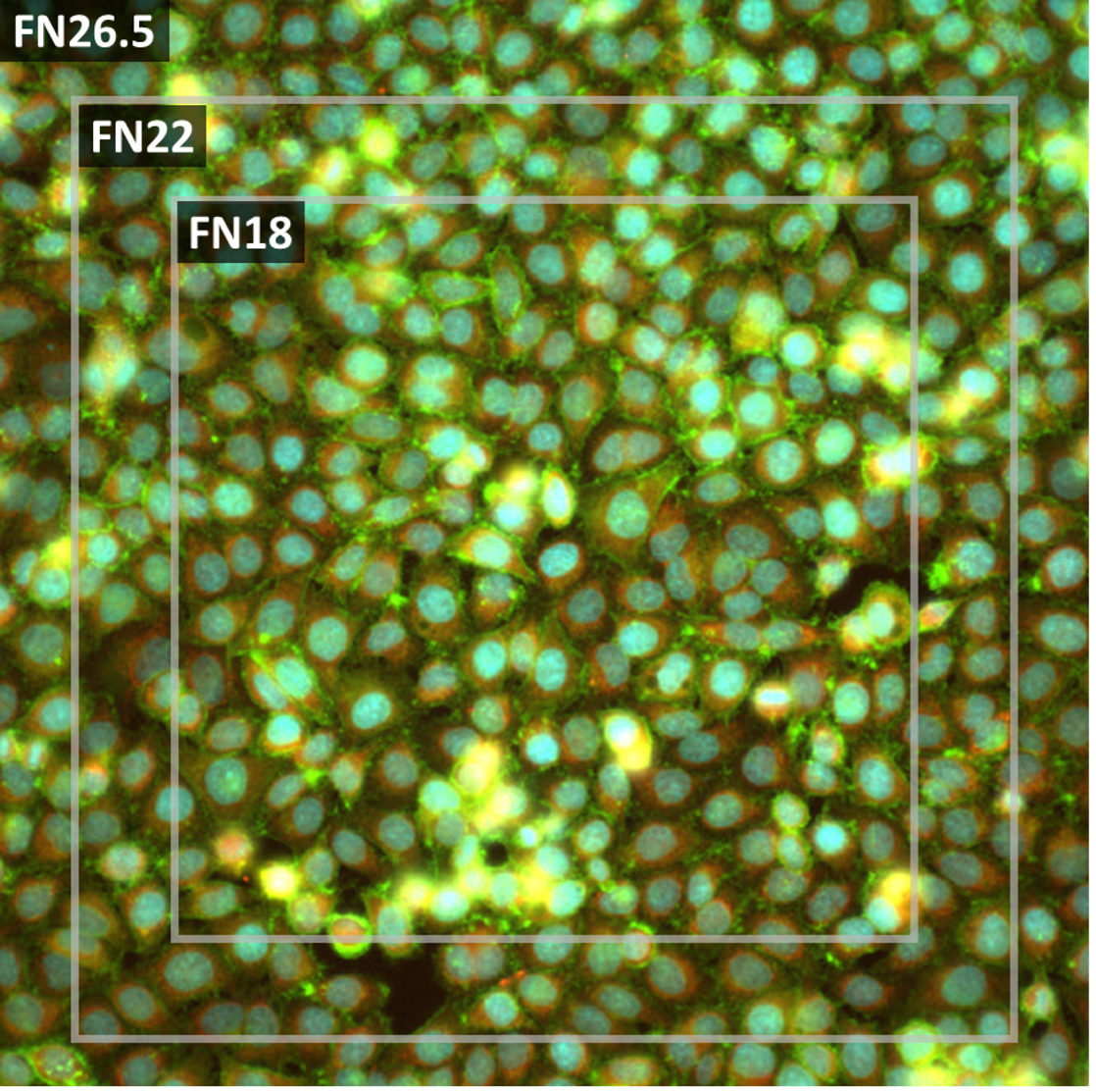
Fluorescence image of cultured HeLa* cells. The field of view of FN26.5, FN22, and FN18.
*To learn more about the origin of HeLa cells, visit henriettalacksfoundation.org.
The Intelligent Shading Correction feature for image stitching is also very useful. When performing experiments to observe the process of wound healing in cultured epithelial cells, we previously had to manually take dozens of images and stitch them together. At that time, the light intensity would drop at the periphery of the images, causing unevenness when stitched, which we had to correct manually. The IX85 automatically corrects for uneven illumination, which is a huge benefit for researchers working with large samples or tissues.
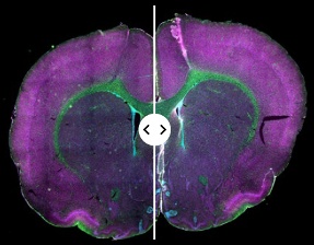
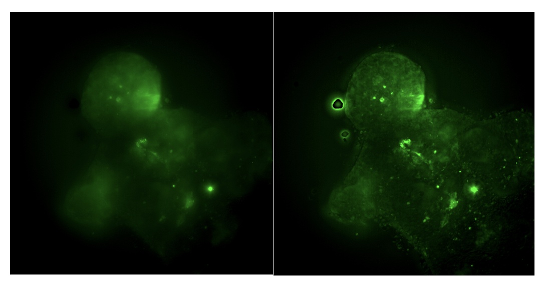
Fluorescence image of a mouse brain slice.
Left: Original stitched image.
Right: Stitched image with Intelligent Shading Correction applied.
Sample provided by EnCor Biotechnology Inc.
In addition to these points, I felt that the microscope was excellent in terms of image quality. Even with cancer organoids that have weak fluorescence expression, applying 3D deconvolution to the acquired images allowed us to clearly visualize intracellular aggregates. I was surprised at how much information we could extract from such dim fluorescent samples.
Fluorescence image of an organoid (GFP).
Left: Original stitched image.
Right: After 3D deconvolution
Images courtesy of Dr. Kazuhiro Aoki.
Q: How did the LUPLAPO25XS objective lens, which uses a silicone gel pad, help with your experiments?
Dr. Aoki: The 25X magnification of the objective is a perfect match for observing the deep tissues—cancer organoids and cysts—that we study. Furthermore, its design, which features a long working distance and suppresses index mismatch with the specimen, makes it a highly useful objective because it allows for clear observation of deep areas of the sample that were previously unobservable.
Additionally, the silicone gel pad, which doesn't use oil for immersion, is a significant advantage for microscope imaging experiments. For example, when searching for samples in a 96-well plate, it's difficult to know where spherical structures, such as our organoids or cysts, are located. This requires a workflow of first searching at low magnification and then observing at high magnification. When using a high-magnification oil immersion objective, you must be careful with the process of wiping and dropping oil, and there's also the risk of introducing bubbles. In contrast, using the silicone gel pad allows for a smooth switch from low to high magnification without contaminating the sample, which significantly streamlines the entire workflow.
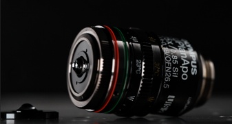
Evident’s LUPLAPO25XS objective lens with silicone gel pad technology.
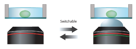
Dry (left) and silicone gel pad (right) objectives can be easily switched.
Q: What new approaches are you hoping to pursue in your future research?
Dr. Aoki: I want to use microscopes to gain a deeper understanding of the diversity of how cell collectives behave. I plan to leverage microscopy technology that allows for high-resolution, wider field, and deep observation to more accurately capture cellular dynamics. I also want to build an automated macro-to-micro workflow that enables us to understand the whole picture at low magnification before observing the details at high magnification.
Disclaimer: The opinions and statements expressed in this interview are those of the individual researcher and do not necessarily reflect the views or claims of Evident. The products and technologies mentioned are intended for research use only and are not designed for clinical or diagnostic applications.
Related Content
New Imaging Possibilities Using the World's First Multi-Immersion Silicone Gel Objective
Intelligent Shading Correction for High-Quality Stitched Images in Advanced Microscopy