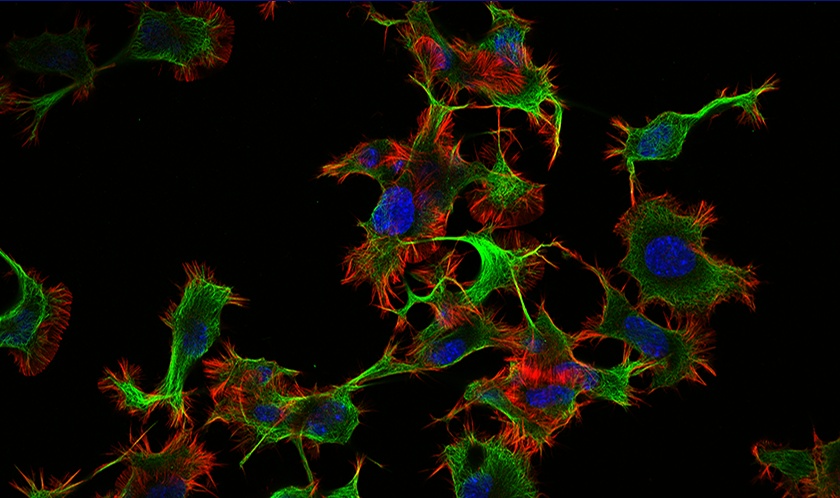
Fluorescence live cell imaging can be tricky since you need to obtain high-quality images without affecting cell viability. But with the right microscopy tools and techniques, you can run a successful experiment and get reliable image data.
Here are six tips for getting good fluorescence live cell imaging results.
1. Find the right microscope for your imaging needs.
When it comes to live cell imaging, not all microscopes are created equal. For instance, inverted microscopes work well for live cell imaging because they image from below the sample. This enables the objective lens to sit closer to the living specimen, which is necessary for most high numerical aperture (NA) objective lenses.
2. Use coverslip glass bottom culture dishes.
Plastic bottom dishes can cause autofluorescence and degrade your signal-to-noise ratio (S/N ratio). For these reasons, it’s important to use coverslip glass bottom culture dishes whenever possible during live cell fluorescence imaging experiments.
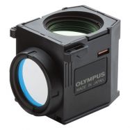
3. Use fluorescence filters with high transmission rates to improve the S/N ratio.One of the most important pieces of hardware for live cell fluorescence imaging experiments is the fluorescence filter cube and the filters within it. Look for filters with high rates of transmission so that you can collect as much fluorescence signal as possible. This enables shorter exposure times that will reduce photobleaching and phototoxicity. Olympus filter cubes also contain a special coating that reduces the amount of stray light within the cube and improves the final images’ S\N ratio. |  Olympus filter cube |
4. Use high NA objective lenses to collect more light and reduce exposure times.
Another great way to reduce exposure times is to use objectives with high NAs. These objectives will collect more light than their lower NA counterparts at the same magnification. This helps to reduce exposure times even further, leading to better cell health during long-term live cell experiments with fluorescence.

Olympus X Line 40X oil objective
5. Optimize incubation models for your experiment needs.
A wide range of microscope incubators are available on the market today. Whether using stage top or full microscope enclosures, be sure that your incubator suits all the needs of your live cells, including the ability to provide humidity and proper gas concentration levels.
Some incubation enclosures even come with blackout panels that prevent the microscope from collecting room light, which helps to improve the S/N ratio.
6. Use focus maintenance devices to reduce the number of Z-frames needed to achieve the correct focal position.
Since live samples move around, it’s important to make sure the focus can be properly captured during time-lapse experiments.
Rather than expose your samples to multiple illuminations per timepoint through Z-stacking, use a drift compensation device like our TruFocus Z-drift compensation module that will help maintain focus during time-lapse experiments.

Olympus TruFocus Z-drift compensator
Related Content
How to Choose the Right Microscope Objective: 10 Questions to Ask
White Paper: X Line Objectives Offer Revolutionary Optical Performance

