Evident LogoOlympus Logo

咨询专家

3D Segmentation for Fluorescence Images: From Qualitative to Quantitative

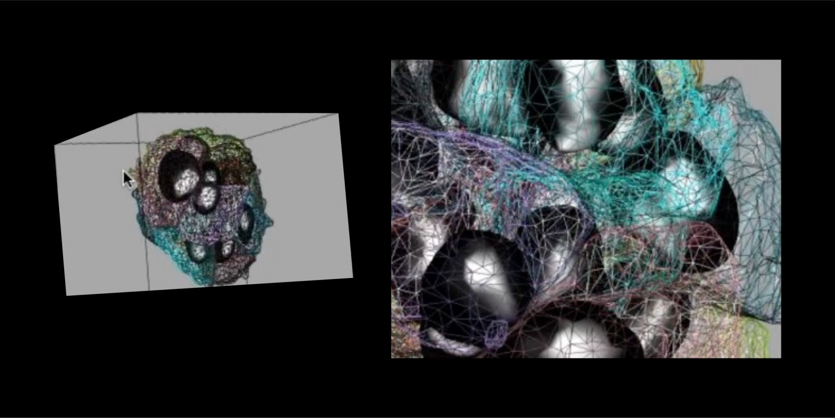
Cells are 3D functional elements in biology science and they are actively moving to perform their functions. Collective cell migration is appreciated as an important model for the understanding of the mechanism governing the cell movement in Vivo and in Vitro. It is a highly kinetic process involved in immune response, wound healing, tissue development and cancer metastasis. Recent decades have seen the fast development of various optical imaging techniques with excellent spatial-temporal resolution, dimensionality and scale. The generation of novel probes have also allowed us to acquire the movies of migrating cells with specific proteins/molecules. However, we lack of advanced solution to analyse such high-content and highly dynamic images/videos.

 In this talk, we will review some important components for 3D image analysis using two cell migration problems:  I). cancer B cell migrating in given Chemokine gradient; and II). border cell cluster migrating in Drosophila egg chamber. The extracted quantitative information provided us new insight on how a cluster of cells coordinates and collectively moves to the target.

Experts
Dr. Yu Weimiao
Head of Computational & Molecular Pathology Lab (CMPL) Agency of Science
Technology and Research

Dr. Yu Weimiao obtained his Ph.D. from the National University of Singapore (NUS) in 2007, majoring in image processing and machine vision. He joined the Agency of Science, Technology and Research (A*STAR) in 2007. He is currently heading Computational Digital Pathology Lab (CMPL) in BII to deepen and extend the R&D with clinical and industrial partners. He is also the joint PI in IMCB leading the Computational & Molecular Pathology Lab (CMPL). His research interests are Computational Biomedical Image Analysis and Quantitative Imaging Informatics. He applied 3D image analysis solution to segment and tracking the cells in 3D to understand the developmental problem and collective cell migration mechanisms. His work was published in Nature Communication, Current biology, Nature Cell Biology, etc. To enhance the applications in clinical diagnosis/prognosis, he co-founded a biotech company, known as A!maginostic Pte. Ltd. He established a world-class joint platform for the immunodiagnosis at the tissue level. The platform allows the researchers, clinicians, and pharma to profile the patient immune signature for diagnosis, prognosis, and drug response study.

3D Segmentation for Fluorescence Images: From Qualitative to Quantitative2024年10月28日
Sorry, this page is not available in your country
Ask the Expert Sign-up

By clicking subscribe you are agreeing to our privacy policy which can be found here.

对不起,此内容在您的国家不适用。

Sorry, this page is not available in your country