Judges 2004

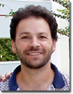
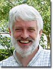
 | Dr. James A. Fadool - A faculty member in the Biological Sciences Department at The Florida State University, Dr. Fadool specializes in novel transgenic technologies for identification of transcriptionally active regions of the genome using the zebrafish as a model organism. His other interests include vertebrate embryology, genetic and biochemical mechanisms regulating development and degenerative diseases of the visual system. Much of Dr. Fadool's work involves transfection and selection of stable clones containing gene products linked to cyan and yellow fluorescent protein derivatives for imaging applications in fluorescence microscopy. Among the optical microscopy techniques employed by Dr. Fadool in his research are widefield and confocal fluorescence, brightfield, and phase contrast. |
 | Dr. Victoria Centonze Frohlich - The Associate Director of the Optical Imaging Facility for the Department of Cellular and Structural Biology at the University of Texas Health Science Center at San Antonia, Dr. Frohlich is a practicing cell biologist with a significant interest in the areas of cell motility and early development. Throughout her career, Dr. Frohlich has applied a variety of optical microscopy techniques to her research, including many of transmitted methods mentioned above. Among the advanced fluorescence techniques utilized by Dr. Frohlich are fluorescence recovery after photobleaching (FRAP), fluorescence resonance energy transfer (FRET), photolysis, and optical trapping. Dr. Frohlich also has experience with transmission and scanning electron microscopy. |
 | Dr. Kenneth N. Fish - An Assistant Professor in the Translational Neuroscience Program in the Department of Psychiatry at the University of Pittsburgh, Dr. Fish uses knockout and genetic mutants that have subtle neuroachitectural abnormalities to study the functional consequences that mispositioning of a subset of neurons during development has on local circuitry. Studies using these mutants may be helpful in understanding aspects of the neuropathology that result in abnormal brain connectivity associated with schizophrenia, autism, and epilepsies. Optical microscopy techniques utilized in this research include confocal and multiphoton fluorescence imaging, as well as total internal reflection fluorescence (TIRFM) along with traditional widefield fluorescence methods. Dr. Fish also has experience with brightfield imaging modes, including phase contrast, differential interference contrast, and oblique illumination. |
 | Dr. Douglas B. Murphy - Professor Murphy is the Director of the optical and electron microscopy laboratory at the new Howard Hughes Medical Institute in Janelia Farm, Virginia. This resource is a core facility that provides equipment and services in fluorescence, confocal, and electron microscopy to laboratories throughout the research campus. Dr. Murphy's research interests include the dependence of microtubules in organelle transport and related microtubule behavior, including polymer annealing and the mechanism of binding microtubule-associated protein (MAP-2) on microtubule surfaces. He has also explored the mechanical and conformational requirements of the motor protein, kinesin. The core facility specializes in advanced fluorescence techniques (FRET, FRAP, TIRFM), confocal, multiphoton, and time-lapse in multiple fluorescence channels. Dr. Murphy has been a significant contributor to review articles and Java tutorials on the Olympus optical microscopy educational websites. |
对不起,此内容在您的国家不适用。