Last updated on September 13, 2024.
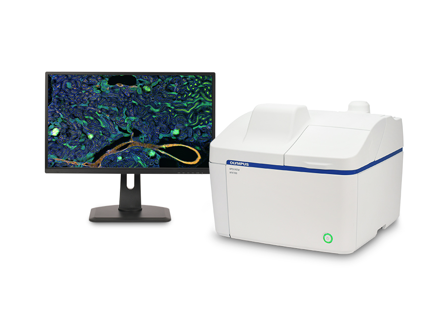
Do you spend a lot of time adjusting your microscope before you begin imaging? Do you wish there was an easy way to capture high-quality microscope images? Are you tired of having to use a darkroom? The APEXVIEW™ APX100 benchtop fluorescence microscope overcomes these common research challenges with one easy-to-use system.
Imaging workflow with a conventional microscope vs. the APX100 benchtop fluorescence microscope
With no need to look through an eyepiece, and a suite of smart features designed to streamline your workflow, the APX100 microscope dramatically reduces time spent adjusting your microscope’s settings for research imaging. The microscope works with our renowned objectives, including our award-winning X Line™ series, so you can obtain publication-quality images in just a few clicks.
Here are some ways the APX100 microscope makes it easy to obtain exceptional images:
1. Smart sample navigator
When you place your sample in the sample holder, the smart sample navigator automatically acquires a macro image, and the built-in AI locates your sample on the slide. The system then automatically centers your sample over the objective and displays it on the monitor. Here you can choose the observation method and immediately start capturing images. Learn more in the video below:
2. Fast autofocus
The system’s autofocus is up to 12x faster than conventional autofocus algorithms. This enables you to quickly find the ideal imaging plane. With coordinated automation, you can spend less time searching for samples and more time collecting data. See how the high-speed autofocus works in this video:
3. Easy to learn and teach to researchers
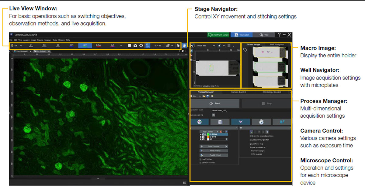
Using the APX100 microscope is simple. Load the sample, close the lid, and click a button. The software’s clear layout and streamlined workflow help you start image acquisition with little training.
Given that teaching multiple imaging modalities to new researchers can be a time-consuming process, the system’s ease of use greatly simplifies and accelerates the training process.
Researchers who are unfamiliar with microscopes can quickly learn to operate the system and capture publication-quality images, while microscopy experts will appreciate the system’s automation and streamlined workflow.
A simple layout makes it easy for researchers to capture images
4. Compact imaging device for labs and core facilities
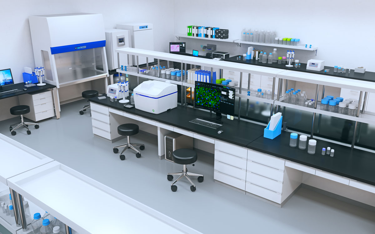
With a small footprint, built-in anti-vibration technology, and shielded optics, you can place the APX100 microscope almost anywhere. Install the unit directly on a lab table to image in parallel with other experiments, even in a brightly lit room. As no dedicated darkroom is required, you can save valuable space in your lab or core facility.
The APX100 system can be installed directly on a lab table to save space
5. Organized images
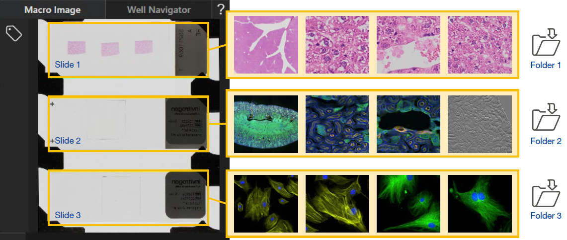
The APX100 microscope has a dedicated system to organize and store your data. When you acquire an image, the software automatically creates folders for each sample and saves the data into the correct one. The consistent indexing keeps your data organized and easy to locate, and prevents you from accidentally saving data in the wrong folder. Further, important settings are saved with each image for future reference.
Image organization on the APX100 system
6. Flexible imaging options for many research applications
The APX100 microscope supports a wide range of research imaging applications for slides, dishes, and well plates. You can use the included imaging methods, such as multichannel, stitching, time-lapse, and Z-stack acquisition, in any combination to fit your research protocol. Also, you can acquire images with brightfield, phase contrast, fluorescence, and gradient contrast.
Developed by Evident, gradient contrast is a unique transmission observation method that enables the acquisition of impressive high-contrast 3D images. Learn more about gradient contrast in this brief video:
With all these imaging options, you can acquire an array of stunning, publication-quality images. Below are some example application images that you can acquire with the APX100 benchtop fluorescence microscope.
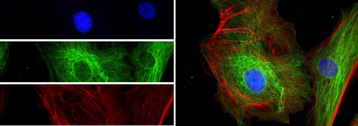
Multichannel images
BPAE cells. Stain: Mouse Anti-α-tubulin, BODIPY FL Goat Anti-Mouse IgG, Texas Red-X Phalloidin, and DAPI.
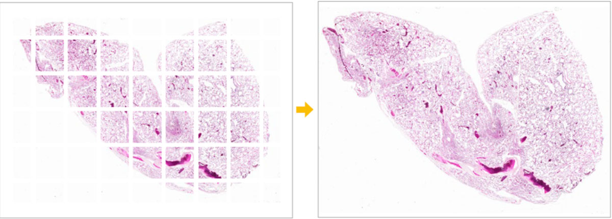
Stitched images
Mouse lung captured with a UPLXAPO4X objective. Stain: HE.
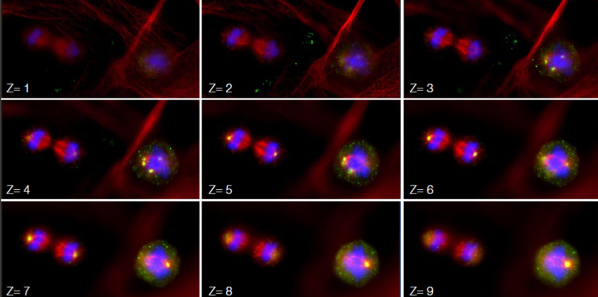
Z-stack images
Subcellular localization of the centrosome protein kendrin/pericentrin. Stain: Pericentrin-green, alpha-tubulin-red, DNAblue.
Image data courtesy of Dr. Kazuhiko Matsuo, Anatomy and Developmental Biology, Kyoto Prefectural University of Medicine.
Time-lapse images
Time-lapse observation of a fertilized mouse egg.
Tip: if you need to use fluorescence resonance energy transfer (FRET), total internal reflection fluorescence (TIRF), time-lapse experiments for longer than 2-days, or super-resolution microscopy, IXplore™ inverted microscopes are the ideal choice. Reach out to an expert to discuss your imaging and research needs.
To learn more about the imaging capabilities of the APX100 microscope and see more example images, visit our APX100 image gallery. Explore all the features of the APX100 microscope online or contact your local sales rep to plan a demo.
Related Content
Brochure: APEXVIEW APX100 Benchtop Fluorescence Microscope
Video: Introducing the APEXVIEW™ APX100 Benchtop Fluorescence Microscope