革新 共聚焦显微镜技术
FLUOVIEW Smart™ 软件介绍 直观与人工智能增强的工作流程 介绍
共聚焦激光扫描显微镜已成为生命科学及医学研究领域不可或缺的工具,能够对各类生物样本(如细胞和组织)进行高分辨率观察及 三维分析。
它们被广泛用于细胞生物学、神经科学,以及发育生物学等领域,作为支撑研究质量和可重复性的核心技术。 为了满足多样化的研究需求,共聚焦激光扫描显微镜不断发展,提供了更加先进的功能和更灵活的成像条件选择。 然而,这一进步也使软件界面变得日益复杂,带来了显著挑战——尤其是对于初学者用户——包括设置耗时、操作错误风险增加,以及研究工作流程效率下降。
复杂共聚焦软件界面带 来的挑战
- 学习曲线高: 操作的掌握过程耗时较长,进而推迟了研究的开展。 初学者常常不知道从哪 里开始,因此经常需要经验丰富的用户或指导人员的帮助。
- 误操作导致效率降低: 由于设置繁多且功能难以查找,工作流程变得不连贯。 错误可能导 致数据丢失或样品损坏,需进行高成本的再次成像或重复实验。
- 共享环境中的问题: 当经验水平各异的多位用户共享同一系统时,未被察觉的设置更改可 能导致非预期的成像条件、样品浪费以及实验需重新进行。
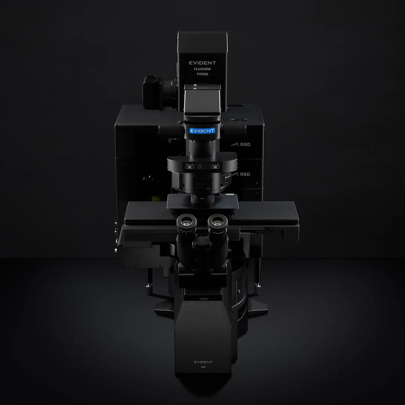
FV5000 是新一代平台,能够以前所未有的速度和便捷性采集更清晰、完全可量化的数据
解决这些问题需要 一个直观、高效、以 用户工作流程为核心的软 件界面。
优化 FLUOVIEW Smart™ 共聚焦软 件的用户体验
Evident 的 FLUOVIEW Smart 共聚焦软件旨在确保不同技能水平 的用户都能自信、高效地操作 FLUOVIEW™ FV5000 激光扫描显微镜。 它采用三项核心方法,以减少操作负担,提高科研效率和 结果的可重复性。
图形用户界面与屏幕引导,降低学习成本
基本的显微镜操作——如 Z 范围设置和物镜选择——不仅仅是配 置,而且是了解当前系统状态的重要信息。 FLUOVIEW Smart以 图形方式呈现这些设置,帮助用户直观理解其含义和影响(见图 1和图2)。
屏幕指南进一步协助用户清晰无误地完成任务,减少了持续监督 的需要,并支持自主数据采集。 受一线研究人员应对科学挑战的启发,FV5000正在革新共聚焦成像技术,使其更智能、更高效。
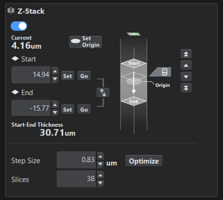
图 1。 Z 范围设置界面。 成像的起始和结束位置以及当前观察位置一目了然。
功能优先布局
FLUOVIEW Smart™ 软件并未将所有功能汇聚于同一屏幕,而是 采用层级结构设计,根据使用频率和重要性进行组织。 关键控 制项(如激光功率、扫描范围和 Z 堆栈设置)始终随时可用,充 分契合自然工作流程(样品检测→条件设置→成像)。 这种设计 减少了查找时间,并显著降低了出错风险。

图 2。 物镜选择界面 用户可以直观地选择所需的物镜,激活的物镜一目了然。
重置与按用途自动布局
为了防止共享环境中出现问题,FLUOVIEW Smart 软件会在启动 时将设置重置为默认设置。 同时,软件允许用户保存和恢复先 前的配置,以实现可重复性。 此外,选择成像目的(如 Z 堆栈、 延时拍摄、多区域拼接)会自动调整界面布局和操作引导,为初 学者和专家提供最佳使用界面(见图 3)。
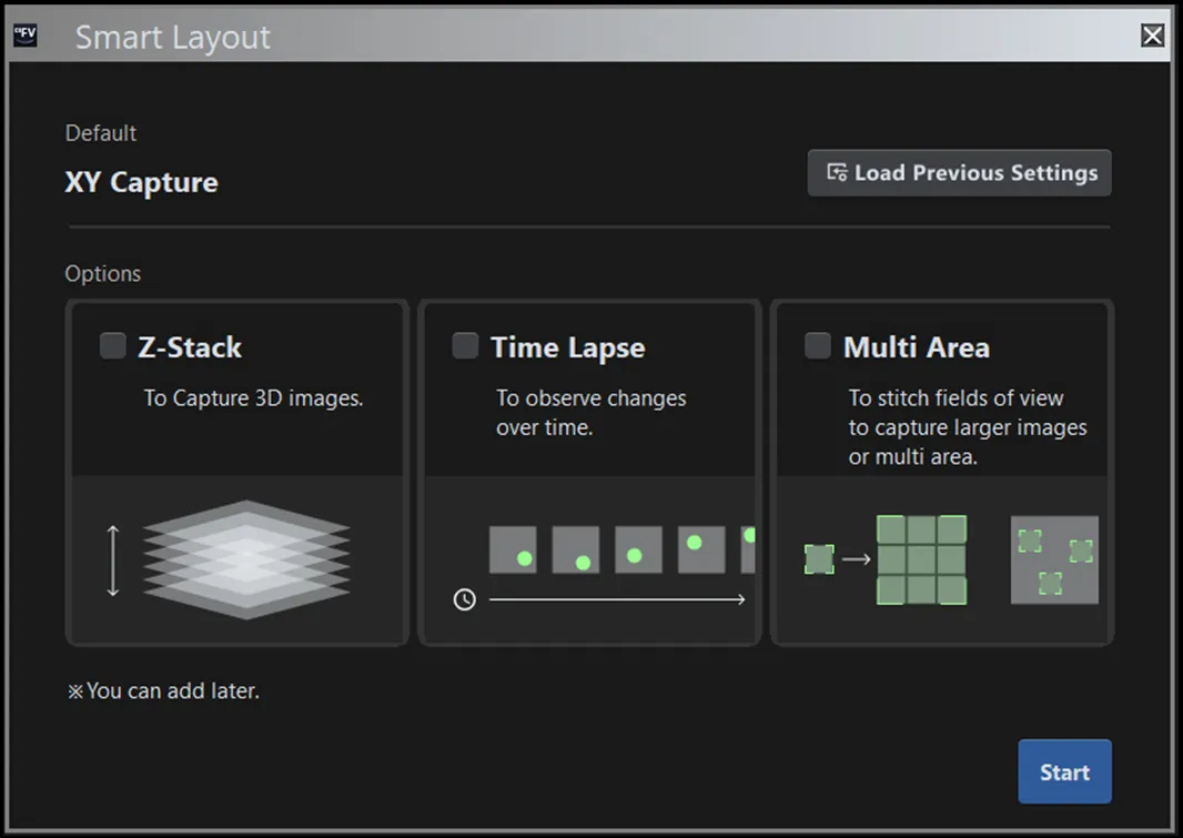
图 3。 用途选择界面。 界面布局和屏幕上的指导会根据所选择的成像目的自动调整。
初学者的AI辅助
除了简化界面之外,FLUOVIEW Smart™ 软件还利用人工智能自动化和优化对新手用户来说特别具有挑战性的步骤。
自动样品检测
由于共聚焦显微镜的光学原理,如果样品在 XY 或 Z 方向上失焦,屏幕可能会显示空白……让新手用户不确定问题是由于设置还是硬 件故障造成的。
为了解决这一难题,Evident与Epistra借助在生命科学人工智能开发方面的深厚经验,联合开发了一种图像识别算法,能够自动将显微 镜切换到最适合搜索样本的最佳状态,并自动定位样本区域。 这显著简化并加快了此前令初学者感到困难的流程(见图4)。
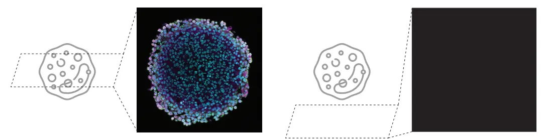
图 4. 在共聚焦激光扫描显微镜中,样品搜索过程的高难度可能导致出现空白屏幕(见右图)
图 5 展示了样本位置估计的整体工作流程。
系统设计为仿照专家用户的操作流程: 使用4×低倍物镜对Z轴和XY轴进行粗略校准,随后用10×高倍物镜进行精细调整。
具体而言,系统依次执行以下四个步骤,以自动确定最佳样本位置。
Z 搜索(4X)
本步骤利用 4X 物镜依次搜索*,粗略地确定搜索范围内的最佳 Z 位置。 在每一个候选的 Z 位置,系统会采集一张图像,并由 AI 评估其聚焦评分。 基于这些结果,系统会自动更新下一个要探索的 Z 位置,并持续迭代,直至获得最高焦点分数的位置。 系统会根据用户在向导中的选择合理设置 Z 搜索范围。
XY-SEARCH (4X)
利用4X物镜采集的图像,调整XY载物台,使局部亮度最高的区域居于显示屏中心。 在此过程之前,系统会检测预定义的伪影(例如,容器边缘)。 如果检测到伪影,系统会弹出警告,并终止该流程。 此时用户可以根据需要手动调整样本的 XY 位置。
Z-SEARCH (10X)
切换至 10X 物镜后,系统会依次搜索以确定范围内的最佳 Z 位置。 Z 搜索范围依据使用 4X 物镜时找到的最佳 Z 位置和 4X 物镜的 Z 分辨率进行设定。
XY-SEARCH (10X)
在 10X 图像下,XY 载物台移动,将局部亮度最高的区域置于屏幕中心。 经过此过程,系统向用户显示 当前显微镜图像,完成 搜索过程。
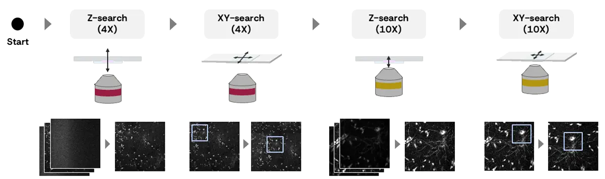
图 5. 样本位置估计的整体工作流程:
Z轴查找(4X)
- 在共焦孔径打开的情况下,对样品沿 Z 轴进行粗略扫描。
- 设置共焦孔径为 1 Airy 单位,并微调 Z 位置以获得最佳聚焦。
XY搜索 (4X)
- AI 异常检测(若未检测到标本或检测到盖玻片/孔的边缘时)。
- 将载物台移动到标本上的较亮区域。
- 在地图上映射4X图像。
Z轴查找 (10X)
- 在共焦孔径打开的情况下,对样品沿 Z 轴进行粗略扫描。
- 使用 1 个 Airy 单位共焦孔径微调 Z 位置,以获得最佳焦点。
XY 搜索(10X)
- 将载物台移至标本上更亮的位置
*顺序搜索 一种通过依次拍摄图像,在规定的搜索范围内定位样本位置的方法。
成像条件优化
在传统的共聚焦激光扫描显微镜中,探测器灵敏度和激光功率之 间存在固有的权衡,需要反复试验才能找到最佳设置。 初学者 常常面临这样的难题:提高灵敏度会引入更多噪声,而提升激光 功率则可能损坏样品(见图6)。 因此,确定适当的成像条件可能 会耗时。
Evident 通过其专有的下一代 SilVIR™探测器技术,有效消除了调 整探测器灵敏度的需求。 减少了用户必须配置的参数数量,从而 使操作更简单。 因此,激光功率的设置以实现适当的对比度,成 为影响图像质量的最关键因素。
FLUOVIEW Smart™ 软件会根据实时图像分析和机器学习预测自 动确定最优激光功率。 只需根据最小化样品损伤和最大化对比 度的优先级选择模式,即可轻松应用适合自身需求的设置。 初学者在提高检测灵敏度时往往会遇到噪 声增加的问题,而提高激光功率则有可能 损伤样品。
激光功率过高激光功率过高会产生较高的光毒性
图 6. 激光功率和图像质量之间的权衡。 | 激光功率过低激光功率过低会导致图像噪声过大
|
总体流程如下:
模式选择
用户可根据样本保护与图像对比度的优先级,选择“柔和”、“平衡”或“高质量”三种模式中的一种。
自动激光功率调节
当用户点击执行按钮时,系统会自动设置最佳激光功率并完成该过程。 系统内部会依次完成以下步骤:
- 系统在不同激光功率水平下捕获四张图像。
- 系统针对每张图像计算 AI 得分,用以评估图像质量(图 7 中的步骤 1-3)。 系统将 AI 评分绘制成图表,并根据图表和所选模式估算最佳激光功率(见图 7 的步骤 4)。
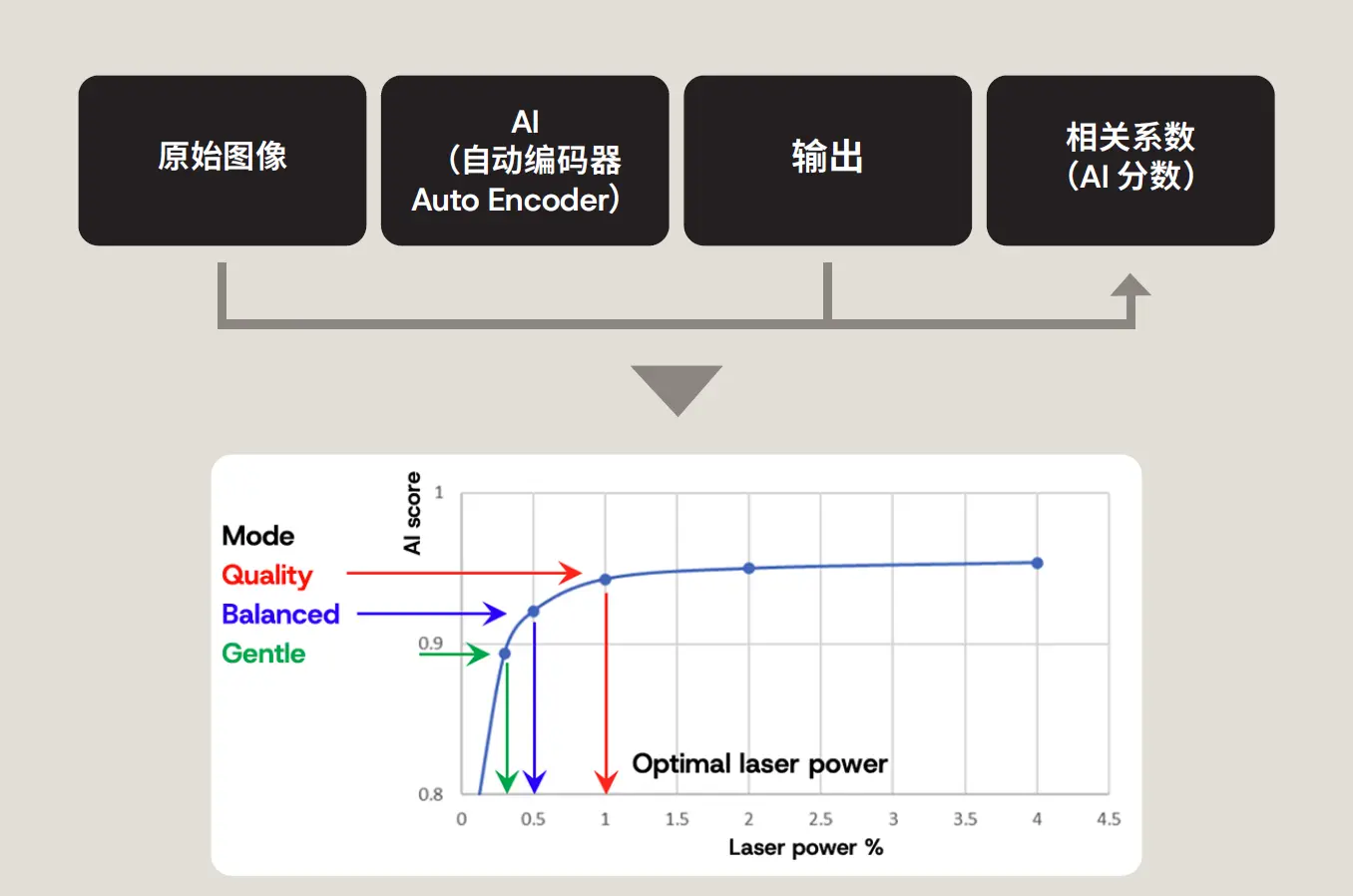
图7. 激光功率估算的概念包括以下步骤: 1: 1.使用 AI 从原始图像预测干净、低噪声的图像。 2.将预测的图像与原始图像进行比较。 3.计算相关系数(AI 得分:得分高表示稳定且噪声低,得分低表示不稳定且噪声高)。 AI分数高表示图像稳定且噪音低,AI分数低表示图像不稳定且噪音高。 4. 根据所选模式估算最佳激光功率
注意: AI 分数与图像信噪比(SNR)相似,但略有不同。 分数会随着激光功率的增加而升高,这反映了噪声降低带来的图像质量提升。 AI 评分曲线的平台区域表示适当的激光功率设置: 在此范围内,进一步增加激光功率对图像质量的提升有限,因此并不高效。
结论
FLUOVIEW Smart™ 共聚焦软件简化了 FLUOVIEW™ FV5000 激光扫描显微镜的用户界面,并通过人工智能技术提升了工作流程(图 8)。
- 复杂的软件界面对新手用户来说是一个显著的阻碍。
- 通过优先功能、基于工作流的操作设计和图形用户界面实现简化。
- AI实现了样品检测和激光功率调整的自动化,大大降低了操作复杂性。
通过结合以用户体验为核心的软件界面和 AI 驱动的智能辅助功能,FLUOVIEW Smart 软件让即使是新手也能迅速获得高质量图像,从而极大提升研究环境中的工作效率和实验结果的可重复性。
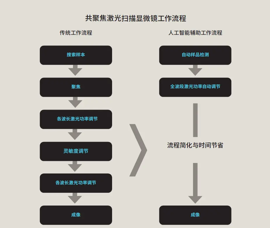
图8。通过AI辅助流程简化共聚焦显微镜观察。
作者
Ryoji Kitamura
生命科学高端成像系统全球产品经理
在北海道大学信息科学技术研究生院取得硕士学位,研究方向为利用多光子显微镜进行体内成像。他在 Evident 开始了自己的职业生涯,担任软件工程师,后来成为 SLIDEVIEW™ VS200 通用全切片成像扫描仪的产品负责人。他还曾担任 IXplore™ IX85 倒置显微镜系统的全球产品经理。现任 FLUOVIEW™ 共聚焦显微镜系列全球产品经理,主导产品战略、规划与开发。
适于这类应用的产品
对不起,此内容在您的国家不适用。