

SLIDEVIEW VS200 for Life Science Research
Extract More Insights from Every Slide in High ResolutionWe’ve combined high-quality multiplex imaging, accurate color reproduction in brightfield, optical clarity in thick samples, and the broadest range of imaging modalities in one slide scanner. The SLIDEVIEW™ VS200 universal slide scanner enables researchers to capture more meaningful data from samples—without added complexity, manual intervention, or the need for multiple instruments. Designed to deliver consistent, repeatable results across users and workflows, the VS200 system helps you unlock more insights from your slides while supporting scalable imaging needs. |
One Scanner. Every Slide. No Limits.
Capture the biological insights you need with a single slide scanner designed for flexible, multimodal imaging.
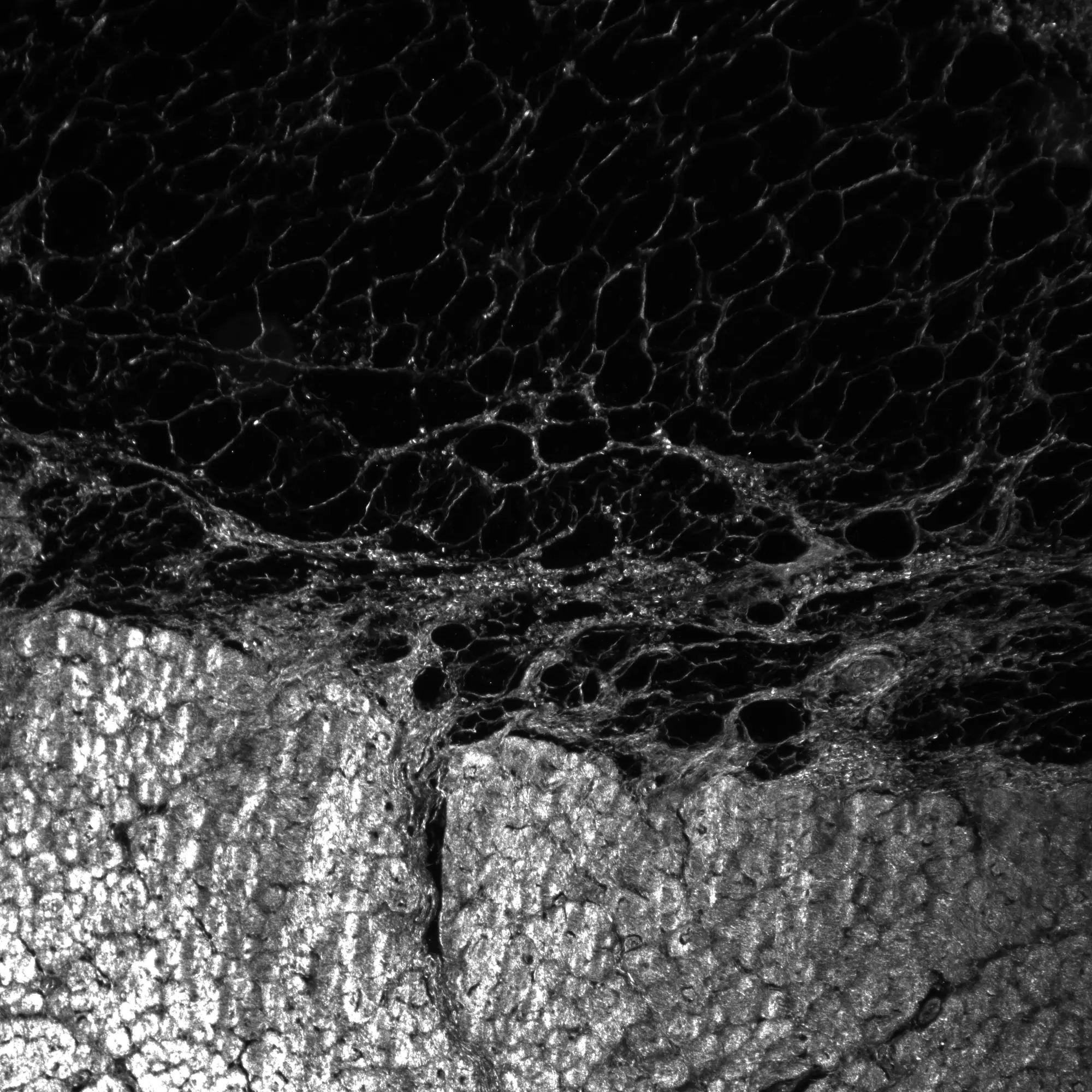
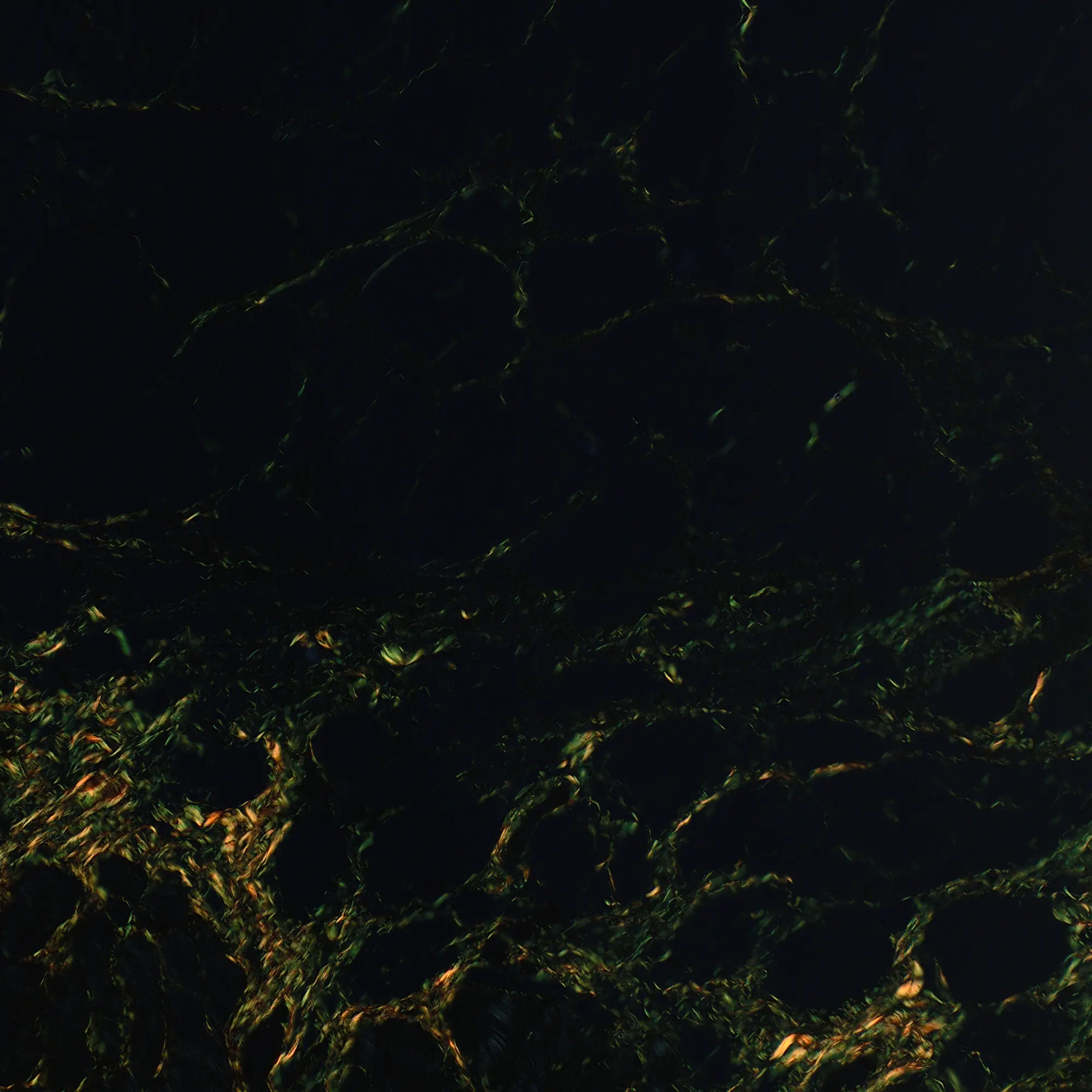
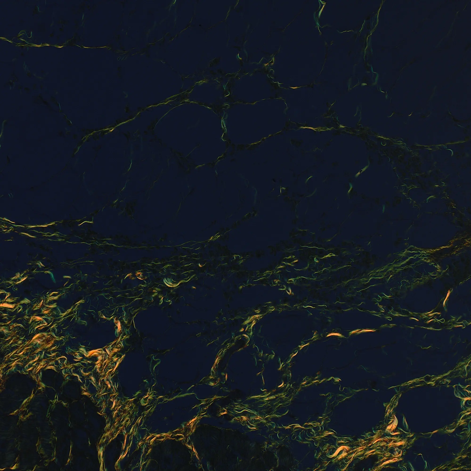
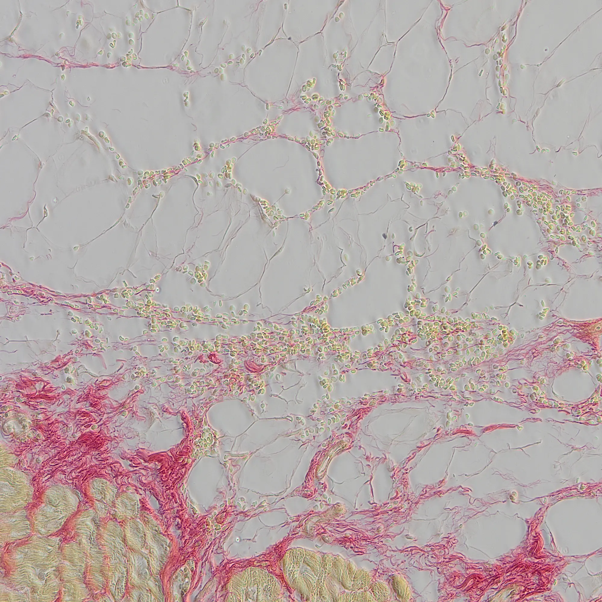
- Ten imaging modes: View structures visible only under certain conditions using up to 10 observation methods, with the option to combine methods
- Geological sample imaging: Precisely control the polarization angle using a manual rotatable analyzer
- Multiplex imaging: Supports up to 10 fluorescence colors, enabling multiplex imaging with high resolution, spatial alignment, and reproducibility
 |  |  |
 |  |  |
 |  |  |
 |
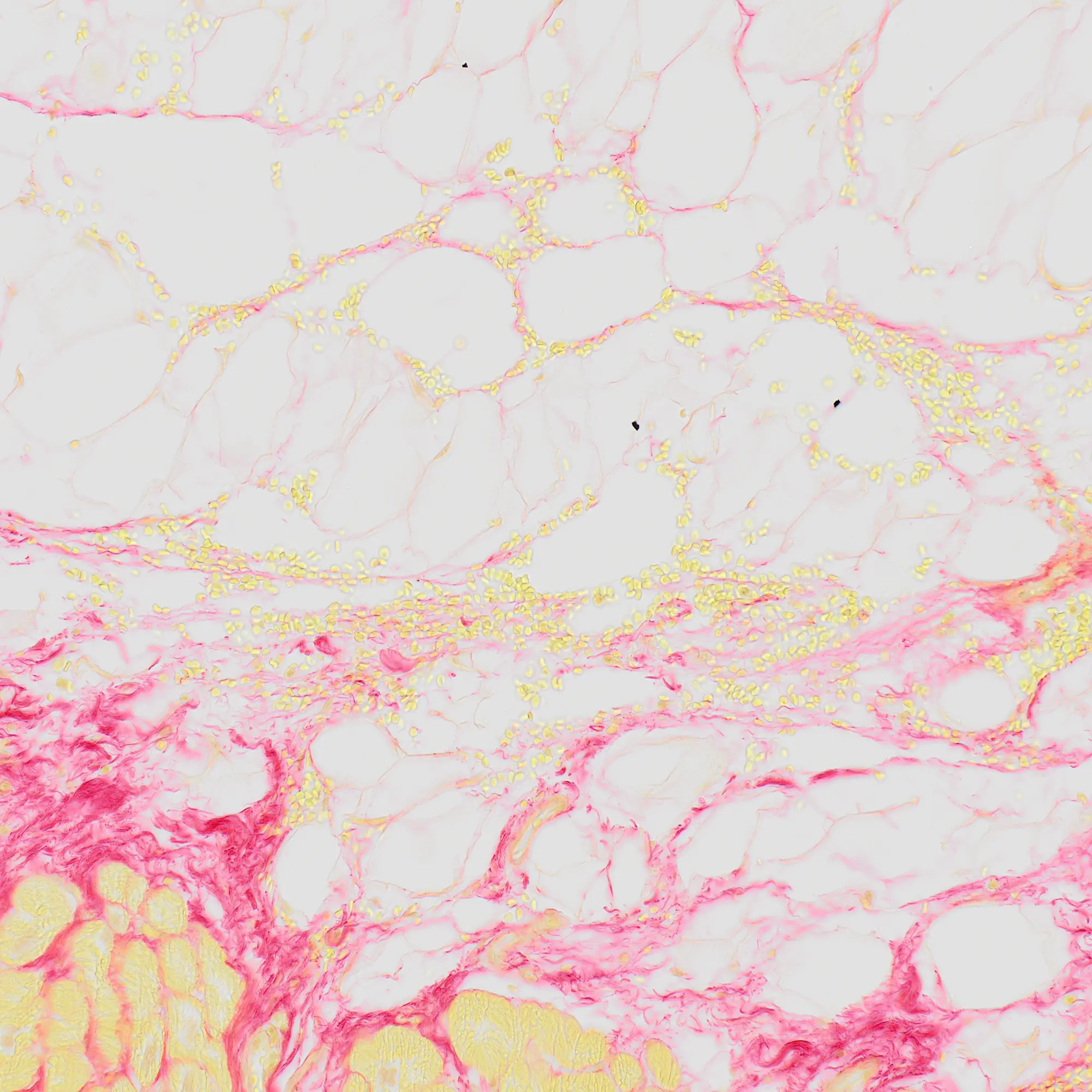
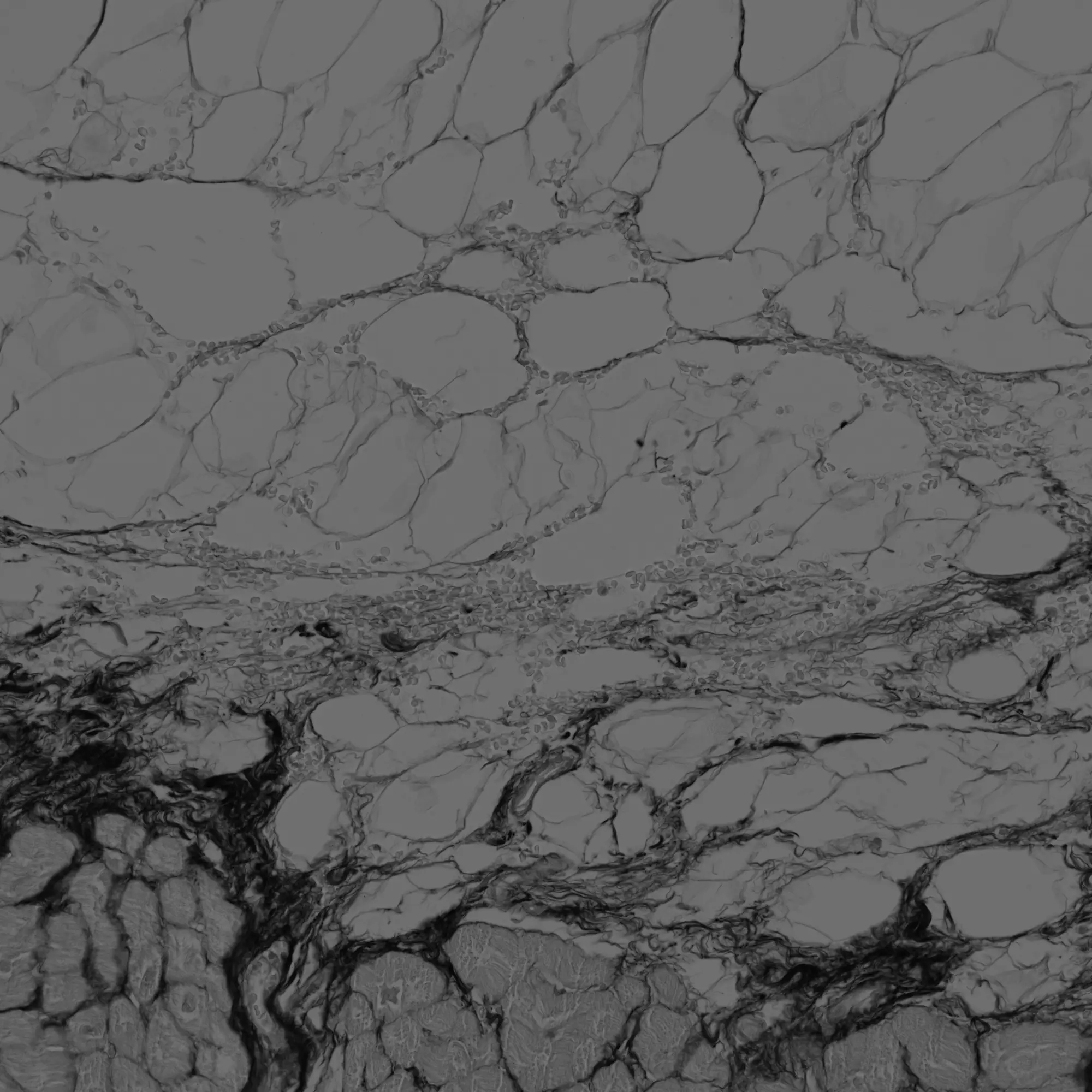
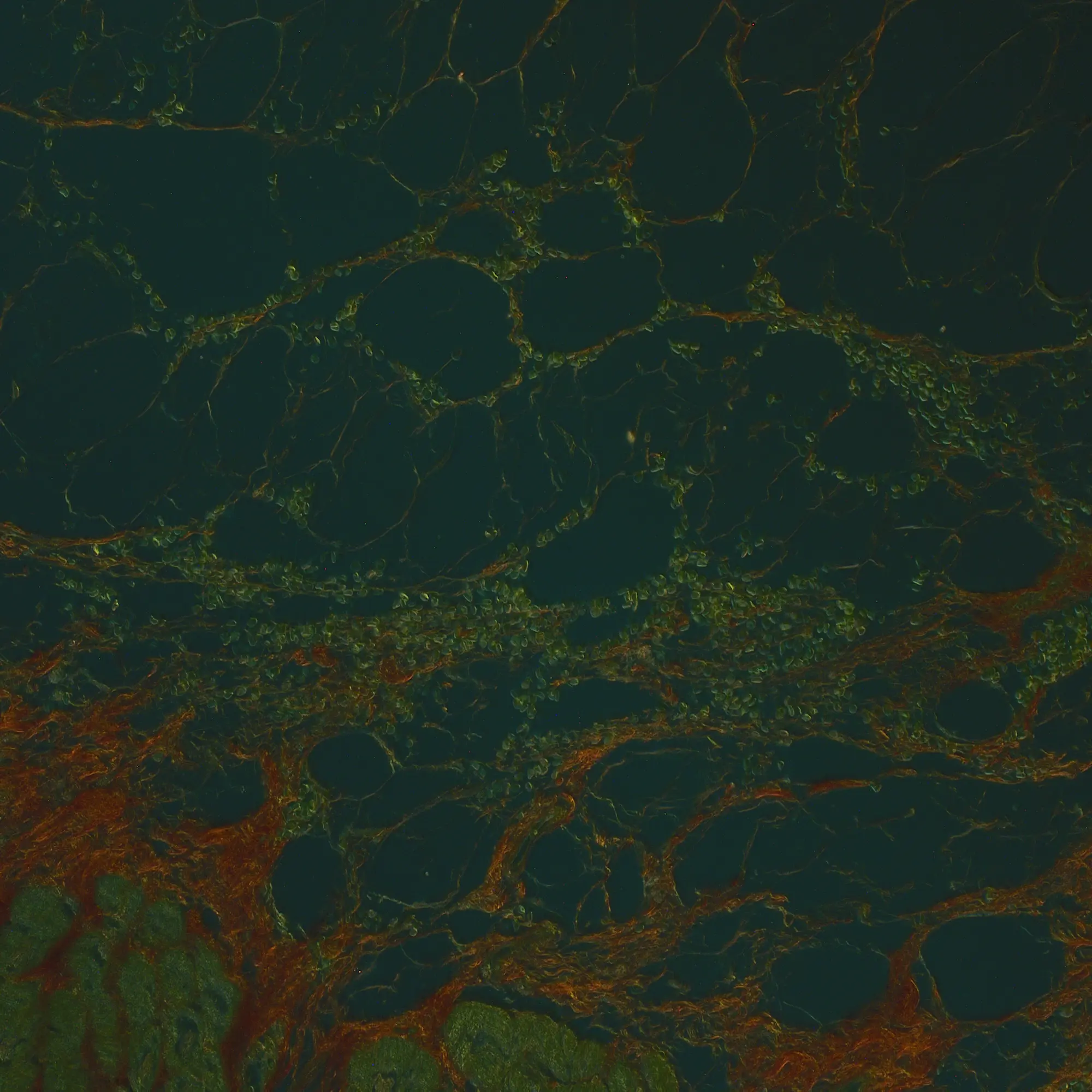
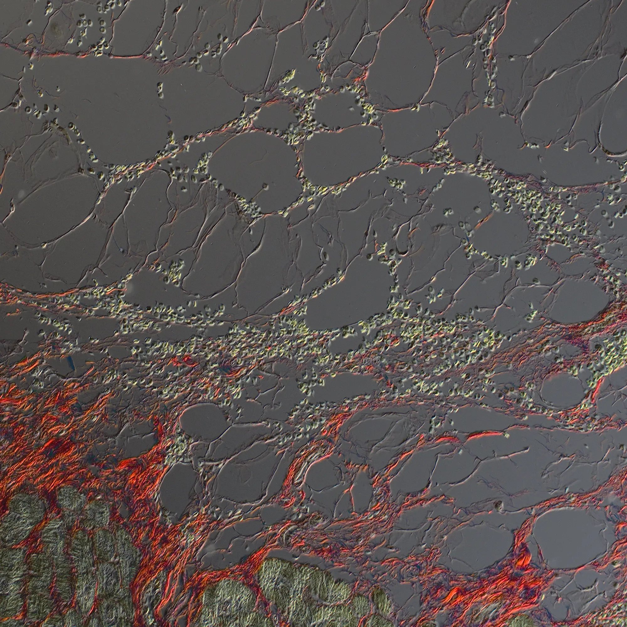
Lung tissue section stained with PicroSirius Red and imaged using 10 different observation modes. Most images were acquired using X Line™ objectives (UPLXAPO20X, NA 0.80). For phase contrast imaging, the UPLAPOPH20X objective (NA 0.70) was used.
 | “Within the slide scanning facility of the pathology department there was a high demand for a whole slide scanning microscope with multiple methods of illumination (brightfield, polarization, and fluorescence) and a diverse range of objectives (air and oil). The SLIDEVIEW VS200 scanner fits our needs as a day-to-day whole slide scanning system for research purposes without compromising on image quality.” Michel Meijer |
Handle Any Slide Format Effortlessly—Because Your Work Deserves No Limits
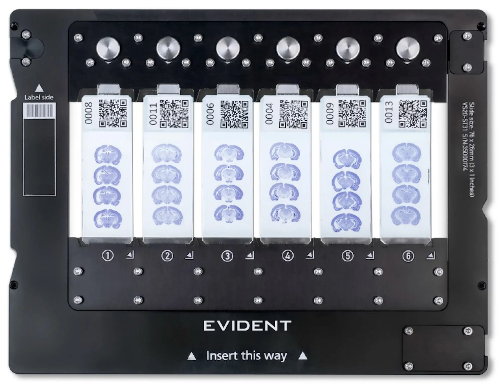
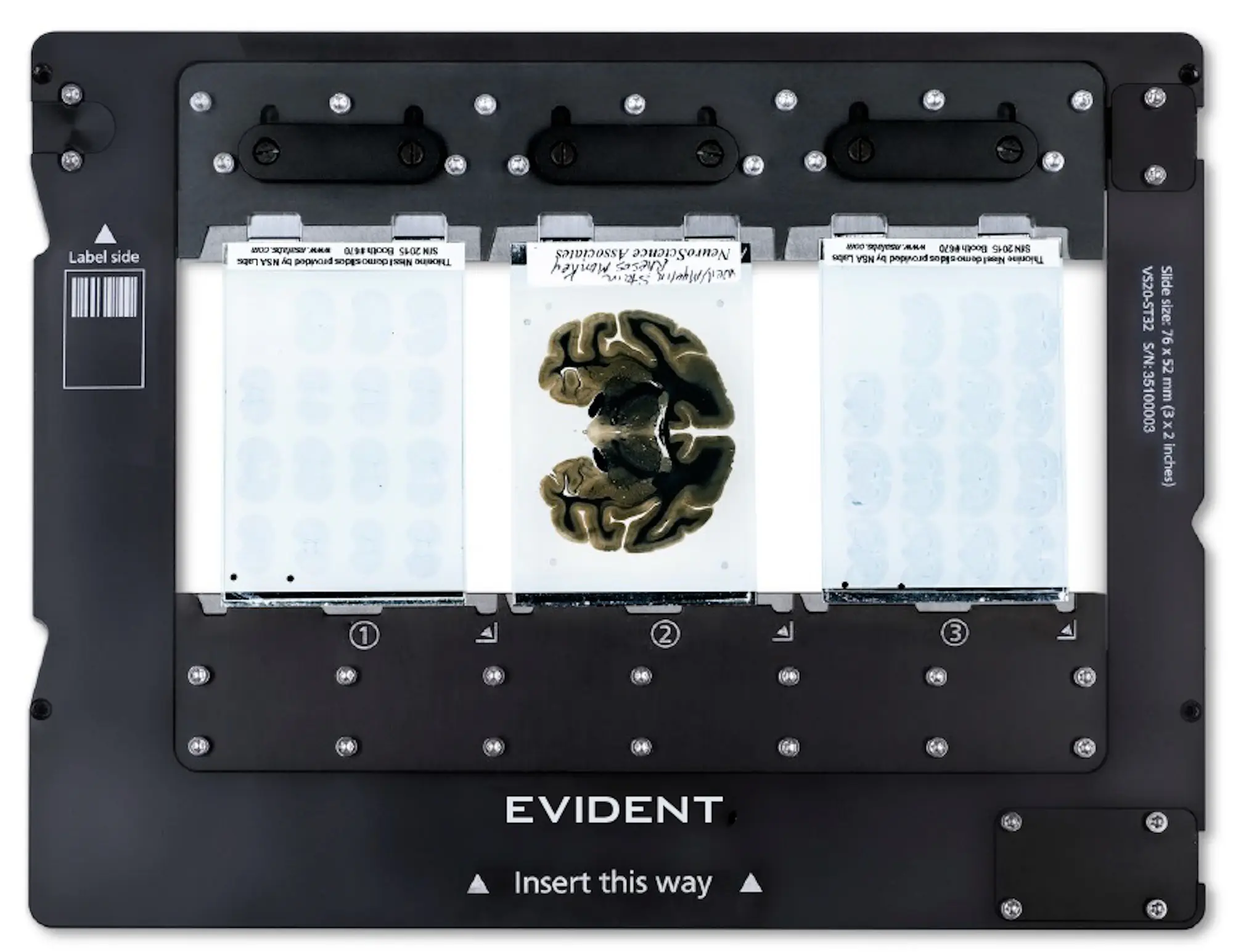
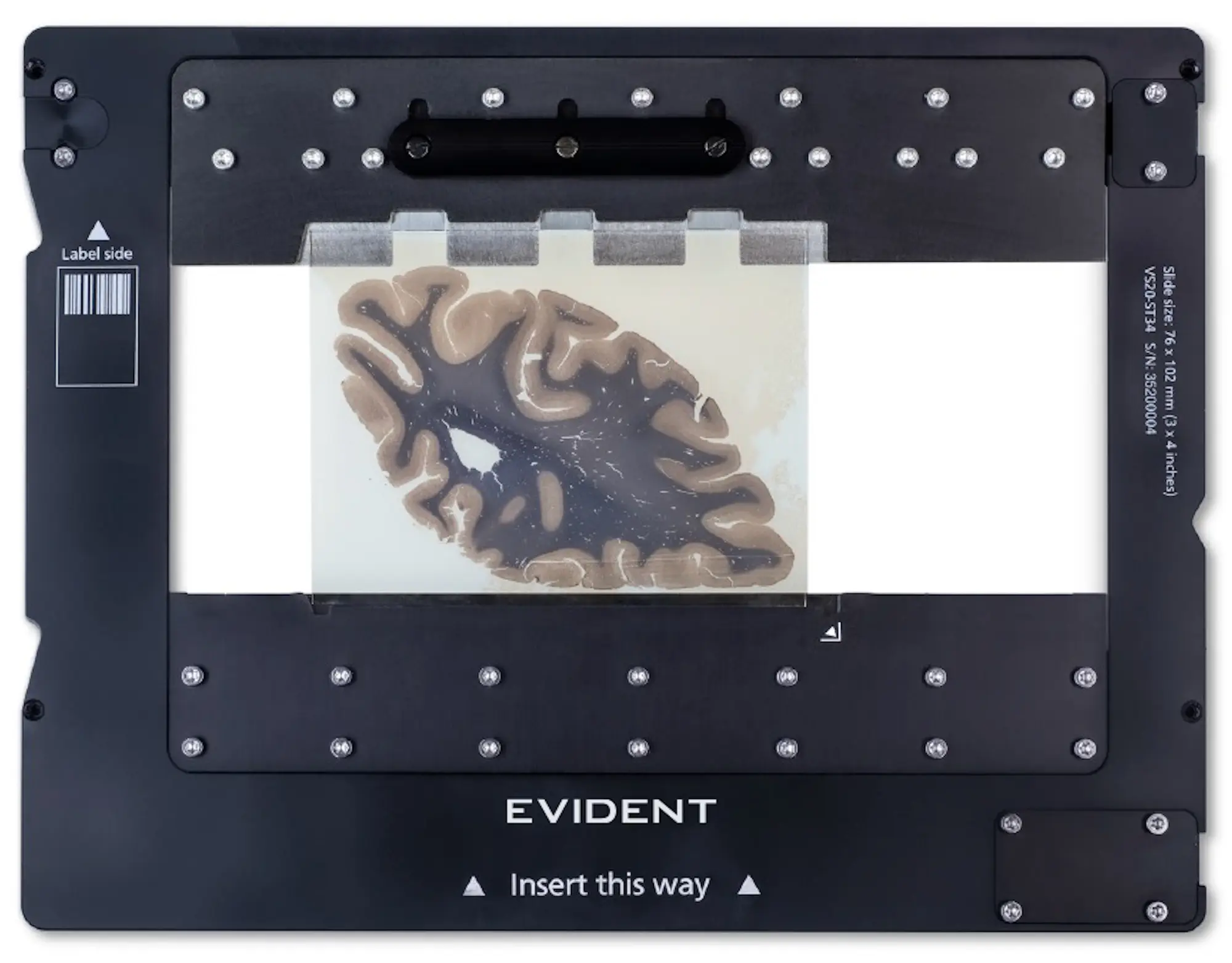
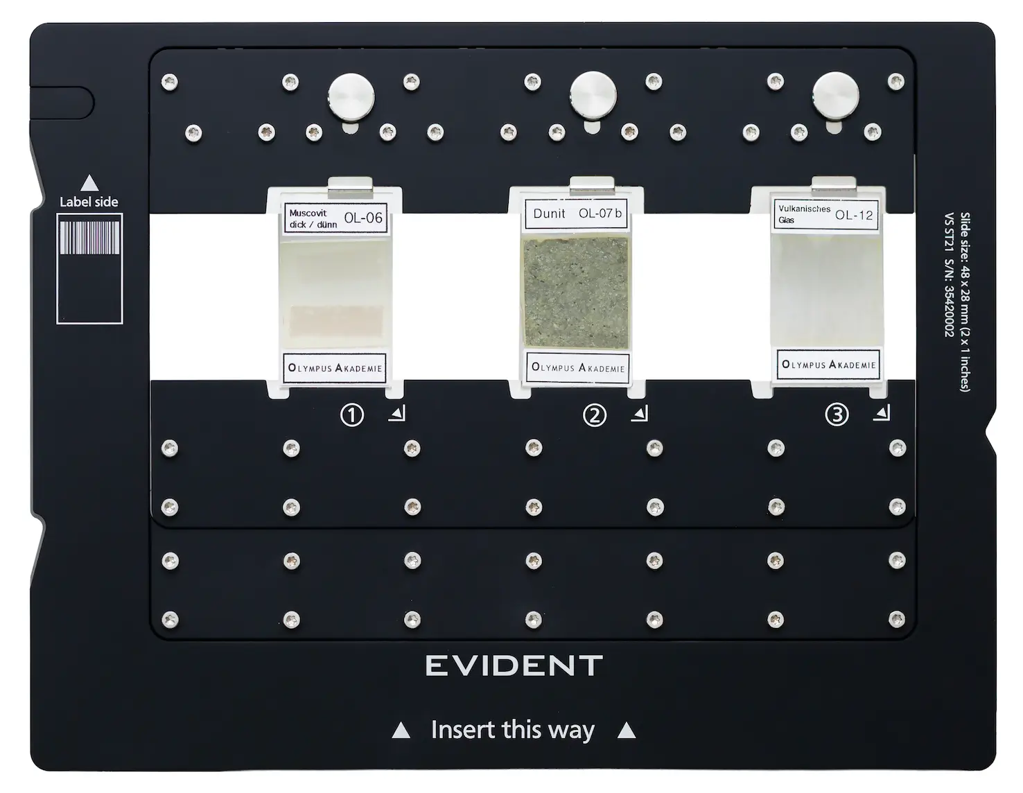
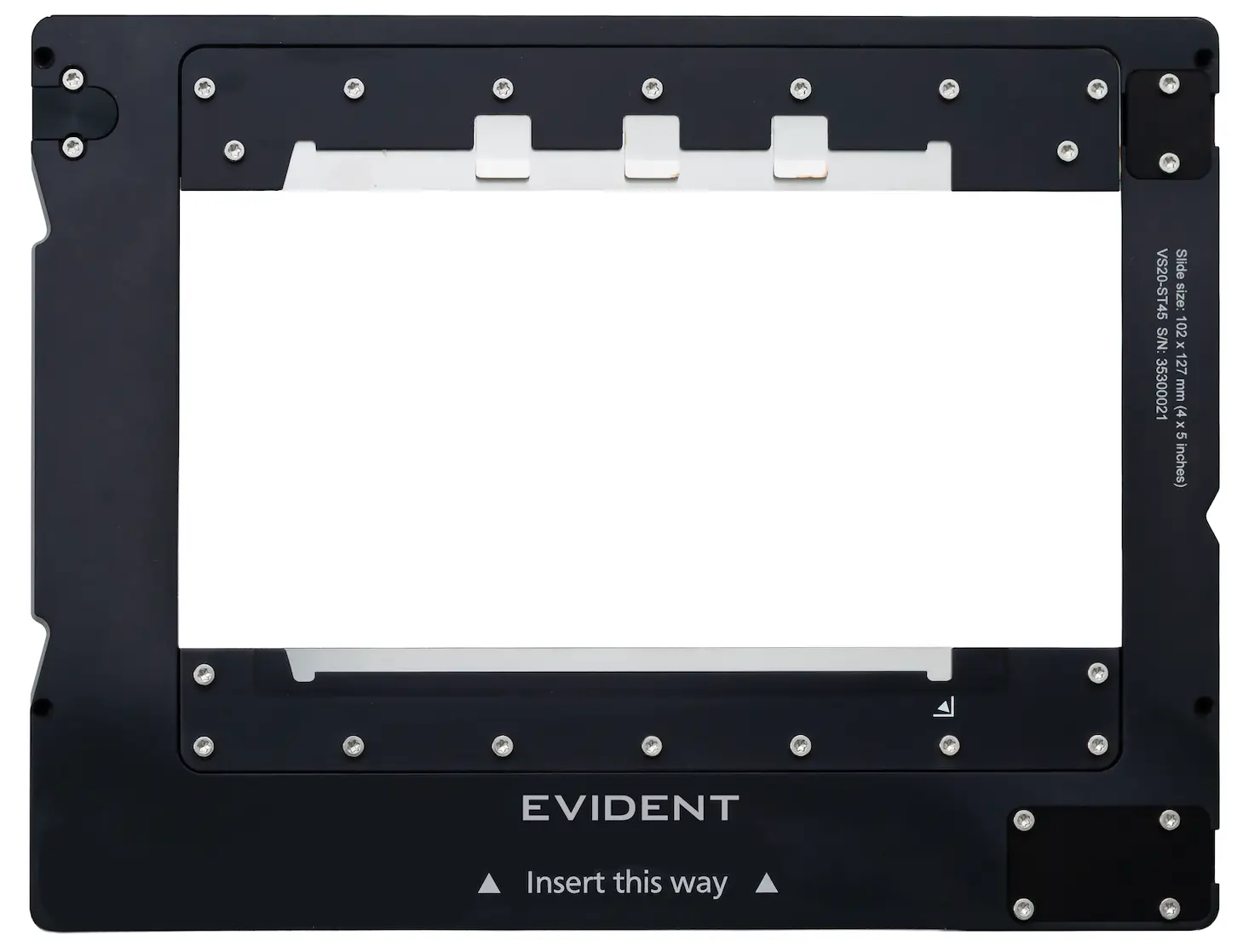
The simple-to-use slide tray supports five different slide sizes:
- 26 × 52 mm (1 × 2 in.)
- 26 × 76 mm (1 × 3 in.)
- 52 × 76 mm (2 × 3 in.)
- 76 × 102 mm (3 × 4 in.)
- 102 × 127 mm (4 × 5 in.)
The VS200 system enables you to mix different slide sizes. Use our new slide insertion tool to save time loading slides into the trays. The type of slide trays, number of slides, and the size of the slides are immediately detected, while the integrated barcode reader automatically captures and records the slide information.
 |  |  |
 |  |  |
Automated barcode reader in the VS200 system.
Adaptable Imaging
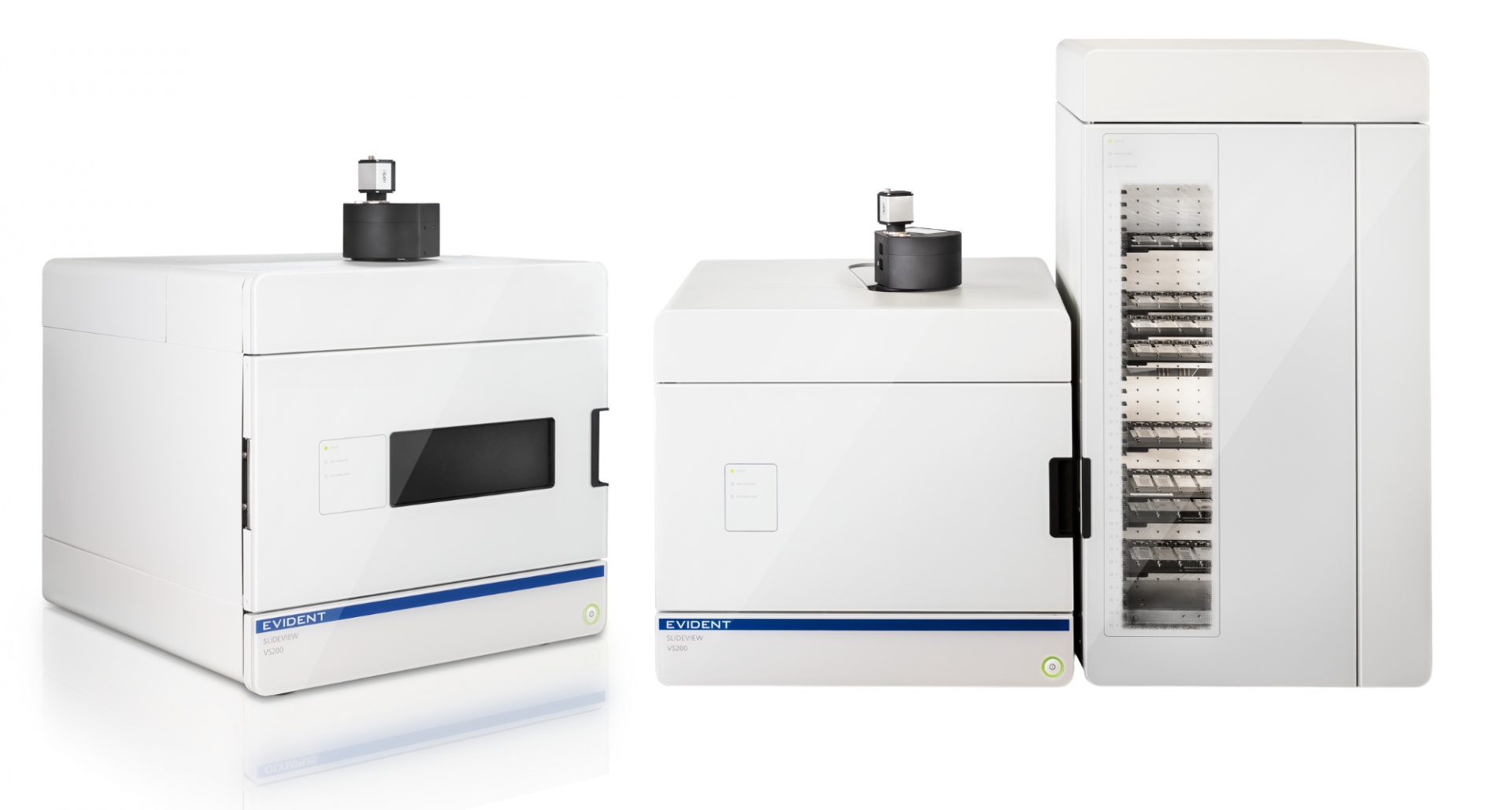
| A System That Grows with Your NeedsImage up to six standard slides (26 × 76 mm or 1 × 3 in.) simultaneously with the single-tray system. As your throughput needs grow, upgrade on-site by adding the loader, which accommodates up to 210 standard slides in 35 trays. The loader’s robotic system moves entire trays—not individual slides—helping keep your samples safe and intact. |
Confidence in Your Data Quality
|
|
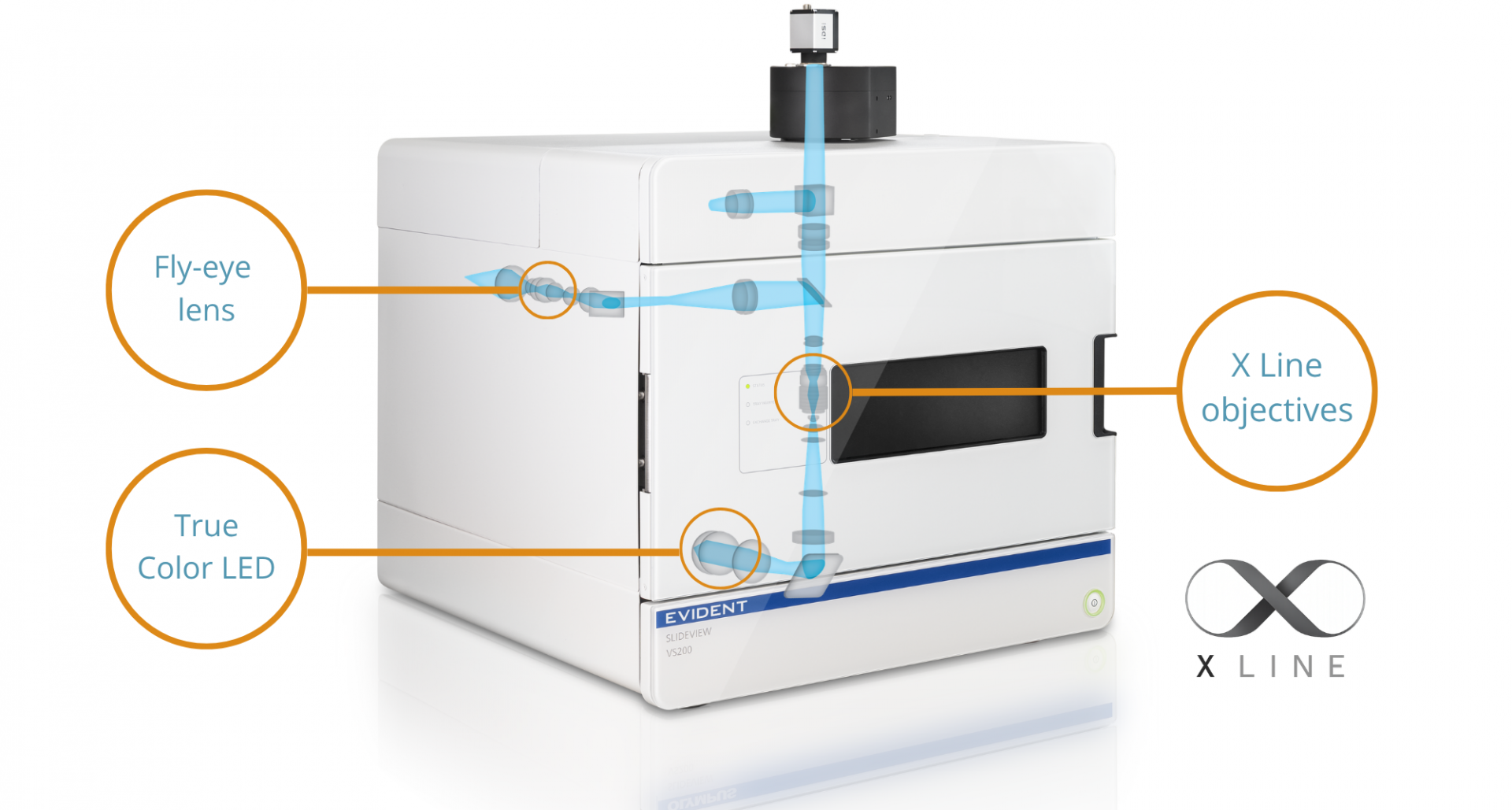
| “The system has been an incredibly user-friendly tool for scanning both brightfield and fluorescent sections. In particular, for fluorescently stained slides, it allows me to scan whole slides or tissue areas quickly and efficiently, providing high-quality whole slide images rather than limited fields." Nadia Nasreddin CRUK Scotland Institute |

View Samples Down to the Subcellular LevelThe automatic liquid dispenser enables batch scanning with high-magnification oil or silicone oil objectives—eliminating the need to pause and manually apply the immersion medium. Combined with the UPLXAPO40X oil objective (NA 1.40), this system achieves high-resolution imaging with up to six times less exposure time compared to a 40X dry objective.
|
| “In our liquid biopsy cancer research, we analyze complex multicolor fluorescently stained tissue and cell samples. These specimens require high-resolution imaging across seven fluorescent channels, multiple Z-layers, and larger areas, which poses technical challenges. Using the SLIDEVIEW VS200 scanner, we achieve detailed whole slide imaging with the precision necessary for our research applications." Amin El-Heliebi Medical University of Graz Evident Discovery Center |

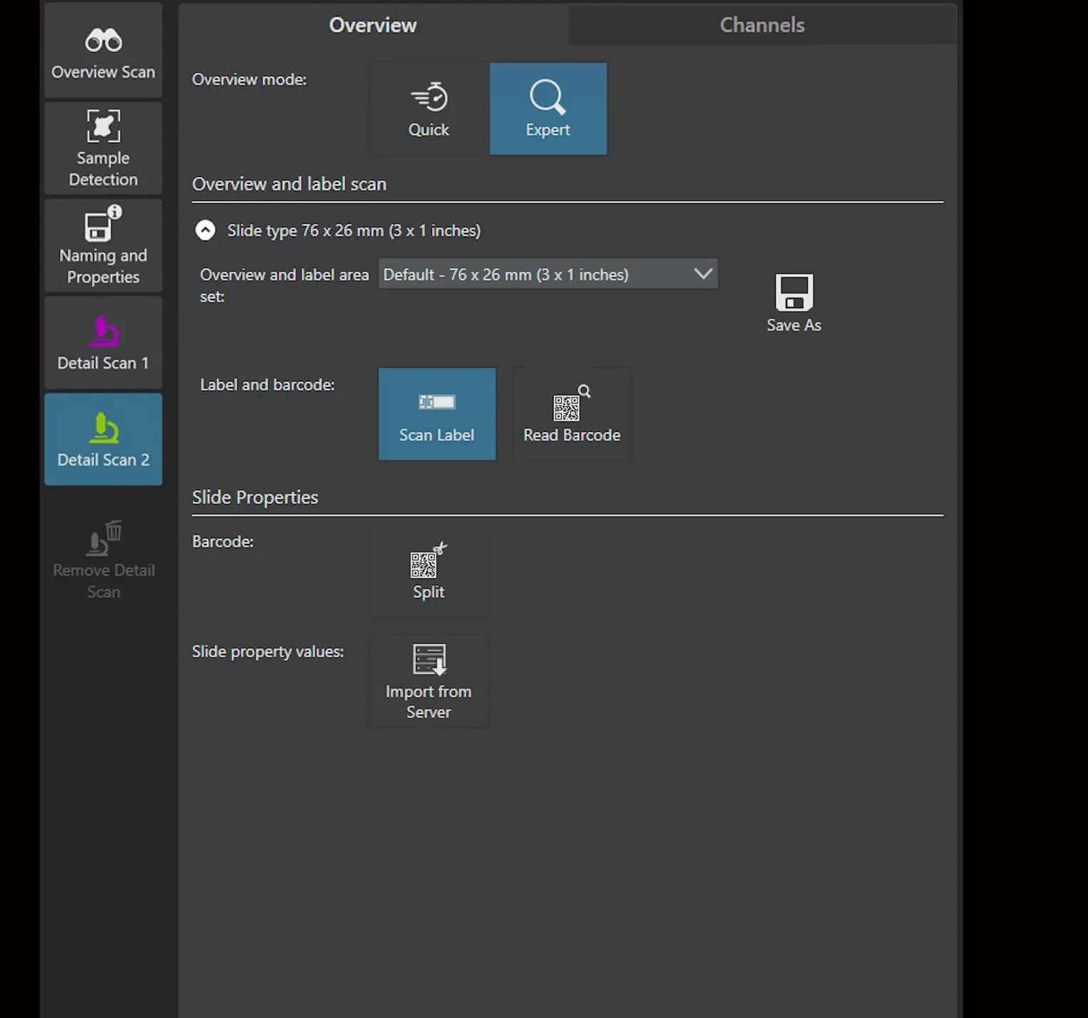
Stay Productive with Software Designed for All User LevelsAn intuitive interface guides the setup process, so users of all levels can produce high-quality data with ease.
|
|
Let Deep Learning Do the Work for You
*Pretrained models are general and will not perform perfectly in every instance. |
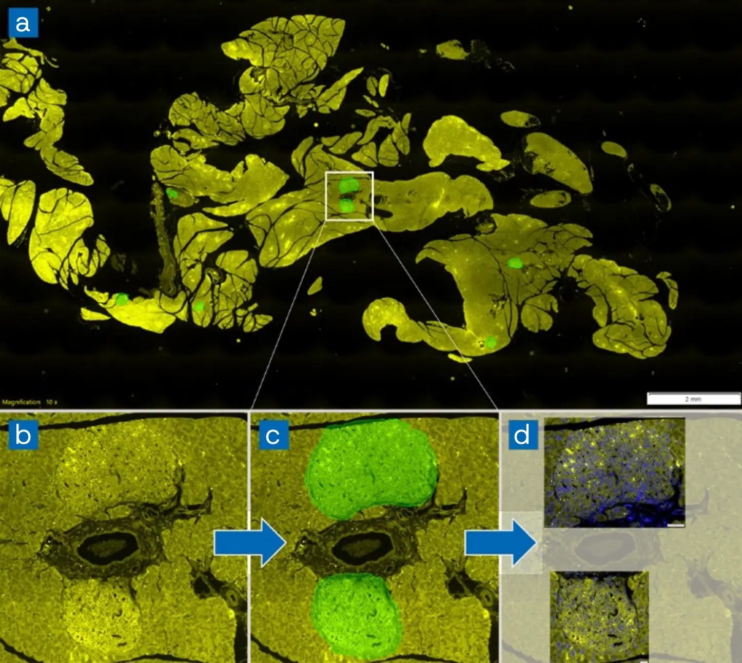
The figure presents an example of selective detection performed using a neural network trained to identify pancreatic islets (PIs). a) Overview of a rat pancreas section stained with fluorescent labels (Alexa Fluor 594, shown in yellow) at 4X magnification. b) and c) Higher-magnification images of two pancreatic islets. d) Final scans (DAPI in blue and Alexa Fluor 594 in yellow) superimposed on the overview image. Only two regions containing pancreatic islets were rescanned at higher magnification (40X). |
Share and Analyze Your Images Easily with Collaborators
|
|

Reveal More Biology in Every Scan
Visualize more biomarkers in one preparation and streamline your spatial biology workflows—without the need for spectral unmixing.
| |
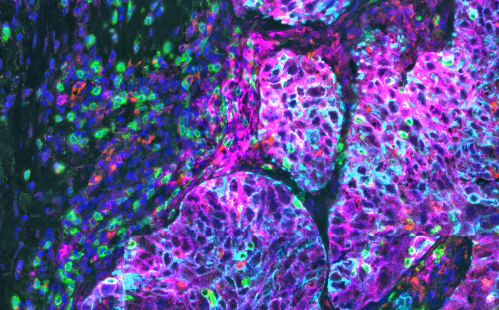 Left: Lung tissue stained with Ultivue’s PD-L1 multiplex kit and imaged with the SpectraSplit 7 filter set and the Excelitas Novem. DAPI: nuclear counterstain, FITC: CD8, TRITC: CD68, Cy5: PD-L1, Cy7: panCK. Imaged at 20X on a VS200 scanner. Image data courtesy of Ultivue, Inc. | 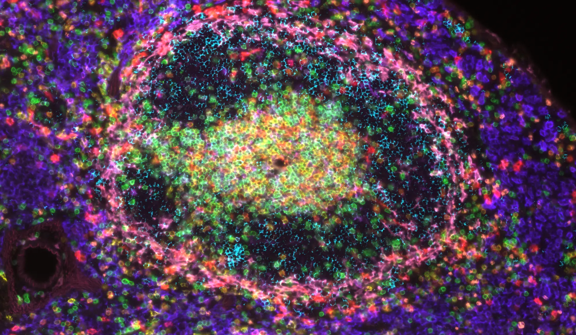 Right: Mouse spleen stained with DAPI and antibodies to F4/80, B220, TCRβ, CD11c, Gr-1, CD4, CD8a, and CD169. Imaged at 20X on the VS200 system with SpectraSplit 10 filters from Kromnigon and the pE-10 from CoolLED. Sample courtesy of Kromnigon. |
 | “The SLIDEVIEW VS200 system has been an invaluable addition to our digital whole slide scanning capabilities, improving HistologiX's operational efficiency and empowering our multiplex assay development and quantitative image analysis services. By reducing administrative workload, it has enabled our scientists to focus on what they do best—applying their expertise in tissue-based analysis to advance our clients' cutting-edge biomedical research. We've been especially impressed with the image quality, which helps us support our clients' research aims." Laura Hubbard |
True Clarity in Thick SamplesCapture sharp images in applications where conventional widefield slide scanners lose detail. The speckle illumination acquisition (VS-SILA) device enables high-quality images of thick samples by removing out-of-focus light through optical sectioning. |
Unlock the full potential of your VS200 scanner with two synchronized light sources delivering uniform, high-intensity illumination. The motorized dual light adapter lets you switch effortlessly between LEDs and lasers—all in one system. From faint fluorescence to dense tissue sections, fine details stand out with clarity. Easily integrated into existing VS200 scanners, the dual light adapter makes on-site upgrades convenient and efficient. |
|
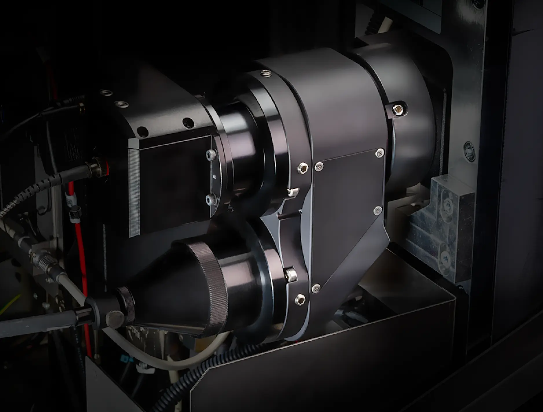
VS-SILACapture sharp images in applications where conventional widefield slide scanners lose detail. The speckle illumination acquisition (VS-SILA) device enables high-quality images of thick samples by removing out-of-focus light through optical sectioning. The compact device easily integrates into the VS200 scanner and software and can be added to both new and existing systems. |
Net Image Server NIS-SQLManaging the large amount of data generated by your VS200 scanner is easier than ever thanks to compatibility with our Net Image Server (NIS) SQL database and OlyVIA™ web programs. You can automatically upload your images to one or more databases, differentiate between users, and take advantage of offline visualization and annotation tools. |
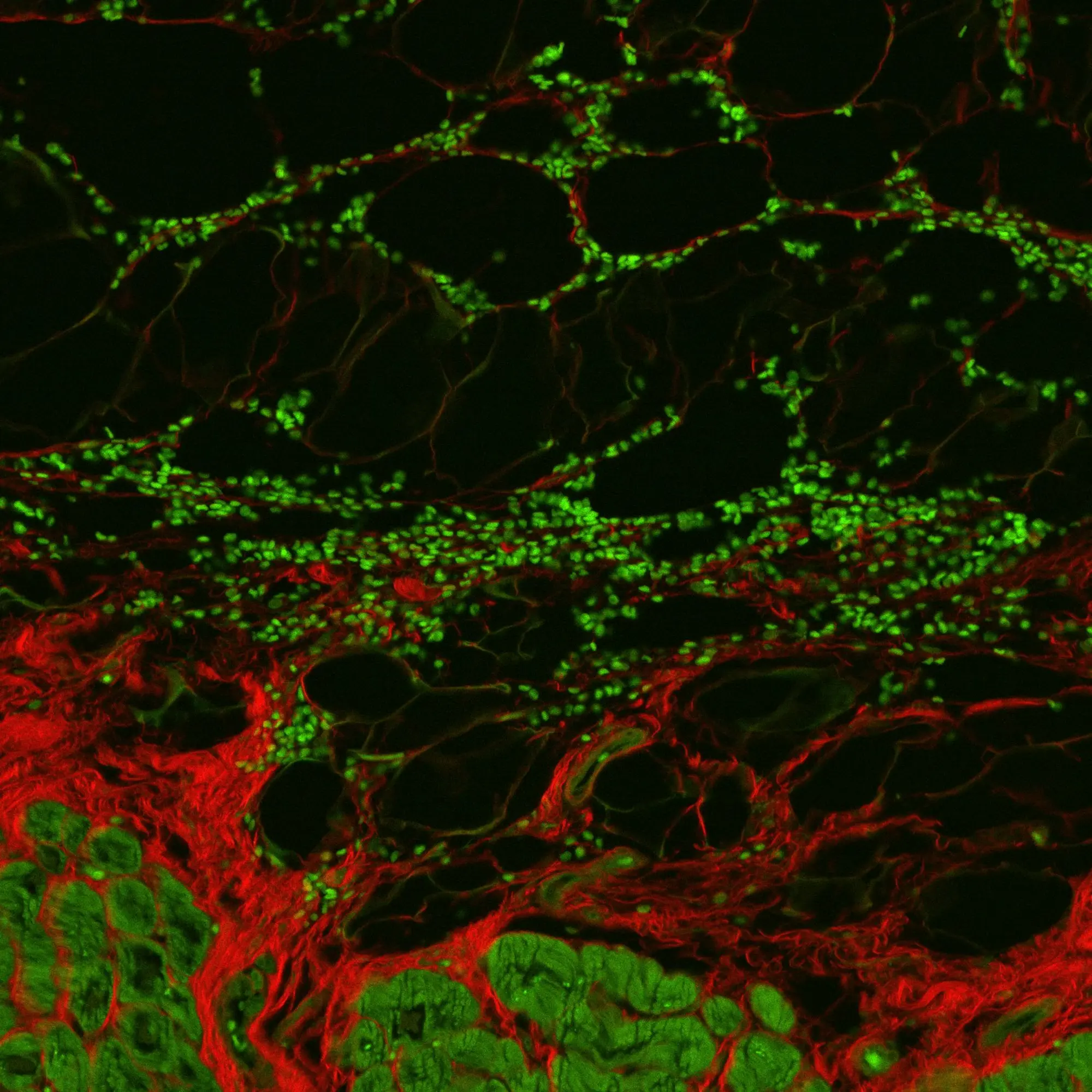
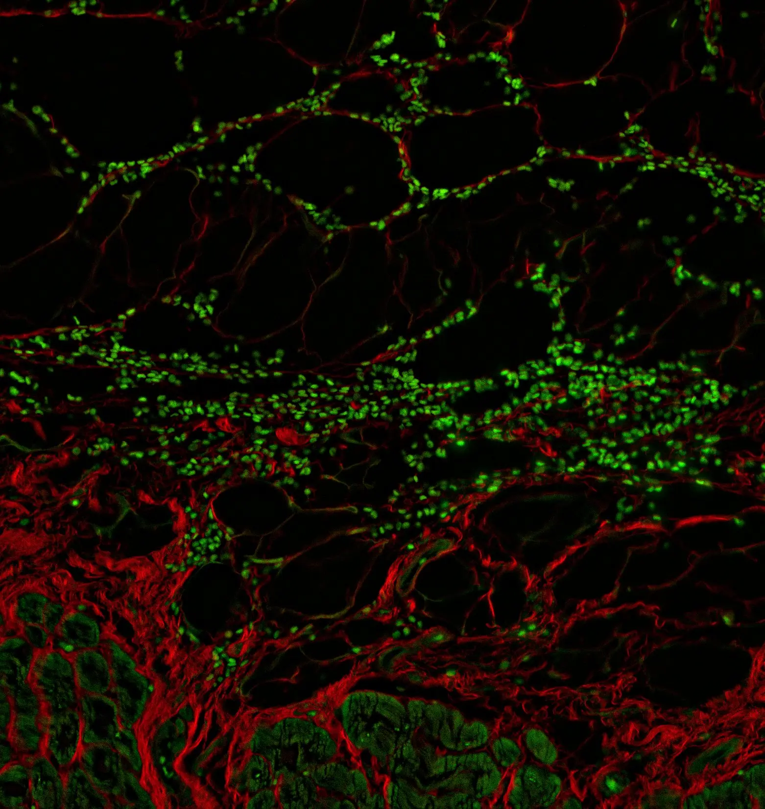
TruAI™ Deep-LearningTruAI technology, powered by deep learning, enhances image analysis accuracy and improves workflow efficiency. Traditional threshold-based methods struggle to extract shape features from samples and accurately identify target areas. With TruAI, users can train the system by selecting pancreatic islet features in a pancreas sample. The system then automatically and precisely detects only the pancreatic islets, allowing for quick and accurate measurement of their number and size. |
X Line Extended Apochromat ObjectivesX Line™ extended apochromat objectives are high-performance lenses that offer simultaneously enhanced brightness, resolution, uniformity, and color with our most advanced lens technology. Our award-winning X Line UPLXAPO series enables consistent quality across applications, from brightfield and fluorescence to confocal and super-resolution microscopy. These lenses are corrected for four wavelengths, exceeding the standard three-wavelength correction of conventional apochromats for sharper images with accurate color across an industry-leading spectral range of 400–1000 nm. Whether used in pathology, laboratory diagnostics, high-end research, or whole slide imaging, X Line objectives help ensure optimal image quality from the center to the edge of the field of view. |



Products Related to This Application
Sorry, this page is not
available in your country.