
Takeo Ogama
Takeo Ogama is a senior product and strategy planner and a product manager for microscope cameras at Evident. He has eight years of experience working in the research and development department for various products, including cameras, and eight years of product planning, marketing, and management experience. He holds a master’s degree in neutrino physics from Osaka University in Japan.

Alket Mertiri
Alket Mertiri, PhD has a background in optics and photonics. He received his PhD in materials science and engineering from Boston University where he worked on developing novel microscopy techniques. From 2018–2019, he was an associate product manager at Olympus, advancing scientists’ research by finding solutions from basic microscopy to advanced technology like total internal reflection microscopy.

Lauren Alvarenga
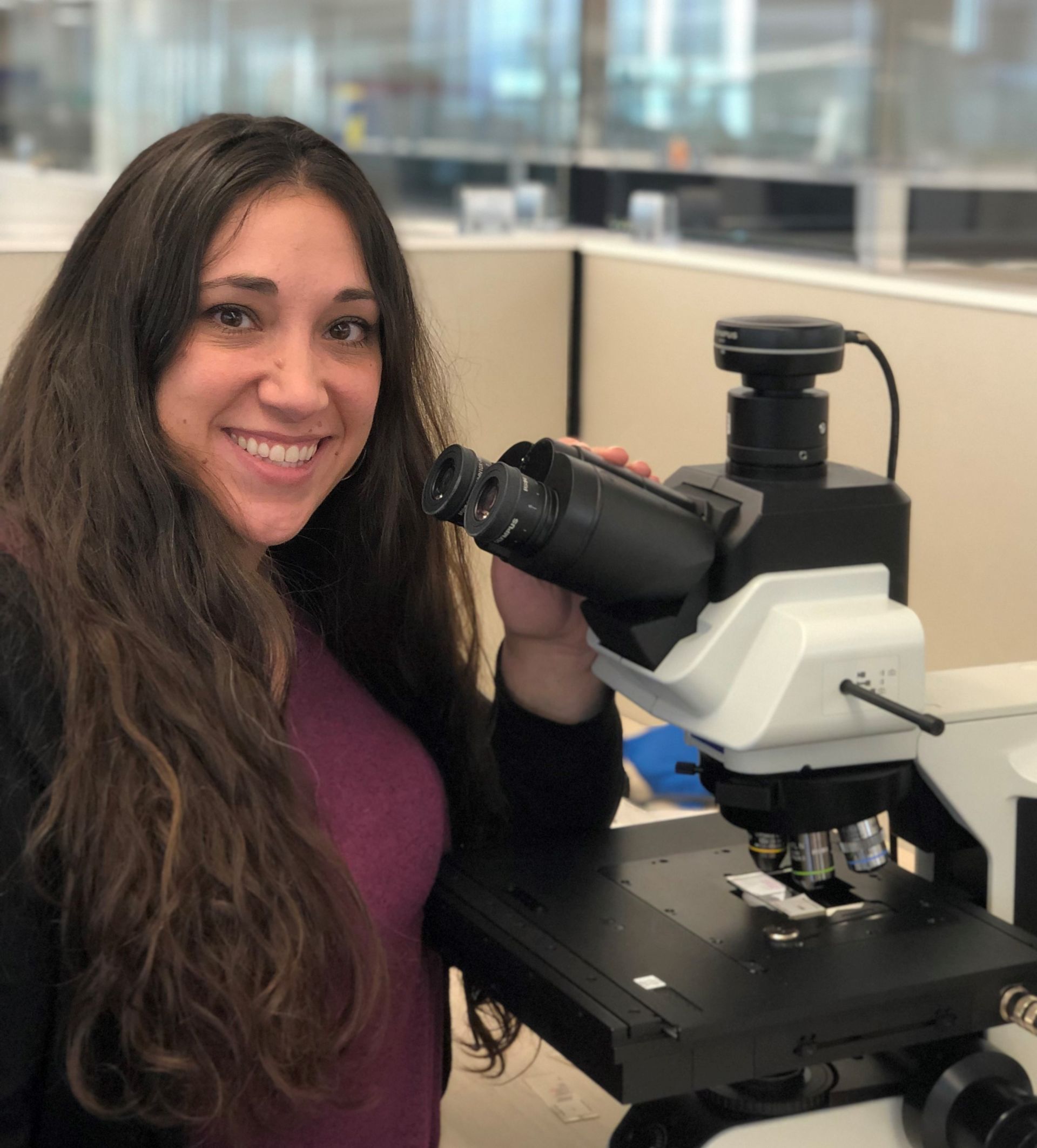
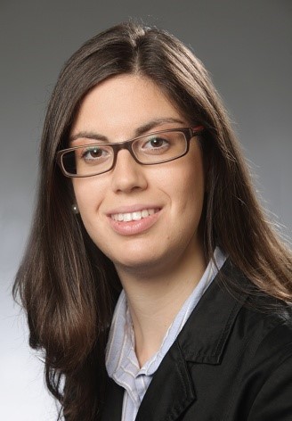
Lauren Alvarenga is a senior product manager for clinical microscopy at Evident. She specializes in objective lenses and imaging software. She holds a Bachelor of Science in biomedical photographic communications from the Rochester Institute of Technology.

Ralf Schaefer
Ralf Schaefer graduated from Hamburg University with a degree in chemistry. He has worked in marketing communications in various technical environments for several agencies and companies. Ralf drove and shaped the visibility of the microscopy and industrial businesses at Olympus in various projects and roles.
Kerry Israel
Kerry Israel is the Manager of Marketing and Communications for Life Science at Evident. She has a Bachelor of Arts degree from Brandeis University, and more than 15 years of experience in all aspects of marketing, from advertising and social media strategy to grassroots outreach.

Alec De Grand
Alec De Grand is a product manager for virtual slide scanning and upright microscopes for life science at Evident. He has been with Evident for over 10 years, during which he has managed clinical products, marketing initiatives, imaging courses, and trade shows.
Carlo Alonzo
Carlo Alonzo, PhD is sales representative for life science microscopy at Evident. He helps scientists identify and understand technologies that support their research goals. His interest in biomedical optics drew him to the academic community in Boston, where he spent several years immersed in research using multiphoton microscopy. He holds a doctorate in physics from the University of the Philippines and pursued postdoctoral training at the Technical University of Denmark and the Massachusetts General Hospital of Harvard Medical School.

Joanna Hawryluk
Dr. Joanna Hawryluk is a product manager for research imaging at Evident Scientific located in Waltham, Massachusetts. She received her doctorate degree from the University of Connecticut within the department of Physiology and Neurobiology. She has been with Evident since 2017 and is currently responsible for laser scanning microscopes, 3D cell analysis software, and lightsheet microscopes.

Laz Amador
Laz Amador is a senior manager with the national applications team at Evident. Laz has 36 years of experience in microscope sales and has been with Evident since 1988. Prior to joining the applications team, Laz was the executive director of technical marketing in Latin America. Laz lives in Weston, Florida, with his wife, Jan.

Rebecca Chandler
Rebecca holds a bachelor's degree in journalism from Endicott College and writes about trends and technologies in science and industry. She works closely with Evident engineers and scientists to write pieces about the latest laser scanning, super-resolution, multiphoton, upright, stereo, and inverted microscope systems, as well as leading-edge optics, cameras, and software. Follow her work to learn about Evident's latest for numerous applications, including cytology, pathology, education, and more.

Dr. James Lopez
James Lopez received his PhD in biomedical sciences from the University of Chicago in 2010. With nearly a decade of experience in calcium imaging, FRET, live cell imaging, and intravital imaging, James joined Evident as a confocal and multiphoton sales representative. He later transitioned to Evident’s Life Science Applications Group supporting confocal and multiphoton systems. Now he manages the Life Science Applications Group in the US, Canada, and Latin America markets.
.jpg?rev=29C8)
Matthew Weitzman
Matthew Weitzman holds a PhD in biological sciences from the University of Delaware and has numerous years of advanced imaging experience. From 2014–2021, he worked with Olympus’ confocal and multiphoton imaging systems to assist with product development and application support.

Janeen Manning
Janeen Manning is a product manager for clinical and educational microscopes at Evident. Before joining Evident, she worked for a medical device company specializing in infectious diseases. She has a Bachelor of Science in biochemistry, microbiology, and molecular biology from the University of Maine and a Master of Liberal Arts in biotechnology from Harvard University.
Christopher D. Higgins
Christopher Higgins is the business development manager for life science at Evident. A 24-year veteran of the microscopy field, he has spent much of his career in product management and applications for a wide variety of biological light microscopes used in both research and clinical applications.
In his current position, Chris works directly with researchers and pathologists on the development of new technologies and applications. Images captured with today’s virtual microscope systems tell the stories of not only individual cells but entire tissues, and are vital for research, education and more. Chris believes the next great frontier is preparing whole slide imaging and artificial intelligence analysis to enter its next era as it moves toward acceptance for wider application in the clinical realm.
A member of the Digital Pathology Association, Chris holds a bachelor’s degree from the University of Miami. He is an avid outdoorsman and accomplished nature photographer, and lives in Lehigh Valley, Pennsylvania, USA.

Cheng-Hao Chien
Dr. Cheng-Hao Chien received his PhD in biophotonics from National Yang-Ming University, Taiwan, and pursued postdoctoral training at the Department of Neuroscience, Tufts University School of Medicine, Boston. With a decade of experience in advanced microscopy and life science research, he worked with Olympus’ multiphoton microscopy and customized solutions from 2020–2021 to provide product and application support.

Brendan Brinkman
Brendan Brinkman spent years working in the stem cell field, including primary mouse neurosphere derivation. He joined Evident after several years as a core imaging facility manager and assisted in building novel systems. In 2017, he returned from 2.5 years with Evident Tokyo, where he helped develop next-generation solutions.

Kathy Lindsley

Manoel Veiga

Heiko Gäthje

Rebecca Bonfig
Dr. Rebecca Bonfig completed her graduate work at the University of Louisville in the Department of Physiology and Biophysics, studying the post-transcriptional regulation of the renal phosphate transporter Npt2a by parathyroid hormone. From 2015–2022, Dr. Bonfig worked with our confocal microscopes, supporting the FLUOVIEW™ series as a product manager at Evident.

Bülent Peker
Flavio Giacobone

Shohei Imamura
Shohei Imamura is a strategic project manager at Evident. He has four years of experience working in scientific microscopy sales and seven years of experience in product planning, strategic project management, and project execution. He has a Bachelor of Commerce degree from Meiji University in Japan.

Daniel Bemmerl

Paul Monnier

Dr. Gulpreet Kaur
Dr. Gulpreet Kaur completed her PhD in Molecular Biology at the University of Wisconsin-Madison, where she studied organelle biology using live-cell spinning disk confocal microscopy.
Dr. Kaur has over six years of experience in microscopy, and has worked on various technologies including confocal, spinning disk, super resolution, and light sheet imaging. She served as a trainer for a microscopy and image analysis core facility at University of Wisconsin-Madison and consulted with scientists on experiment design and optimization of imaging conditions.
From 2018–2022, Dr. Kaur helped scientists apply Evident’s microscopy and imaging technologies in their research projects.

Dr. Nicolas Bourg

Sarah Williams
Sarah Williams worked for nearly a decade as a researcher and copywriter in the broadcast media industry. Now Sarah applies her skills as a writer and editor to produce compelling, high-quality material on topics related to Evident’s wide range of life science solutions. She writes about the latest laser scanning, multiphoton, super-resolution, stereo, upright, and inverted microscope systems, as well as the optics, cameras, and software that support them. She also explores their applications and contributions to cytology, pathology, education, and other fields. Sarah works at the office in Quebec City, where she resides with her partner, David, and her three children, Sophie, Anouk, and Éloi.

Dr. Mike Woerdemann
Mike Woerdemann earned his PhD in optical tweezer technology based on structured light fields. He worked as an application specialist supporting high-content screening systems for many years, and during that time he gained in-depth, practical knowledge of the field. Mike is now a product manager for high-content screening stations and deep-learning products developed at the EVIDENT Technology Center Europe.

Chunsong Yan
Chunsong Yan is a business development manager for life science at Evident in Australia and New Zealand. He is currently responsible for confocal, multiphoton, light sheet, and slide scanning systems. Since joining Evident, Chunsong has worked in various roles while always striving to offer the best imaging solution to customers.
Minju Kim

Junsung Kim

Ganesh Kadasoor

Wei Juan Wong

Srivats Hariharan

Stefan Marawske
Bunryu Arashi
Bunryu Arashi works as an OEM manager at Evident in Europe. With 12 years’ experience in microscope product development, Bunryu has designed objectives and camera adapters, as well as illumination optical systems for BX™ and CKX™ series microscopes. He has a master's degree in engineering from Osaka University in Japan.

Craig Rappaport
Craig Rappaport is a senior microscope sales representative for Evident Life Science. He has 26 years of microscope sales experience, along with a dozen years in scientific and technical marketing. Since joining Evident, Craig has worked on market analysis, voice of customer surveys, and new product research. He has been with the sales team since 2012 covering San Diego and Imperial Counties in Southern California. Craig earned his Bachelor of Arts at UC Santa Barbara and his MBA at Duke University Fuqua School of Business.

Marcus Krauel
Marcus Krauel is an OEM manager at Evident in Europe. With 12 years’ experience working with optics, he helps our OEM customers across Europe on applications ranging from industrial to medical. Marcus holds a degree in physics and optical engineering.

Klaus Willeke
Klaus Willeke is a product marketing manager at Evident Life Science. Klaus has worked at Evident for more than 22 years. He started as a sales representative for industrial and life science microcopy in Germany for 17 years and later joined the European Product Marketing team, where he is responsible for upright clinical and research microscopes and X Line™ objectives.

Yu Kikuchi
Yu Kikuchi is an optical engineer for component products and microscope products at Evident. He has experience in optical design and evaluation of microscope products. Yu has focused on developing customized products, matching specifications to customer requirements, and on-site testing and alignment. He holds a master’s degree in molecular biology from Tohoku University in Japan.

Stephan Fliegel
Stephan Fliegel earned his university degree in physical engineering with a focus on laser application technology at the Münster University of Applied Sciences in 2007. He initially worked as an application specialist, then as a product and project manager in the industrial lasers field focusing on the design layout, development, and planning of customer-specific laser systems. He is now a product manager for light microscope cameras at Evident.

Dr. Daniel Göttel
Daniel Göttel earned his PhD in human genetics at the German Cancer Research Center in Heidelberg, Germany. He worked as an application specialist for virtual slide scanning systems for many years and gained in-depth, practical knowledge of the field. Daniel is now a sales support manager for the SLIDEVIEW™ VS200 research slide scanner and Net Image Server (NIS) SQL at Evident.
Lee Botes
Lee Botes has 24 years of sales experience with Olympus microscopes. Bringing a background in microbiology, Lee began her microscope sales career in South Africa. As part of the sales team at Evident in New Zealand, she is responsible for our industrial, clinical, and research microscopes in Wellington and the South Island, as well as confocal microscopes and slide scanners in New Zealand.

Masato Yabe
Masato Yabe works in the Optical Development division at Evident, where he has been involved in the development of microscope products for 19 years, designing optical systems for objectives and autofocus units. He holds a master’s degree in engineering from Wakayama University in Japan.

Shodai Hosono
Shodai Hosono is a member of Evident’s mechanical engineering department. He has been involved in developing microscope products for over a decade. As a mechanical engineer, he has designed various microscopes and modular microscope assemblies. He holds a bachelor’s degree in engineering from Saitama University in Japan.
Kei Sato
Kei Sato works in the electrical engineering department at Evident and has been developing microscope products for nine years. As a product leader, Kei is in charge of electrical design for industrial microscopes and microscope components.

Joanna Hawryluk
Dr. Joanna Hawryluk is a product manager for research imaging at Evident Scientific located in Waltham, Massachusetts. She received her doctorate degree from the University of Connecticut within the department of Physiology and Neurobiology. She has been with Evident since 2017 and is currently responsible for laser scanning microscopes, 3D cell analysis software, and lightsheet microscopes.

Dr. Hunter Hines
Dr. Hunter Hines received his PhD in 2019 while registered in a joint international collaboration with Bournemouth University (UK) and Harbor Branch Oceanographic Institute (HBOI) at Florida Atlantic University. He conducted research into microbial communities found in tropical aquatic and terrestrial ecosystems in Florida, focusing on the biogeography and biodiversity of ciliates.
Today, Dr. Hines continues his research at HBOI, where he examines microbial communities and the impact they have on ecosystems. He is also the senior scientist on the Amoray Marine Conservation Team at the Amoray SCUBA Resort in Key Largo, Florida.
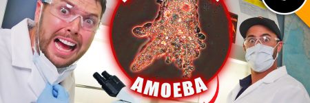
On social media, Dr. Hines plays a significant science outreach role for microbiology and ecology. During his PhD, he created the Instagram account Microbialecology, which has over 140,000 followers. He is a frequent collaborator with the popular YouTube channel Brave Wilderness, assisting with everything microbial and featuring in several episodes.

Kenji Ono
Kenji Ono works in the Optical Component Department at Olympus. Since joining the company, he has helped to plan and develop optical systems for digital cameras in our imaging business. Applying his knowledge to the optical field, he is now in charge of marketing activities for OEM microscopes. Kenji earned his bachelor's degree at Kitasato University in Japan.
Kristina Mayer
Kristina Mayer is a product marketing manager for inverted microscopes in the European Product Marketing team at Evident. She has worked at Evident for more than 10 years and is responsible for the IXplore™ inverted microscope series to support research imaging, including total internal reflection fluorescence microscopy (TIRF) and spinning disk confocal microscopy within the EMEA region. Kristina holds a PhD in cell biology from Jacobs University Bremen.

Benjamin Compans, Ph.D.

Yuan Yue
Yuan Yue works in the market strategy department at Evident where she is mainly in charge of digital marketing. She has nearly six years of experience in marketing communications at Evident.

Shogo Usui

Dr. Anne Beghin

Dr. Xiaotong Cui

Dr. Kasmira Wilson

Dr. Dan Zhu

Dr. Graham Wright

Mr. Srivats Hariharan

Ms. Gency Gunasingh

Dr. Dong Gao

Dr. Yu Weimiao

Dr. Motoki Takagi
Dr. Ningbo Wu
.jpg?rev=47A5)
Mr. Hiroya Ishihara

Marie Kawasaki
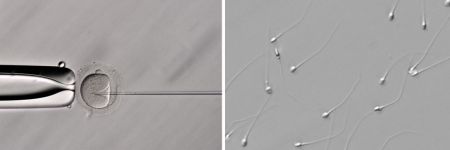
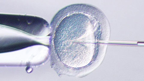
Marie Kawasaki works in the Life Science Marketing division at Evident. She has been involved in the marketing of microscopes related to clinical research for seven years and is currently in charge of global sales promotion for the fertility treatment research (intracytoplasmic sperm injection) industry.
Wei Juan Wong
Wei Juan Wong is an application specialist for digital slide scanning systems at Evident. She started as a product specialist in Singapore to support our Southeast Asia customers using widefield microscopes, including the SLIDEVIEW™ VS200 research slide scanner. She later moved to Germany to join the EVIDENT Technology Center Europe as an application specialist for digital slide scanning systems, where she provides application and marketing support to customers worldwide. Wei Juan has a degree in physics and has worked in a biophysical research laboratory as well as a microscopy core facility.
Samuel M Lawrence
Samuel M. Lawrence is the chief executive officer and cofounder of CytoViva, Inc. He led the development of the core, patented optical illumination technology that is the foundation of CytoViva’s products, as well as played a key role in the development of the patented CytoViva 3D imaging technology. He continues to lead all product development efforts and actively supports the company’s sales efforts both domestically and internationally.

Manoel Veiga
Manoel Veiga earned his PhD in physical chemistry at the University of Santiago de Compostela, Spain, working with picosecond and femtosecond time-resolved spectroscopy. Following two postdocs at the Complutense University of Madrid and the University of Münster, he worked for PicoQuant as a senior scientist in the fields of time-resolved spectroscopy, fluorescence lifetime imaging microscopy (FLIM), and fluorescence correlation spectroscopy (FCS). Manoel now works as a global application specialist at Evident in Germany, where he focuses on high-content analysis (HCA) and deep learning.

Dr. Manfred Kässens
Dr. Manfred Kässens leads the Technical Product Information department at the EVIDENT Technology Center Europe and is a member of the Global Marketing Communications department at Evident. He holds a PhD in physics from the University of Münster, Germany, and has worked at Evident since 1997.
Sara Quiñones Gonzalez
Sara Quiñones González has a degree in biotechnology. She worked in several research and clinical laboratories before joining the EVIDENT Technology Center Europe as a product manager. She is responsible for the SLIDEVIEW™ scanner series, including the VS200 research slide scanner.

Dr. Thomas Bauer-Jazayeri

Maria Ada Prusicki
Maria Ada Prusicki received her PhD in biology from the University of Hamburg in 2019. During her doctoral studies she focused on live-cell imaging techniques to track plant cell divisions. While pursuing a postdoc, she continued to deepen her knowledge in microscopy techniques. In 2022, Maria became the application specialist for our SLIDEVIEW™ VS200 research slide scanner. Working out of Evident’s Munster, Germany office, Maria provides VS200 application support to our customers worldwide as well as Evident’s global sales and marketing teams.
Taichi Yoshikura
Taichi Yoshikura leads the Global Life Science Marketing Communication team at Evident in Tokyo, Japan. He has a Bachelor of Arts from Waseda University and has worked at Evident since 2016.
Takuma Saito
Takuma Saito has been part of the industrial microscope sales team at Evident in Tokyo, Japan, since 2011. After developing a strong relationship with electronic component manufacturers in Japan, he currently leads the sales development projects for our laser scanning microscope and digital microscope products.

Dr. Yongjie Wang
Dr. Yongjie Wang is an application specialist for Evident Life Science in Beijing, China. She currently focuses on the scientific applications of advanced microscopy. Dr. Wang received her doctorate degree from the department of Chemistry and Chemical Engineering at Nanjing University.

Koji Nakagawa
For 12 years, Koji was involved in the development of digital cameras and oversaw optical system design for imaging lenses. Currently, he works in Evident’s Optical Development division where he develops microscope products. He holds a master’s degree in engineering from the University of Yamanashi in Japan.

Atsuya Toda
Atsuya Toda is an assistant manager for Global Life Science Marketing at Evident. He has more than fifteen years of experience in life science microscopy sales, sales planning, and marketing in Japan. In 2021, he moved to the Global Life Science Marketing team where he is the marketing representative for the APEXVIEW APX100 all-in-one microscope. He holds a Bachelor of Economics degree from Doshisha University, Japan.

Hikaru Mukai
Since joining the company, Hikaru has been responsible for supporting confocal and super-resolution microscopy products, and she has been a member of the Life Science Marketing department since 2022. Hikaru holds a Bachelor of Science degree from Tokyo University of Science, Japan.

Jake Jones
Jake Jones, PhD is an associate product manager for research imaging at Evident, working with inverted imaging systems, total internal reflection fluorescence (TIRF), fluorescence recovery after photobleaching (FRAP), luminescence, and multiphoton microscopy. As a member of the product management team, Jake works to identify the needs of scientists and researchers and helps provide imaging solutions that match the growing needs of the imaging community. He holds a doctorate in biomedical engineering from the University of Arkansas, where he pursued projects involving biomedical applications of multiphoton microscopy, confocal microscopy, in vivo imaging techniques, and neural network-based image processing.
.jpeg?rev=9FF0)
Avi Smith
Avi Smith is an associate product manager for research microscopy at Evident. He currently supports the product lines for cell culture, confocal spinning disk, and high-content screening software. Before joining Evident, he spent 10 years working in tissue engineering where he focused on developing skin models for drug discovery and development. Avi holds a master’s degree in engineering management from Tufts University.

Bülent Peker
Bülent Peker is an expert on laser scanning microscopy. He first developed an interest in microscopy and photonics during his PhD in physical chemistry, where he worked on time-resolved two-photon microscopy, and this passion has been with him ever since. Bülent has been with Evident for over 18 years and has helped the team introduce leading-edge laser scanning microscopes. He’s particularly driven by how advancements in scanning technology, detectors, and software are continuously redefining the boundaries of resolution, speed, and experimental versatility in modern microscopy.
Kaori Hirayama
Kaori Hirayama currently works in the Life Science Marketing department at Evident where she is in charge of marketing customize products. She has over 10 years of experience working in confocal microscopy support. She holds a Bachelor of Hygienic Technology degree from Kitasato University, Japan.

Dr. Peter Su

Michel Biocco

Chloé Savard
Chloé Savard, also known as @tardibabe on Instagram, is a microbiologist from Montreal who discovered the world of microscopy three years ago. Formally trained as a musician (she plays the drums), Chloe decided to study microbiology to broaden her horizons and to answer some existential questions she had since childhood. On her @tardibabe Instagram channel, Chloé strives to transform the imperceptible into art while educating and raising awareness about the fragility of microscopic ecosystems. She also loves to experiment with various foods and everyday items.

Makoto Kuwano
Makoto Kuwano has been in charge of microscope product development for 12 years at Evident. He currently works in the Optical Development Division, where he is involved in optical design of objective lenses, development of component products, and development of measurement techniques for product performance. He holds a Master of Science degree from Tohoku University in Japan.

Dr Grace Yuan
Dr. Grace Yuan earned her PhD at the Shanghai Institute of Plant Physiology and Ecology, Chinese Academy of Sciences. She previously worked as a senior scientist in the field of imaging cytometry. Zhenhuan brings her imaging expertise to Evident as an application specialist with a focus on confocal microscopy, multiphoton microscopy, and high-content analysis (HCA).

Shyam Rathod
Shyam Rathod holds a bachelor’s degree in electrical engineering from the Veermata Jijabai Technological Institute (VJTI) in Mumbai, Maharashtra, India. He works as the deputy executive engineer at the Maharashtra State Electricity Transmission Company (MSETCL) under the Government of Maharashtra, India. He is passionate about photomicrography and has been pursuing this unique art form consistently using his own limited resources. His efforts have earned him international recognition, and he is committed to popularizing this unique blend of art and science through consistent and diligent efforts in refining his images.

Amin El-Heliebi
Masatoshi Dehari
After working in sales for biological microscopy in top research fields in Japan, Masatoshi brought his expertise to Evident in 2023. As part of the Life Science Global Marketing team, he is responsible for innovative products such as the APEXVIEW™ APX100 all-in-one microscope and the CM30 incubation monitoring system. He holds a master’s degree from the University of Tokyo.

Kazuhiko Hosono
Kazuhiko Hosono has a background in biophysics and a Master of Science degree from Waseda University. He joined Evident in 2004 and has over six years of experience in optical development and customized solutions, followed by five years of technical support as a development representative in Hamburg, Germany. After returning to Japan, he joined the global marketing department, where he has been involved in the planning, introduction, and sales expansion of a wide range of life science research products, including confocal laser scanning microscopes, high-performance microscope objectives, and inverted microscope systems.

Gianluca Franco
As territory manager for Evident’s Life Science Center, Italy, for over four years, Gianluca Franco is a skilled specialist with a strong understanding of optical microscopes and analysis techniques. His interest in this field began during university while working on fluorescence microscopy for his master’s thesis in bioengineering and biomedical engineering, and it has persisted ever since. Gianluca provides outstanding support to the team for microscope sales by delivering demonstrations and presentations of advanced imaging systems. He provides support for the growth of the optical microscopy business by identifying and developing new business opportunities.
Britta Frenzel
Britta Frenzel has a strong commercial and technical background with a master's in biomedical engineering from Clemson University and several years of experience in the medical device industry. From 2022–2024, Britta supported Evident’s line of benchtop fluorescence microscopes, fixed stage microscopes, and macro zoom fluorescence microscopes on the product management team, where she worked to identify the needs of researchers and provide imaging solutions that match the growing needs of the imaging community.
.jpg?rev=5CD9)
Akira Saito
Akira Saito studied veterinary medicine at Tokyo University of Agriculture and Technology, Japan, and graduated in 2007. Shortly after, he joined Evident as an application specialist responsible for in vivo imaging systems, high-content analysis systems, and laser confocal systems to support customers in Japan. In 2013, he took over sales promotion for all Evident life science products. In 2018, he moved to Singapore to provide marketing and application support for the APAC market, then moved back to global product marketing in 2023.

Enrico Poege
Enrico Poege is the Global Marketing Communications Lead for Material Science, based in Hamburg, Germany. He holds a diploma in Business Administration from the University of Leipzig and has more than 15 years of experience in marketing and communications.

Dr. Laura Lleras Forero
Dr. Laura Lleras Forero is an accomplished product marketing manager for the EMEA region at Evident, with a strong academic background in biology and a PhD from King’s College London. With more than 10 years of professional experience, she has been with Evident since 2021, where she plays a key role in supporting advanced cell culture microscopy solutions, including the SLIDEVIEW™ VS200 research slide scanner and the APEXVIEW™ APX100 benchtop fluorescence microscope.
Hélie Pigeaud
Hélie Pigeaud is the Export Manager of Argolight, the French biotechnology company leading quality control and quality data monitoring solutions for fluorescence-based systems. With over a decade of experience and commitment to innovation, Argolight has developed cutting-edge technology that combines both hardware and software to provide seamless experience for its users. After graduating with a degree in law and business management and a master's in international business development, he joined Argolight in June 2020 to oversee business development in North America and Asia-Pacific.
Over the past years, his role has been to develop Argolight's activities from France and the United States of America through a network of business partners and partnerships with fluorescence system manufacturers worldwide.
Ryoji Kitamura
Ryoji Kitamura earned his master’s degree from the Graduate School of Information Science and Technology at Hokkaido University, where he focused on in vivo imaging using multiphoton microscopy. He began his career at Evident as a software engineer and later became the product leader for the SLIDEVIEW™ VS200 universal whole slide imaging scanner. He also served as the Global Product Manager for the IXplore™ IX85 inverted microscope system. Currently, he is the Global Product Manager for the FLUOVIEW™ confocal microscope series, leading product strategy, planning, and development.

Bülent Peker
Bülent Peker is an expert on laser scanning microscopy. He first developed an interest in microscopy and photonics during his PhD in physical chemistry, where he worked on time-resolved two-photon microscopy, and this passion has been with him ever since. Bülent has been with Evident for over 18 years and has helped the team introduce leading-edge laser scanning microscopes. He’s particularly driven by how advancements in scanning technology, detectors, and software are continuously redefining the boundaries of resolution, speed, and experimental versatility in modern microscopy.

Yuichiro Imai
Yuichiro Imai is a senior manager in the Product Management team at Evident, responsible for overseeing all life science products in the Japan region. After graduating from the Faculty of Economics at Chiba University in 2005, Yuichiro joined Evident and began his career as a sales representative in the life sciences field, where he helped customers solve complex challenges. Following this, Yuichiro held managerial roles in Japan’s sales planning and marketing, leading strategic initiatives to strengthen market presence and customer engagement. In 2024, Yuichiro transitioned to the global Product Management team, where he now leads the development and execution of product strategies tailored to the Japanese market.
Posts by this author
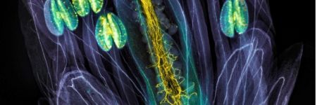
Visualizing Pine Needles with Multimodal Imaging: From Brightfield to Fluorescence
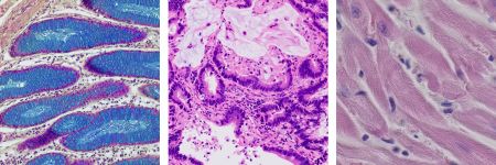
Studying Infectious Diseases in Farm and Companion Animals: From Slides to Analysis
Visualizing Cell Fate Decision with Advanced Microscopy: Interview with Dr. Aoki

Using Microscopy in Translational Research with Liver Organoids: Interview with Dr. Yoneyama
Meet the Microscope Performance Monitor: The Ultimate Tool for Core Facility Managers

Studying C. Elegans Under the Microscope: Evident Partners with BioBus on Science Education
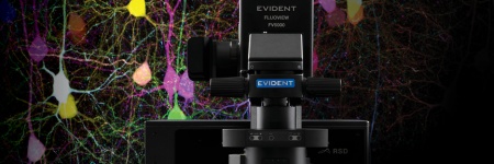
Innovations in Microscopy: FLUOVIEW™ FV5000 Redefines the Boundaries of Confocal and Multiphoton Imaging


Imaging the Retina as a Model Organism in Drug Discovery: From Vision to Insight

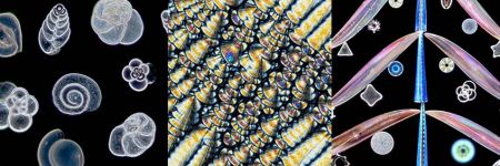
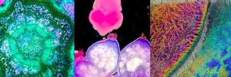
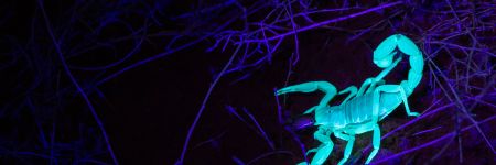
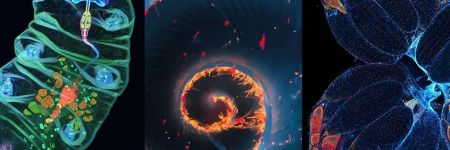
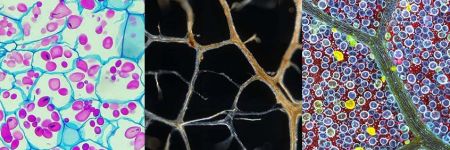
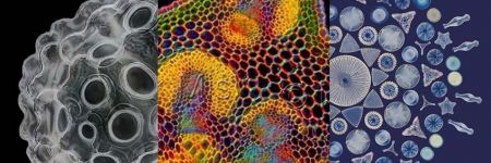
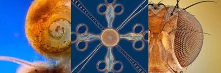
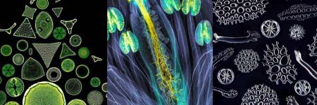
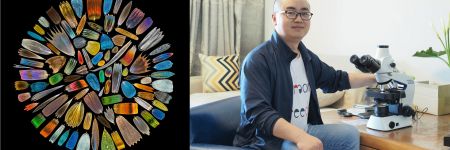
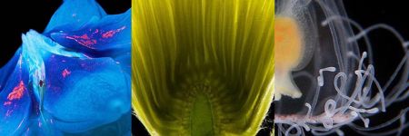
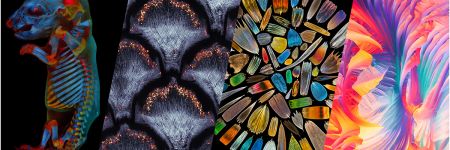
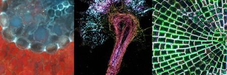
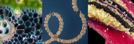
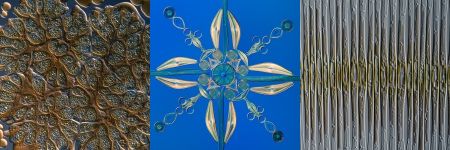
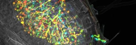
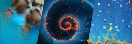
The Revelation of New Perspective—The 5th Annual Image of the Year Award Americas Winner
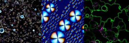
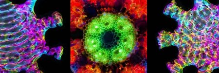
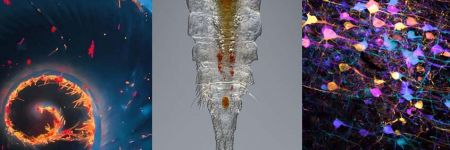
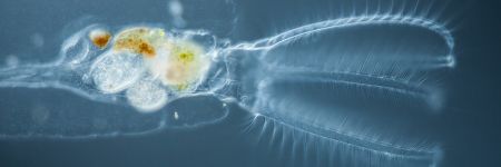
Capturing Beauty of Cosmic Proportions—The 5th Annual Image of the Year Award Global Winner
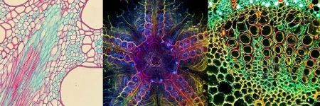
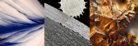
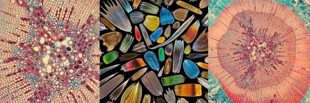
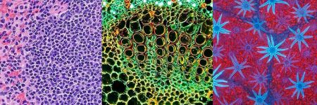
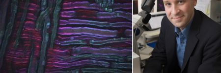
Focusing on What Ties Us Together—The 5th Annual Image of the Year Award Materials Science Winner
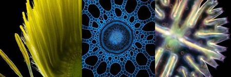
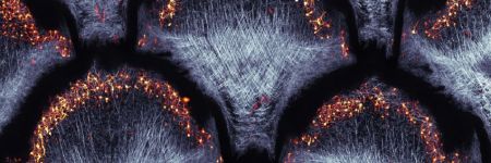
Uncertain Beauty Becomes Crystal Clear—The 5th Annual Image of the Year Award Video Winner
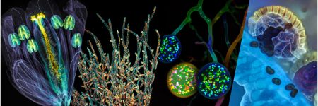
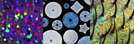
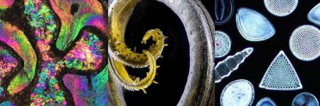
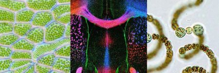
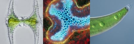
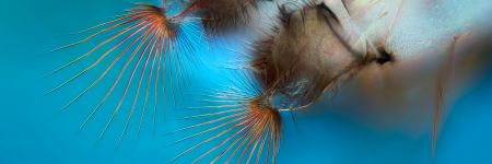
Showcasing Nature’s Glass Gems—The 5th Annual Image of the Year Award Asia-Pacific Winner
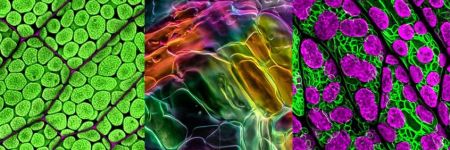
Nature as the Ultimate Artist—The 5th Annual Image of the Year Award EMEA Winner
Frequently Asked Questions about the FV4000 Laser Scanning Confocal Microscope


Nanomaterials Brighten Up Life Science—Bioimaging Applications of Fluorescent Nanoprobes
Dr. Hana Polasek-Sedlackova, Award-Winning Scientist, Reveals the scanR System’s Role in Resolving a DNA Paradox

Crystal Art Photomicrography: Inside the Art of Imaging Crystals with a Microscope
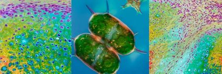


Magnificent Microorganisms—Our Most Popular Microscope Images for September 2023


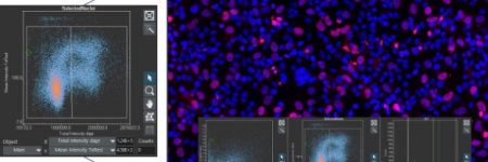
Flow vs. Image Cytometry: Comparing Techniques to Evaluate Large Cell Populations

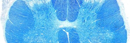
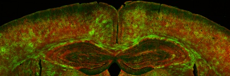
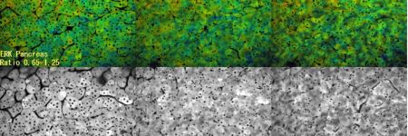
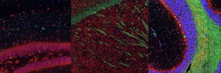
Spinning-Disk Confocal Microscopy Advances Brain Myelin Research for Alzheimer's Disease
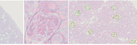
Whole Slide Imaging and Artificial Intelligence (AI) Accelerates Lupus Nephritis Evaluation
Multimodal Solution Supports Whole Slide Scanning and High-Content Screening in Neuroscience

From Tinkerer to Prize Winner: Meet the 2021 Recipient of the IOTY Award for Asia

A Giant Leap in Automated Image Acquisition―Slide Scanning Driven by TruAI™ Deep-Learning Technology
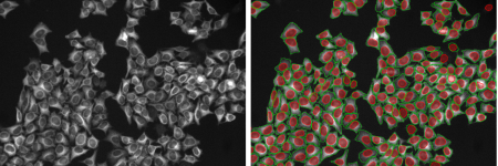
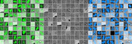
20 Examples of Effortless Nucleus and Cell Segmentation Using Pretrained Deep-Learning Models

Start-Up Scales for the Future Using Olympus’ SLIDEVIEW™ VS200 Research Slide Scanner


Helping Curb the Spread of COVID-19 with Faster, More Sensitive SARS-CoV-2 Detection

Spooky and Spine-Chilling: Our Most Popular Microscope Images for October 2021
Spreading His Wings: IOTY 2020 Asia-Pacific Winner Shares the Beauty of the Microworld
Making the Invisible Visible to Improve Patient Outcomes: A Conversation with Dr. Conor Evans
Brains, Bugs, and Bacteria: Our Most Popular Microscope Images for September 2021
Microscopy Meets Microfluidics—Creating an Adaptable Imaging System for Modern Research
Protists, Plants, and Tigers: Our Most Popular Microscope Images for August 2021
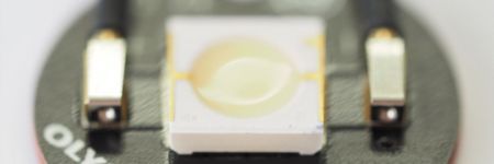
A Bright Idea—Developing LED Components with High Color Rendering for Microscope Devices

Biofilms in Space—Confocal Laser Scanning Microscopy Helps NASA Research Experiment Take Off
Oceans, Organs, and the Outdoors: Our Most Popular Microscope Images for June 2021


Revealing the Beauty of the Microscopic Scale—IOTY’s 2019 Asia-Pacific Regional Winner
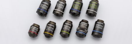
Why Objectives with Wavefront Aberration Control Are Essential for Good Microscope Design

Bugs, Butterflies, and Birds—Our Most Popular Microscope Images for September 2020
Combining Passions for Science and Art—Meet the 2019 IOTY Americas Regional Winner

See What the Buzz Is About: Our Most Popular Microscope Images for August 2020

From Multimode Imaging to Multiplexing: Your Essential Guide to Extracting Rich Data
On the Road to a Breakthrough—Trailblazing Bioimaging Is Transforming Cancer Research
Collaboration across Borders: Medical Centers Join Forces to Advance Cancer Research


Ask the Experts

Investigating Tumor Dissemination by Spatial Transcriptomics


Transforming Precision Imaging: Meet the FLUOVIEW™ FV4000 Confocal Microscope


Introduction to the APEXVIEW™ APX100 Digital Imaging System

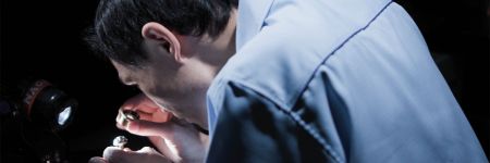
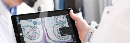
How Polarized Light Can Assist Embryologists in Clinical Routines


Unveiling Nanoscopic Realms: A Journey into Super-Resolution Microscopy

Multiplexing and Deep Tissue Imaging with NIR Confocal Laser Scanning Microscopy (Encore Edition)
Good Cell Culture Practice—How to Improve the Reproducibility of Your Experiments


EVIDENT Organoid Conference 2023 - Looking Deeper, Capturing Complexities

Exceptional Imaging Made Easy: Meet the APEXVIEW™ APX100 All-in-One Microscope


Technology Evaluation: Deciphering Cell-Cell Interactions in a 3D Microenvironment at a Single-Cell Resolution


FV3000 Red Near-Infrared (NIR) Solutions for Confocal Microscopy | 2 p.m.

FV3000 Red Near-Infrared (NIR) Solutions for Confocal Microscopy | 10 a.m.

Olympus Organoid Conference 2021

Olympus Bioimaging Conference: Exploring New Dimensions | 3-Day Virtual Event | March 9–11, 2022
Whole-Brain Functional Calcium Imaging Using Light Sheet Microscopy


Product Demo: SLIDEVIEW™ VS200 Research Slide Scanner


Product Demo: SLIDEVIEW™ VS200 Research Slide Scanner


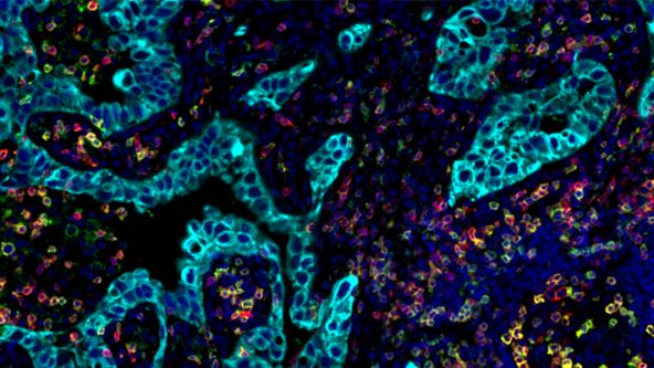
The Use of Multiplexing in Microscopy for Better Understanding the Skin Immune System in the Context of the Tissue

Recent Advances in 3D Imaging and AI-Driven Data Analysis

Now You Have the Power to See More


Metabolic Imaging in Langerhans Human Islets with MPE and FLIM

Product Demo: IXplore™ SpinSR Confocal Super Resolution System

In-Vivo Tracking of Harmonic Nanoparticles by Means of a TIGER Widefield Microscope


Hyperspectral and Brightfield Imaging Combined with Deep Learning Uncover Hidden Regularities of Colors and Patterns in Cells and Tissues

Product Demo: FLUOVIEW™ FV3000 Confocal Laser Scanning Microscope

Product Demo: FLUOVIEW™ FV3000 Confocal Laser Scanning Microscope

Evolution of Scientific Digital Imaging Technologies and their Applications


Deep Learning Approaches to Automated Phenotypic Profiling

Deconvolution of 3D Image Stacks


Confocal Microscopy and Its Use for a Spaceflight Experiment


Accelerating Image Analysis with TruAI™ Deep Learning Technology


A New Way of Thinking—Object Detection with Deep Learning

ICSI - How to improve your technique
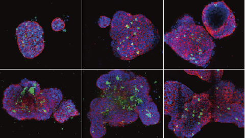
NoviSight™ Demonstration: 3D Image Analysis and Statistical Software for Organoids and Spheroids

Study the Function of Stromal Cells through Intestinal Organoid Co-Culture Technology

An In Vitro System for Evaluating Anticancer Drugs Using Patient-Derived Tumor Organoids

3D Segmentation for Fluorescence Images: From Qualitative to Quantitative

Prostate Cell Lineage Hierarchy and Plasticity

Investigating Spheroid Architecture Using the FV3000 Confocal Microscope

Advances in 3D Optical Imaging Technologies: An Overview

3D Microscopy: Understanding the Give and Take on Instrument Performance to Enable Informed Decisions

Tissue Optical Clearing Imaging: From In Vitro to In Vivo

Utilizing Tumoroids to Explore Anti-Tumor Immunity in Rectal Cancer

Converting from 2D to 3D: Bio-Techne Solutions for Your 3D Culture


Culture and Quantitative 3D Imaging of Organoids: Challenges and Solutions


A Smarter Approach to Culturing and Nurturing Your Cells


Modern Slide Scanning: Single-cell Phenotyping on Fixed Samples (Encore Edition)


Olympus Discovery Summit: Advance Your Imaging | October 26–27, 2021
.jpg?rev=53C5)
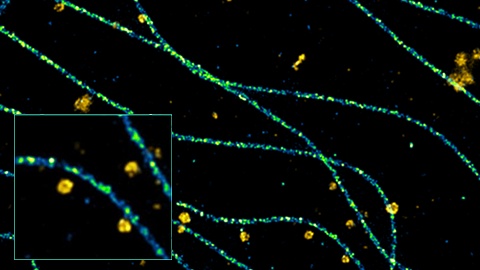
To the Diffraction Limit and Beyond: The Nanoscale Organization of Axo-Axonic Synapses | 2 p.m.


To the Diffraction Limit and Beyond: The Nanoscale Organization of Axo-Axonic Synapses | 10 a.m.

Light Sheet Microscopy – New multi-resolution and -color imaging methods


Olympus Organoid Conference: Think Deep, See Deeper | 3-Day Virtual Event | September 7-9, 2021

Create a Smarter Cell Culture Workflow


Digital Image Processing: Point and Local Operation Filters (Encore Edition)
Depth Matters: Transforming Biology for More Realistic and Meaningful Pursuits

.jpg?rev=53C5)
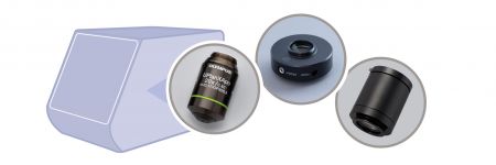
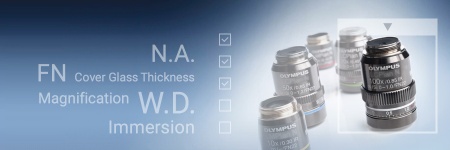
Microscope Objectives—Where the Magic Happens (Encore Edition)


Three-Dimensional High-Throughput Image Analysis of Patient-Derived Organoids and Spheroids

Olympus Discovery Summit—Looking Forward: A New Era of Research
.jpg?rev=53C5)
Digital Image Processing Part 2: Advanced Image Processing Filter
ICSI—Past, Present & Future

Microscope Objectives—Where the Magic Happens

.jpg?rev=53C5)
Microscope Objectives—Where the Magic Happens
High-Content Screening: Customized Analysis Made Easy


Think Deep, See Deeper with Near-Infrared Laser Scanning Confocal Microscope

Digital Image Processing: Point and Local Operation Filters


Live Cell Super Resolution Imaging: The Big Picture of Small Things
Light Sheet Microscopy for Deeper Insight into Life
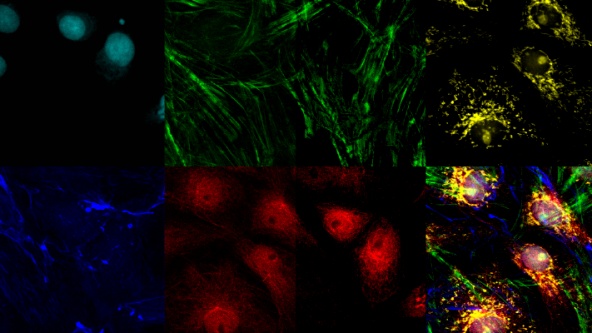
Imaging Multiple Subcellular Structures at the Nanometer Scale in 3D


FluidFM: A Novel Approach to CRISPR Gene Editing by Direct Intra-Nuclear Delivery
3D Assays: Intelligent Software, Insightful Analyses

Modern Slide Scanning: Single-cell Phenotyping on Fixed Samples
Multiplexing and Deep Tissue Imaging with Near-Infrared Confocal Laser Scanning Microscopy

Deep Learning: Opening Doors to New Applications
