Not Available in Your Country
Sorry, this page is not
available in your country.
Overview
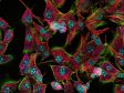
 2K x 2K resonant volumetric imaging. High SNR with the SilVIR detector enables high-quality results with minimal averaging. Four-channel multiphoton dataset of cleared mouse intestine. | Deeper Discoveries and Advanced MultiphotonThe FV5000MPE enables quantitative imaging deep within thick, scattering samples, combining SilVIR detectors, TruSight™ deconvolution, and TruAI noise reduction for outstanding signal-to-noise and clarity. MPE-optimized objectives, TruResolution auto correction collar, and automated IR laser alignment maintain sharp focus throughout the imaging volume. The compact fiber-pigtailed laser system offers an affordable, easily deployed solution for routine multiphoton imaging and rapid installation. For advanced applications, the fully tunable MPE laser configuration provides broad excitation flexibility and precise wavelength control for demanding experiments. One-, two-, or three-line simultaneous MPE laser excitation delivers clarity and reproducibility millimeters deep. |
From Compact to Advanced MPE SetupsConvenient Multiphoton ImagingThe FV5000MPE addresses some of the primary challenges associated with multiphoton excitation (MPE)—costly traditional pulsed tunable IR lasers and strict environmental conditions—with a new generation of compact single wavelength fiber-pigtailed IR lasers that are cost-effective, easy to handle, and designed to bring MPE within reach of a broader range of users. Tunable Infrared Laser Solutions for Advanced Multiphoton StudiesFor more advanced applications, the FV5000MPE supports the latest tunable infrared pulsed lasers for deep multiphoton imaging across 680–1300 nm. These IR lasers provide two laser lines—one tunable and one fixed at 1040/1045 nm—for efficient excitation of far-red fluorophores. | 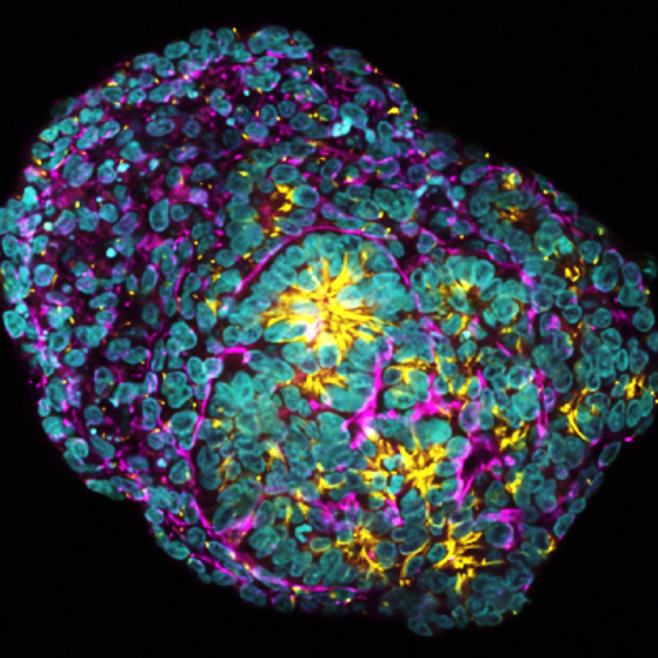 Human kidney organoid. Captured with single wavelength fiber-pigtailed IR lasers at 920 nm and 1064 nm for simultaneous 3CH multiphoton imaging with a LUPLAPO25XO objective lens. |
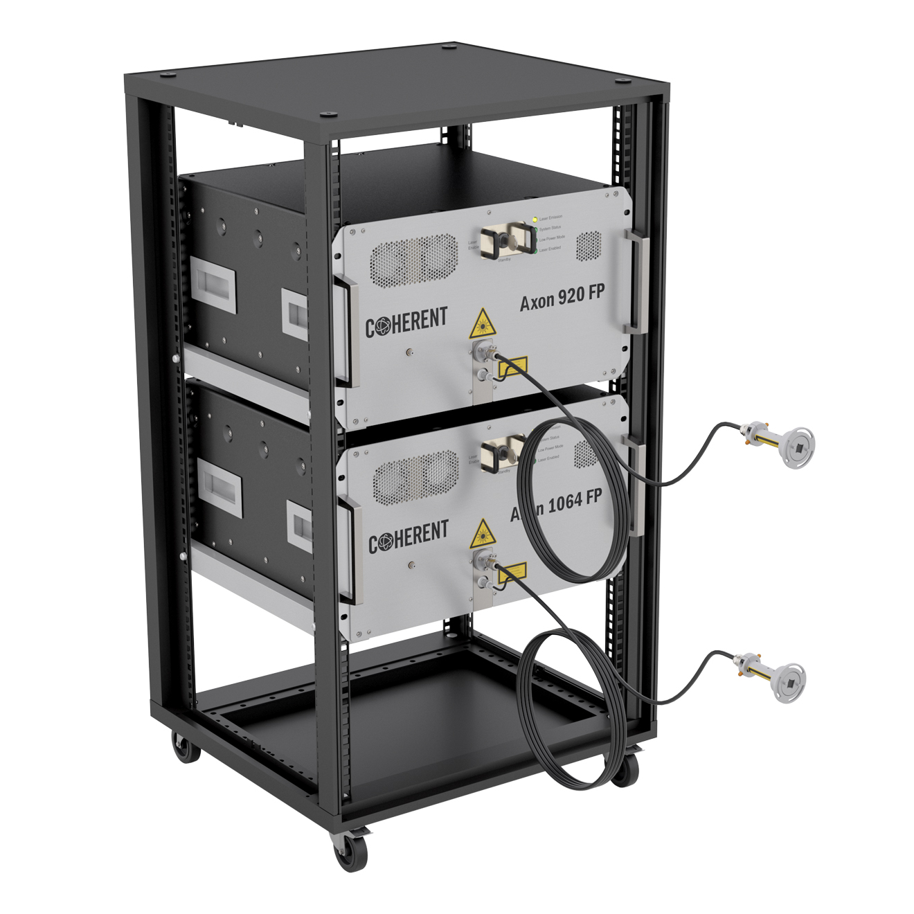 Axon FP lasers at 920 nm and 1064 nm installed in a 19-inch rack. | 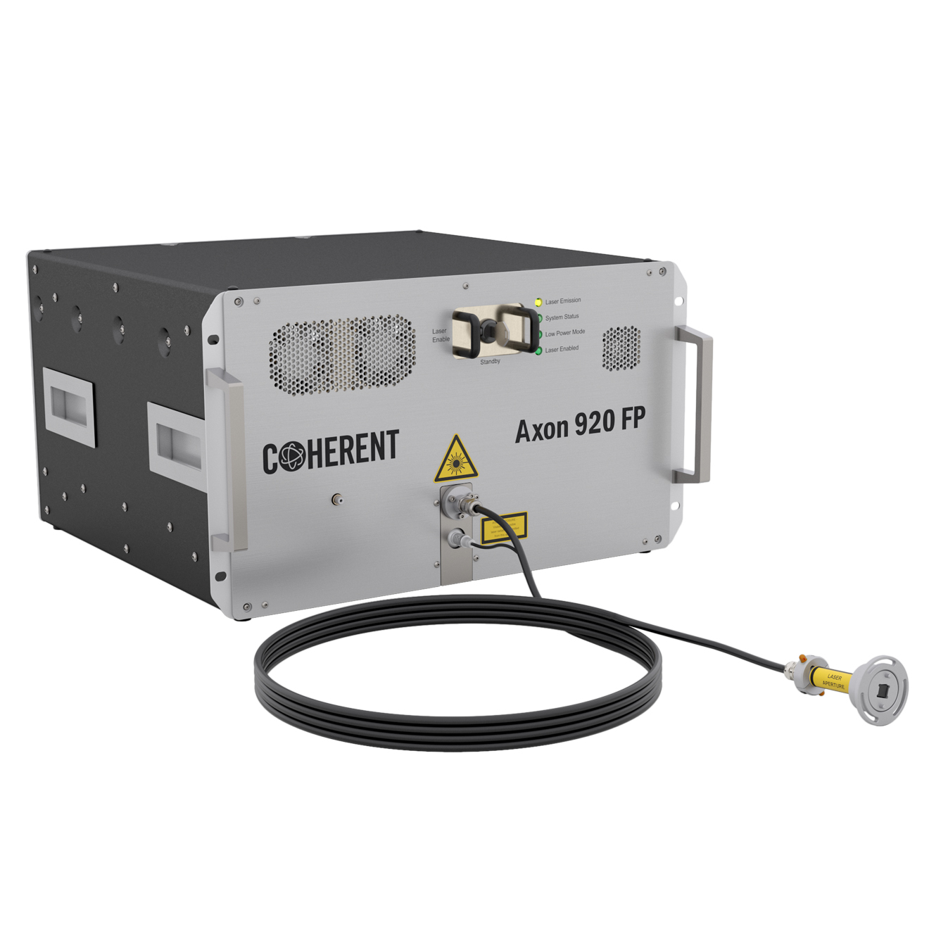 Single-wavelength fiber-pigtailed laser (Axon FP). |
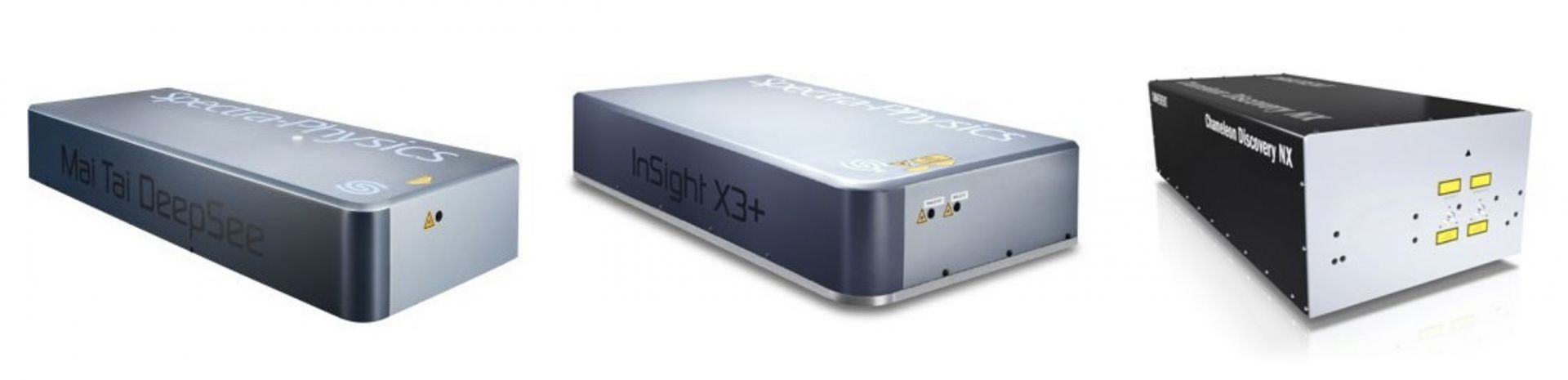 Tunable infrared laser solutions for advanced multiphoton studies from SpectraPhysics and Coherent. |
More Colors and More InformationCapture more colors and more information in one image with the FV5000MPE’s advanced multiplexing features. The FV5000MPE offers simultaneous multicolor imaging with up to six channels at once to capture more information in less time. Make the most of the SilVIR detector’s high sensitivity to quickly produce outstanding images at depth. | 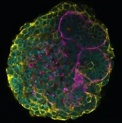 Human iPSC-derived kidney organoids with membrane-GFP. Captured with single-wavelength fiber-pigtailed IR lasers at 920 nm and 1064 nm for simultaneous 3CH multiphoton imaging at 2K resonant imaging. |
Optimized Performance
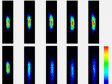
Optimized Performance and ResultsAuto Laser AlignmentThe FV5000MPE’s four-axis auto laser alignment system simplifies maintenance by keeping the excitation beam precisely aligned within the scanner unit, even when wavelength tuning, temperature changes, or other factors cause drift. The system adjusts beam position and angle to maintain optimal laser power and consistent pixel registration. In dual-laser configurations, auto alignment also preserves beam co-alignment, minimizing channel co-registration errors for accurate multi-line imaging. When needed, users can perform manual fine-tuning directly through the software interface. | Left: Pixel shift. Right: Correction of pixel shift. |
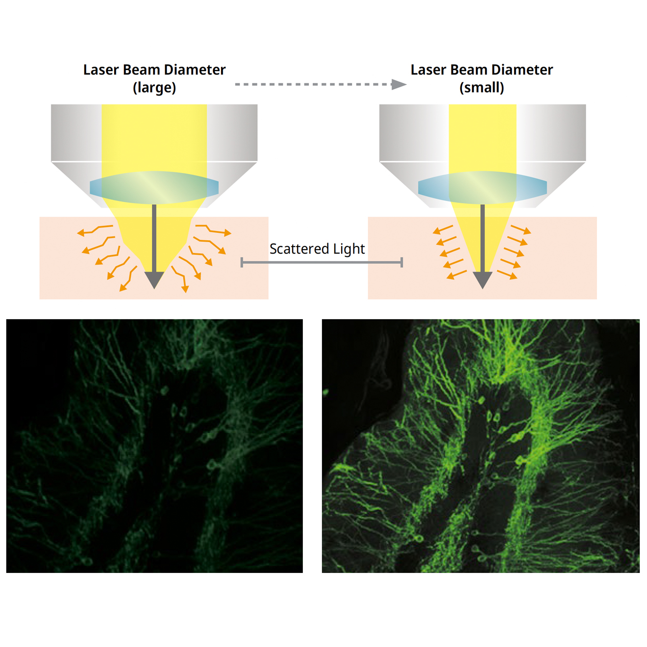 Left: Normal image. Right: Deep Focus mode image. | Maximize Signal with Deep Focus ModeDeep Focus mode adjusts the laser beam diameter according to the sample’s scattering properties. In highly scattering in vivo specimens, the beam can be narrowed to allow more excitation photons to penetrate deeper into the tissue, resulting in bright high-resolution images even at greater depths. |
TruResolution™ MPE Objectives | ||
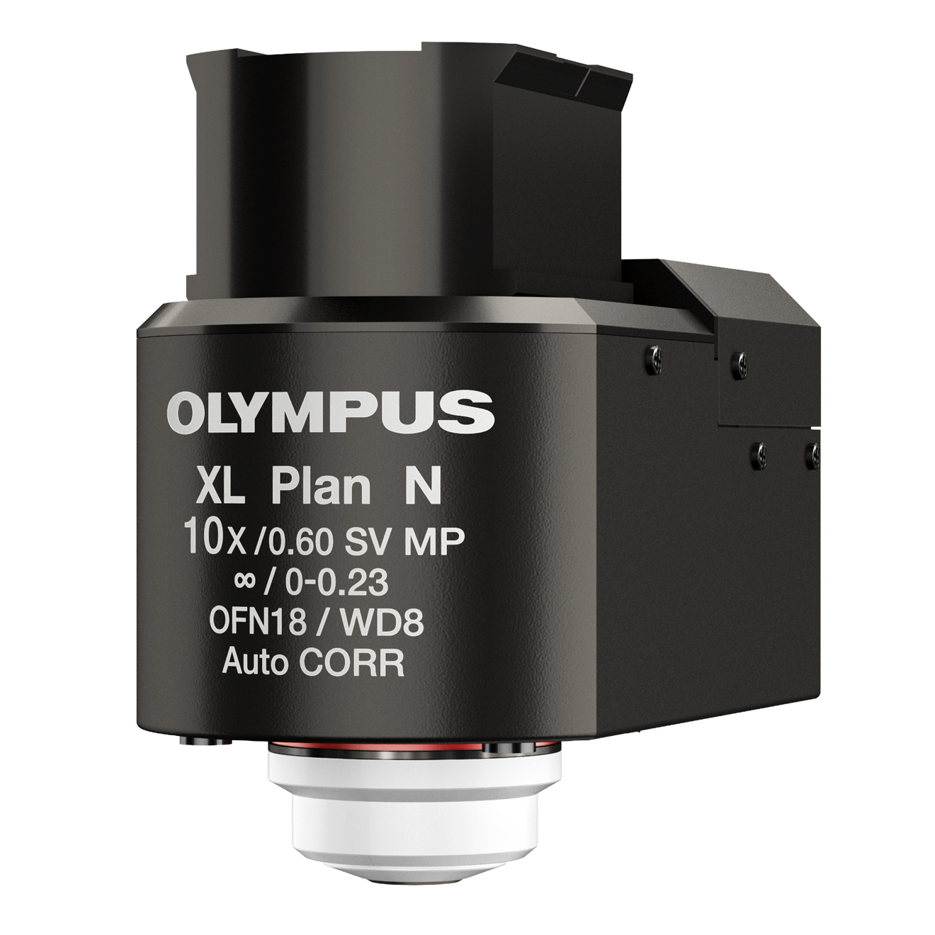 FV30-AC10SV Objective Lens | 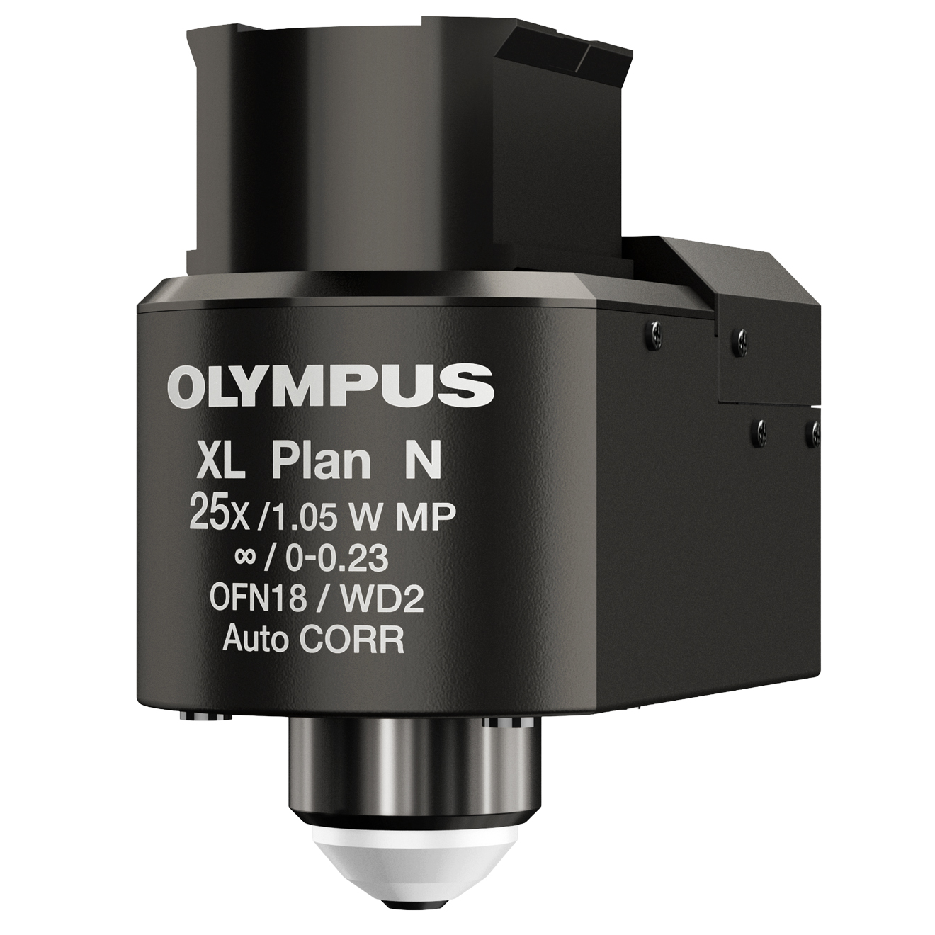 FV30-AC25W Objective Lens | 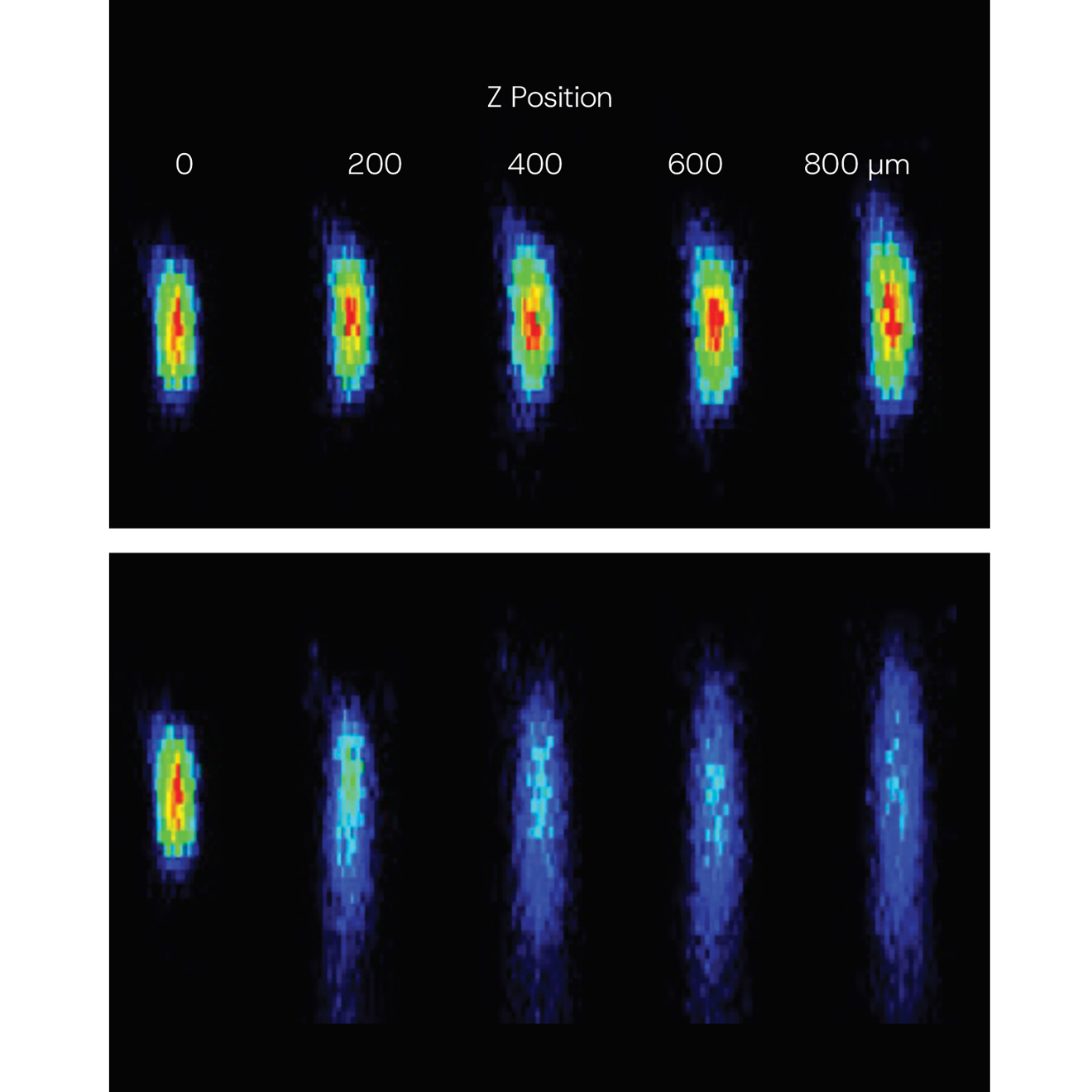 Top: With TruResolution. Bottom: Without TruResolution. XZ images of 0.2 μm fluorescent microspheres in scattering gel (RI=1.36) at various depths acquired using the FV30-AC25W objective lens. |
MPE Objectives
Objectives Designed for DepthSharper Imaging, Deeper InsightsEngineered for multiphoton excitation, A Line™ MPE objectives deliver high-precision imaging of biological specimens at depths of up to 8 mm, supporting both in vivo and transparent samples. These objectives are optimized for performance and versatility, offering:
| 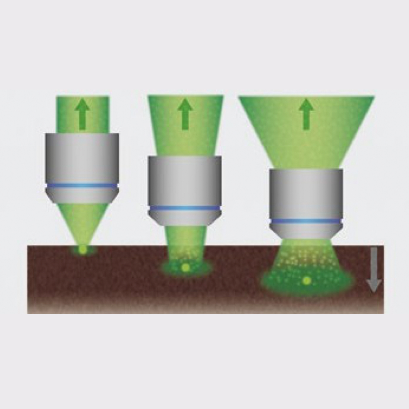 Wide fields of view enable these objectives to efficiently collect scattered fluorescence photons and generate brighter images from deep within specimens. |
| Dedicated Multiphoton Objective | NA | WD (mm) | Immersion Index |
| XLPLN10XSVMP | 0.60 | 8.0 | 1.33–1.52 |
| XLPLN25XWMP2 | 1.05 | 2.0 | 1.33 |
| XLPLN25XSVMP2 | 1.00 | 4.0 | 1.33–1.40 |
| XLSLPLN25XSVMP2 | 0.95 | 8.0 | 1.33–1.40 |
| XLSLPLN25XGMP | 1.00 | 8.0 | 1.41–1.52 |
 Example of 1600 coating technology with the XLPLN25XWMP2 objective lens. | Optics with Outperforming IR CoatingOur innovative IR coating (1600 coating) for multiphoton objectives and scanner optics further refines deep observation quality. |
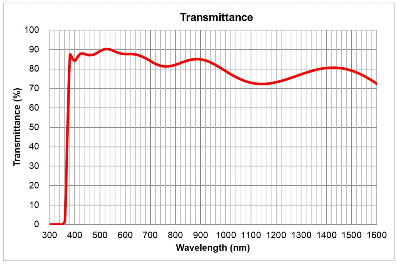
Reliable Tissue ImagingFor upright microscope systems, our newly developed 16X multi-immersion objective (NA 0.8, WD 3.0 mm) offers an excellent balance between resolution and field of view—an ideal choice for broad tissue imaging applications. It has high transmittance from the VIS to NIR range, and enables seamless switching between multiphoton and confocal imaging. | 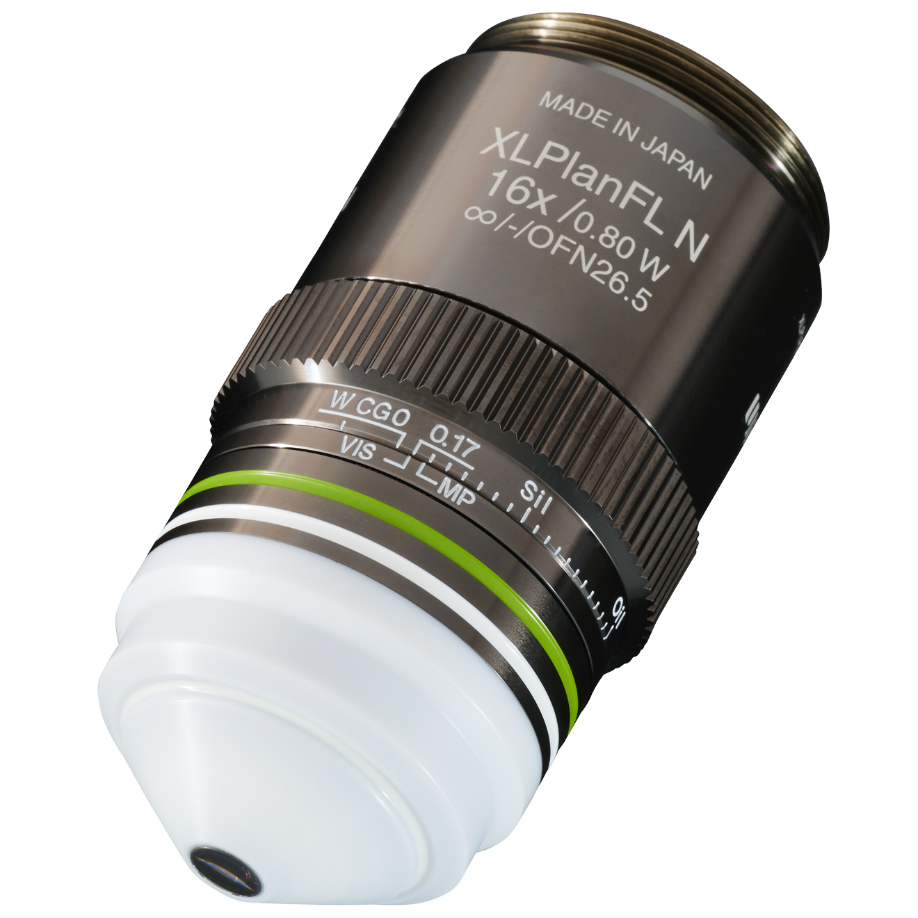 XLPLFLN16XW / NA 0.80 |
Configurations
Built on the Versatile PlatformThe FV5000MPE is part of the modular FV5000 family—engineered to cover every imaging need, from single-cell dynamics to deep-tissue and small-animal studies. It can be configured as a dedicated multiphoton (MPE) system or as a combined confocal + multiphoton setup for seamless multimodal imaging. Optimized designs support thick-tissue, organoid, and in vivo applications, while up to six SilVIR detector channels can be assigned to confocal and six to multiphoton detection—enabling up to 12 simultaneous SilVIR detector channels across modalities. Learn more about the FV5000 confocal configuration here. |
Multiple Frame OptionsDepending on your application, you can choose between upright, gantry, or inverted microscope frames. |
%20WC_white.jpg?rev=152E) Upright Microscope System A large focus stroke accommodates a range of specimens, from tissue slices to live mice and other small animals. | 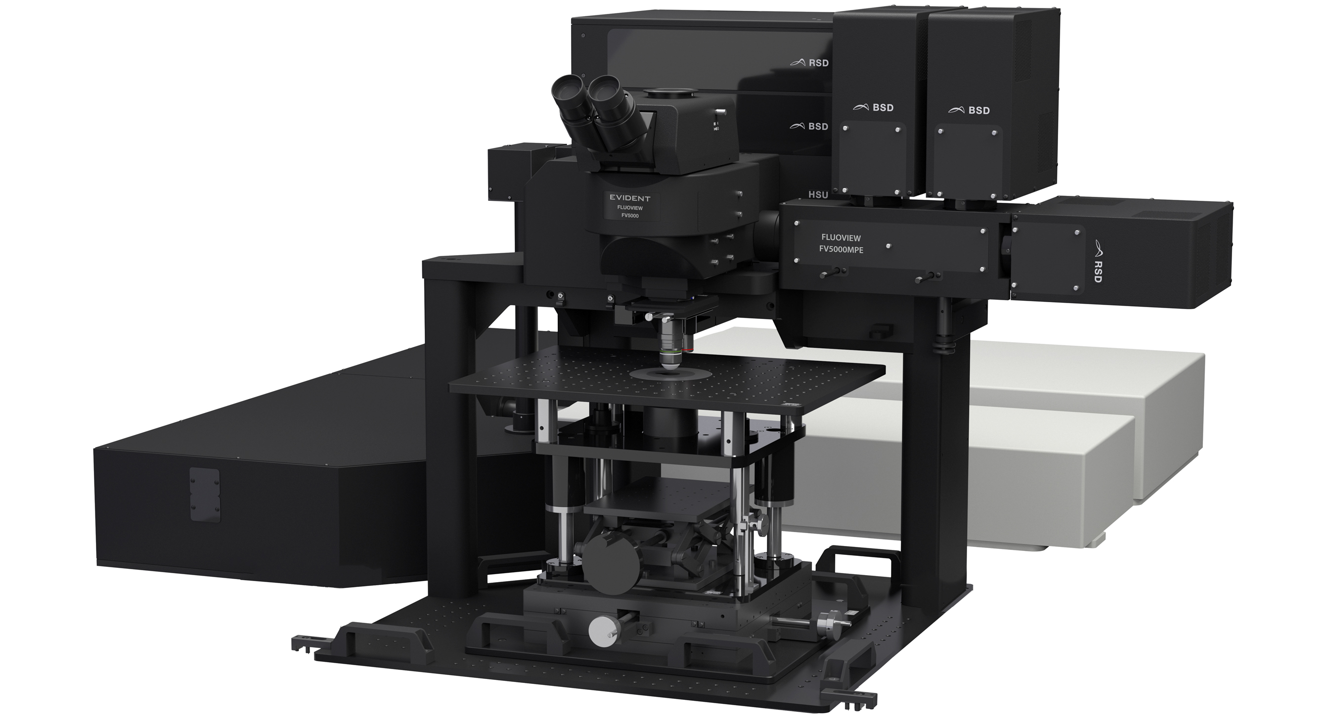 Gantry Microscope System The frame maintains a large working space below the objective, making it easier to position experiment equipment. | 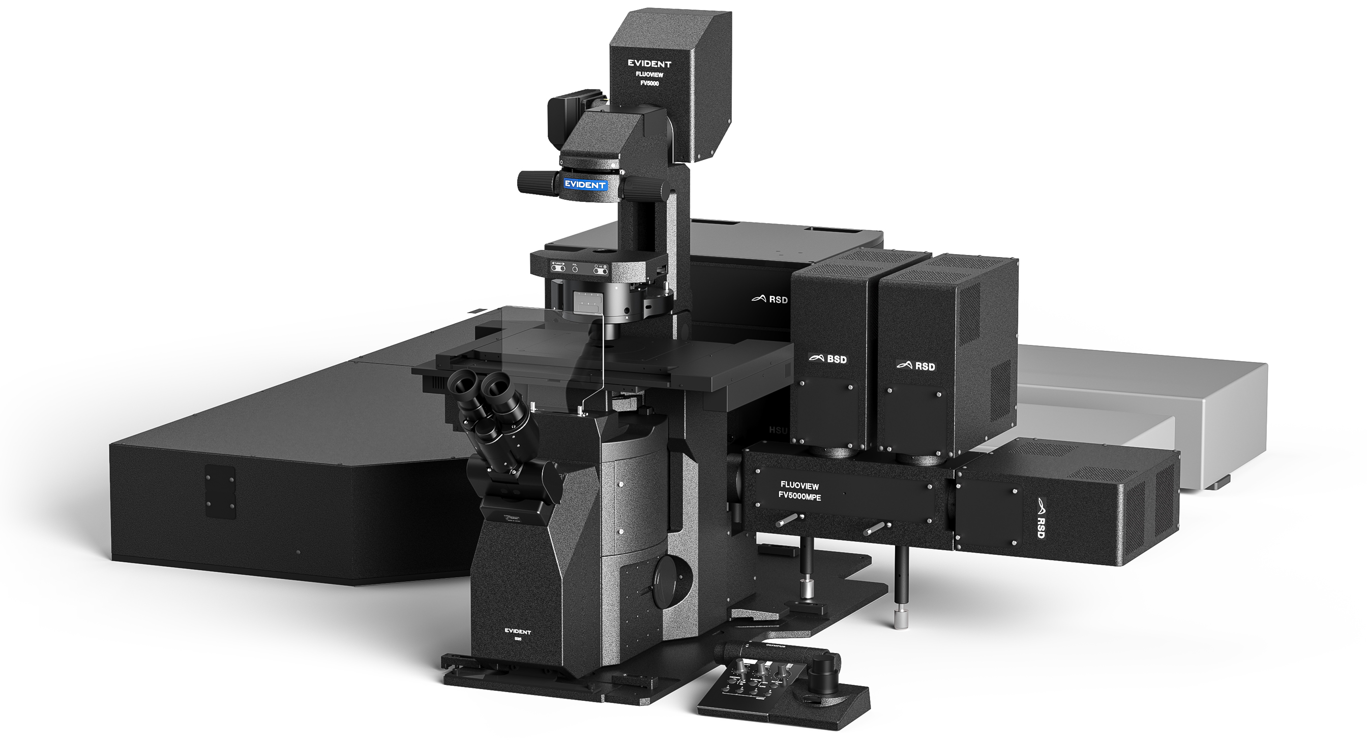 Inverted Microscope System The frame supports observation of 3D cultures and multicellular clusters that are difficult to image using an upright frame. |
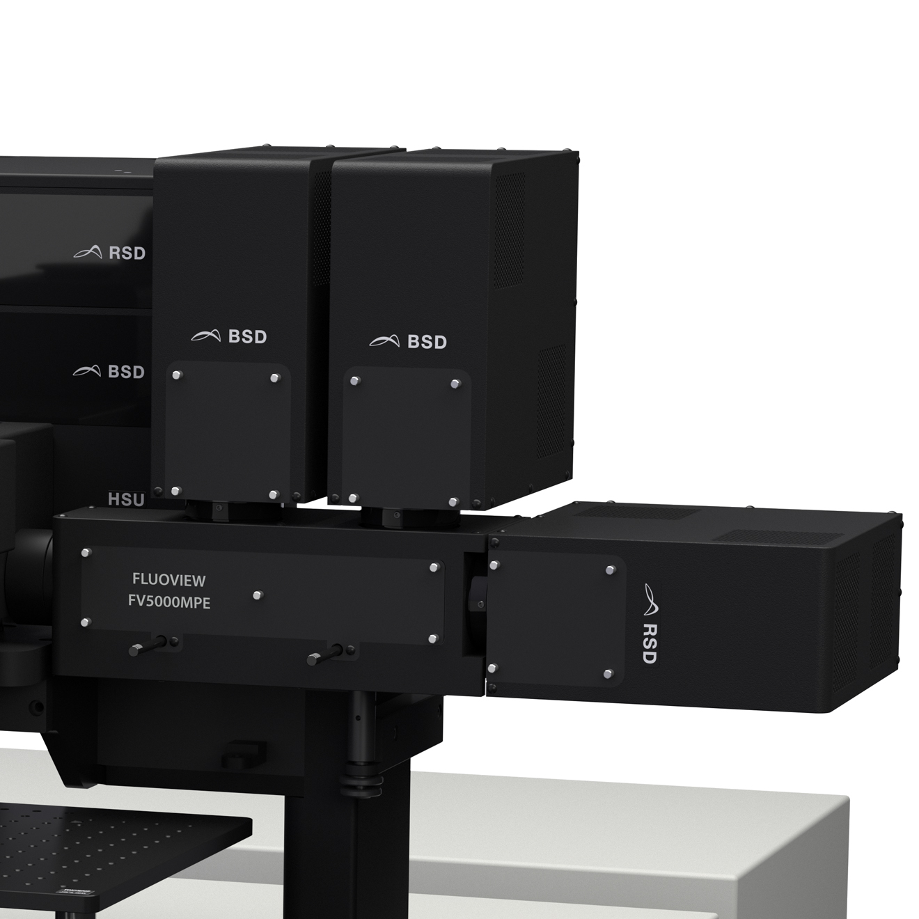 Six-channel non-descanned detection (NDD) module of the FLUOVIEW FV5000MPE. The modular SilVIR detector units enable simultaneous multicolor imaging and deep-tissue acquisition with exceptional sensitivity and unmatched dynamic range. | Create Your Own Custom ConfigurationTake system customization to the next level by integrating your own optical components. For example, you can add a custom laser to an available port to enable advanced techniques such as three-photon or specialized imaging methods. As your research evolves, your imaging system should evolve with it. The FV5000MPE platform supports specialized solutions* that extend the capabilities of standard configurations, helping you adapt to new applications and experimental requirements. *Not available in some countries or regions. |
Support and Service You Can Count OnWhen it comes to protecting your investment and the integrity of your research, your needs come first. We stand behind our products with a commitment to prompt service and technical support to help you achieve your goals. Available in three convenient tiers—Maintenance, Protection, and Performance Plus—our FV5000MPE Service Plans* include priority support to help minimize downtime, regular scheduled maintenance to keep your equipment in peak condition, predictable repair costs to eliminate unplanned expenses, and direct, efficient solutions when you need them most. *Regional variations in service offerings may apply. |  |
FV5000MPE Service Plans |
| Maintenance | Protection | Performance Plus | |
|---|---|---|---|
| Priority remote support | ✓ | ✓ | ✓ |
| Preventative maintenance | ✓ | ✓ | ✓ |
| Repair coverage (parts, labor, travel) | 10% discount | ✓ | ✓ |
| Fast on-site response | - | - | ✓ |
Specifications
FV5000MPE / FV5000MPE-RS Specifications |
| SPECIFICATIONS | FV5000MPE | FV5000MPE-RS | |
|---|---|---|---|
| Scanner | Galvanometer Scanner | 64 × 64 – 8192 × 8192 pixels, 0.2 μs/pixel – 1000 μs/pixel | |
| Resonant Scanner | 512 × 512 pixels, 1024 × 1024 pixels, 2048 × 2048 pixels | ||
| Field Number | 20 (for both scanner types) | ||
| Spectral Confocal Detector | Detector | SilVIR detector (cooled SiPM, broadband type/red-shifted type) | |
| Maximum Channels | Six channels | ||
| Spectral Method | VPH, detectable wavelength range 400-900 nm | ||
| Non-Descanned Detector | Detector | SilVIR detector (cooled SiPM, broadband type/red-shifted type) | |
| Maximum Channels | Six channels | ||
| CW Laser | VIS Laser | 405 nm, 445 nm, 488 nm, 514 nm, 561 nm, 594 nm, 640 nm | |
| NIR Laser | 685 nm, 730 nm, 785 nm | ||
| IR Pulsed Laser | Tunable Laser |
One-laser system, dual-laser-line system, two-laser system
Excitation wavelength: 680-1300 nm Four-axis auto alignment, auto beam expander | |
| Single Wavelength Fiber Pigtailed Laser | 920 nm, 1064 nm | ||
| Image | High dynamic range photon counting (1G cps, 16-bit) | ||