CV1000 Disk Confocal Microscope
Discontinued Products

The CellVoyager CV1000 provides renowned Yokogawa spinning disk confocal imaging technology in an easy to use, incubated bench-top solution for live cell imaging. With its microlens enhanced dual Nipkow disk scanning technology, phototoxicity and photobleaching are drastically reduced, making it ideal for use in observing highly delicate life processes such as iPS/ES cell generation and embryogenesis. From multiple 35mm dishes to multi-well plates, the CV1000 has the throughput to handle some of your most demanding time-lapse research.
CellVoyager is a registered trademark of Yokogawa Electric Corporation
Features
Wide-area Imaging of Tissue Sections
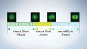
Interkinetic Nuclear Movement in the Cerebral Cortex of Chicken
Sensitive EM-CCD camera acquisition allows rapid collection of multi-channel Z-stacks. Multiple adjacent image sets may be collected and stitched into large area 3D data sets that can be played back over time. Multiple stitched acquisition locations within a well, or in different wells or 35mm dishes, can be easily programmed into the software to be acquired and compiled into separate data sets.
Renowned Olympus Objectives for Live Cell Imaging
Olympus boasts some unique and superior objectives for live cell imaging. The Silicone Oil Objectives, 30 X 1.05 NA and 60 X 1.30 NA, have proven performance in live cell applications, significantly improving performance over both water and oil objectives in many cases. The 30 X objective has a very large working distance of 800 microns, while the 60 X has a remarkable 300 microns at 1.3 NA. Please contact your local Olympus representative for optimized options for your research.
Powerful yet Easy to Use CellVoyager CV1000
- Medium to high throughput live cell imaging with renowned Yokogawa spinning disk technology.
- Up to 3 fluorescent channels for either confocal or wide field fluorescence, plus brightfield.
- Dedicated intuitive software to minimize training time.
- Easy map view allows for highly flexible large field scanning.
Dual Disk Option to Optimize for up to 6 Objectives
- Motorized disk changer allows for changing pinhole size with one mouse click.
- Up to 6 objectives can be mounted for varying magnification.
Accessories
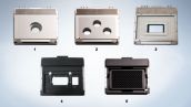
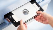
From high-end multi point, long-term time lapse imaging to single shots of fixed cells select the attachment that best meets your requirements. Use an attachment together with the stage incubator (for 35 mm dish, 35 mm 3-dish, glass chamber, or micro-plates) to keep cells healthy during time lapse imaging.
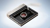
Multi-day incubation achieved through humidification and precise CO2 control.
Please sample in CV1000 for imaging. Multiple inserts can be purchased for pre-incubation.
Specifications
| Main Unit | Type | 3-Color Model | 2-Color Model | Single-Color Model | Basic Model | ||
|---|---|---|---|---|---|---|---|
| Confocal Scanning Method | Microlens enhanced dual Nipkow disk scanning | ||||||
| Scanning Speed | 1,500 - 5,000 rpm (Max 1,000 fps)*1 | ||||||
| Excitation Laswer Wavelength (nm) | 405, 488, 561 | 488, 561 | 488 | 488 | |||
| Brightfield Imaging | LED transimission | - | |||||
| Camera | Type | Back-illuminated EMCCD | Cooled CCD | ||||
Camera | 512 x 512 | 1344 x 1024 | |||||
| XY-Stage |
High-precision auto XY stage
Designated resolution: 0.1 μm | ||||||
| Z-axis Control |
High-precision auto XY stage
Designated resolution: 0.1 μm | ||||||
| Auto Focus | Detection of glass surface with laser + offset | ||||||
| Objective Lens |
[Standard] Dry:10 X
[Option] Up to 5 lenses can be added Dry: 10 X, 20 X, 40 X Oil: 20 X, 40 X, 60 X Water: 60 X LWD: 20 X, 40 X | ||||||
| Stage Incubator*2 | High-precision temperature controllable incubator | - | |||||
| Temperature |
Range: 30 - 40 °C
(Room temperature +5 °C or higher) Designated resolution: 0.1 °C | ||||||
| Humidity Control | Forced humidification with a water bath unit | ||||||
| CO2 Mixing Unit | CO2 5 % gas cylinder*3 CO2gas | ||||||
| External Dimensions (mm) | 580 (W) x 835 (D) x 532 (H) | ||||||
| Weight (kg) | 93 | ||||||
| Utility Box | External Dimensions (mm) | 319 (W) x 368 (D) x 518 (H) | 319 (W) x 368 (D) x 346 (H) | ||||
Utility Box | 16 | 10 | |||||
| Control Software |
Sets conditions for imaging, camera, time-lapse, environments*2, 3D imaging, map view acquisition, multi-color imaging, and multi-point imaging.
Functions include image display. Output file type: 16bit TIFF. | ||||||
| Work Station | Controller work station, Display (24" 1920 x 1200) | ||||||
| Operating Temperature | 15 - 35 °C (When operating temperature is over 30 °C, water cooling of the camera is required.) | ||||||
| Operating Humidity Level | 20 - 70 % RH (no condensation) | ||||||
| Power Consumption | 100 - 240 VAC/ 50 or 60 Hz 1500 VAmax | ||||||
| Option | Pinhole change unit | 50 μm/25 μm Switching time: 2 sec | |||||
Option | Effective no. of pixels: 1024 x 1024 | ||||||
Option |
For Single 35 mm dish with Stage incubator*2*4
For Triple 35 mm dishes with Stage incubator*4 For Micro well plate For Slide glass*5 | ||||||
*1 fsp: frame per second frame rate (Actual frame rate depends on the specification of the camera.)
*2 Option for basic model
*3 CO2 gas cylinder not included with CV1000 system
*4 When you use stage incubator, CO2 mixing unit is required
*5 Option for 3-color model, 2-color model and Single-color model
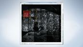
Applications
96-Well Plate Imaging | GFP Transfected 293F Cells | Plant Imaging |
Wide-area Imaging of Tissue Sections | In Vivo Imaging of White Blood Cells Patrolling Zebra Fish | Following the Injection of Mouse Embryos with mRNA |