Not Available in Your Country
Sorry, this page is not
available in your country.
Overview
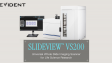
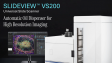
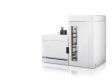
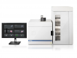
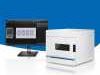
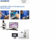
 | SLIDEVIEW VS200 Universal Slide ScannerMultimodal imaging for advanced research and specialized digital pathology. The SLIDEVIEW™ VS200 system is designed to capture a wide variety of samples and stains with multiple magnifications, flexible slide sizes, and up to 10 imaging modes from brightfield to fluorescence. High-resolution imaging of whole slides empowers your lab to unlock more insights from your samples. |
|---|
Choose Your Application
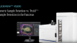
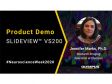
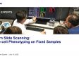
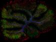
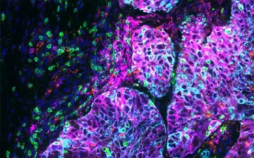
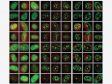
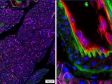
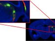
SLIDEVIEW VS200 for Life Science ResearchThe SLIDEVIEW VS200 system offers high-resolution slide scanning for advanced research applications, such as neuroscience, cancer and stem cell research, and spatial biology. With the flexibility of up to 10 imaging modes, multiplexing capabilities, and multiple magnifications up to 100X, the system delivers outstanding image quality for quantitative analysis. |
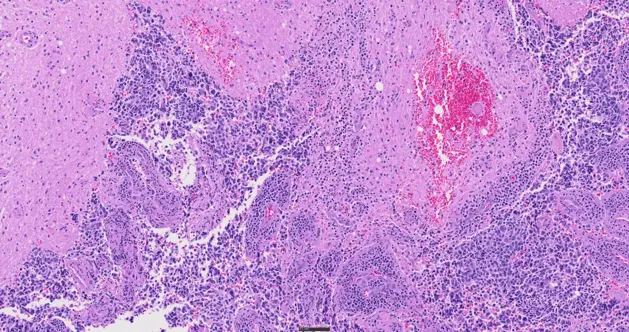
SLIDEVIEW VS200 for Digital PathologyAs a flexible yet intuitive scanner, the SLIDEVIEW VS200 system enables your team to digitize a wide range of sample types with minimal training. The VS200 system helps you achieve a fully digital workflow for pathology with versatile slide scanning, outstanding image quality, and flexible integration options. Get the evidence you trust for standard and specialty samples to make breakthrough decisions with confidence. |
Key Benefits
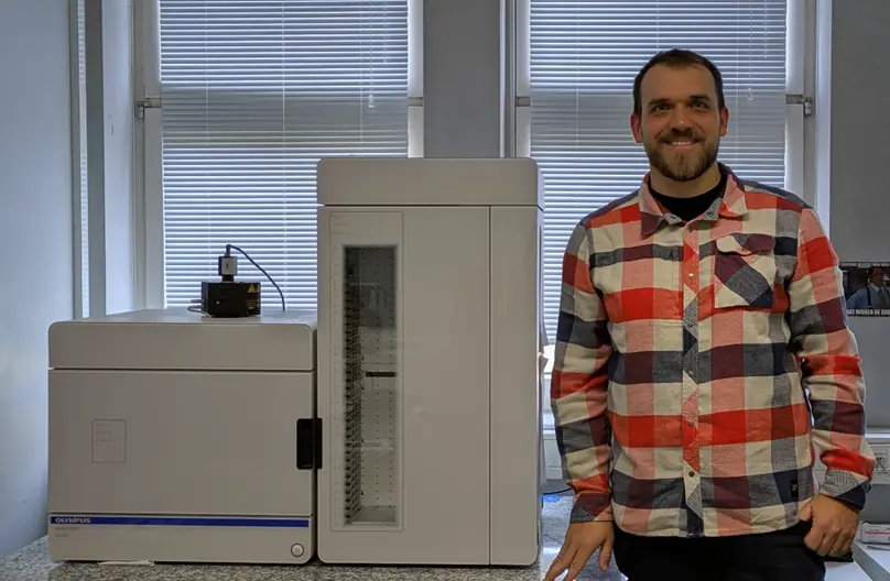
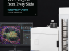
More Insights from Every SlideOne scanner. Every slide. No limits. Capture the biological insights you need with a single slide scanner designed for flexible, multimodal imaging. | Melissa González |
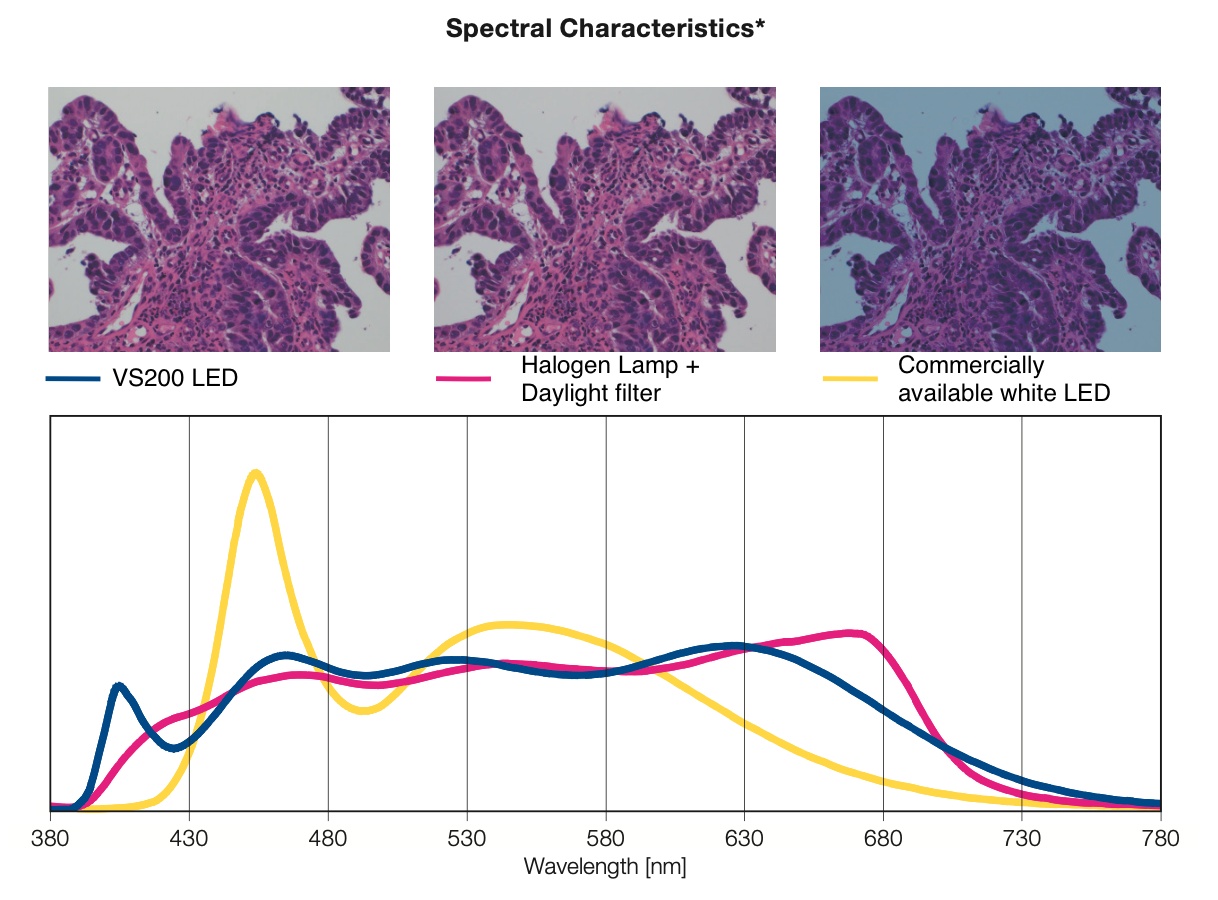 *This graph shows the spectral characteristics of each light source normalized with the luminosity curve. It does not compare the strength of light for each light source. | Confidence in Your Data QualityAdvanced optical innovation and automation make high-quality imaging routine and reproducible, reducing setup, variability, and user dependence across experiments and users. |
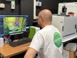
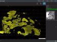
Smarter Imaging and WorkflowAccelerate your path from slide to results with an intuitive interface, enhanced batch-scanning capabilities, faster workflows, and AI-based segmentation tools. Reduce human error, manual effort, and have more confidence in your results. | |
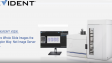
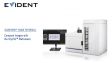
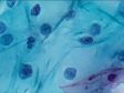
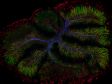
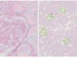
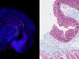
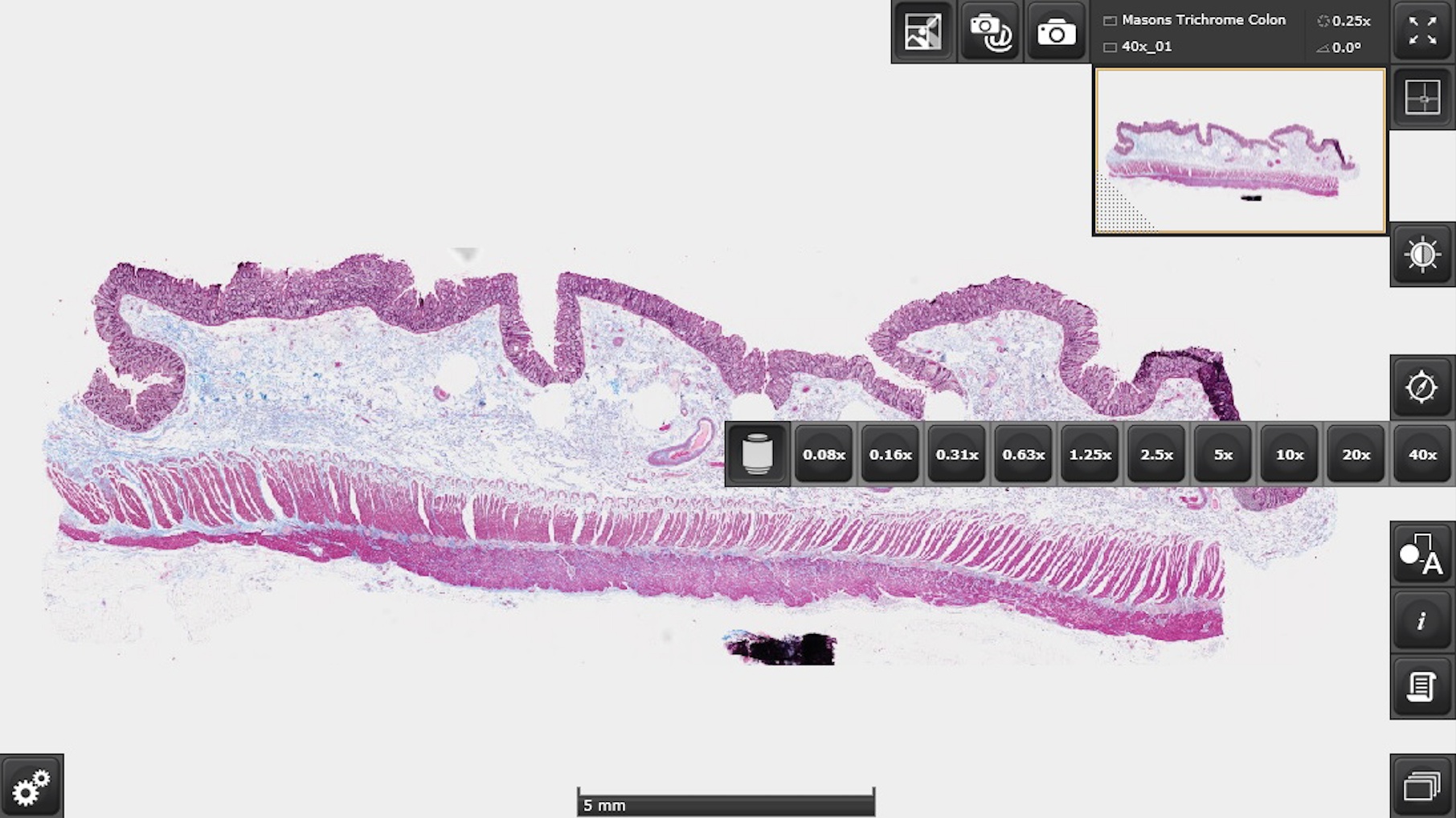 Colon stained with Masson’s trichrome. | Centralized Image AccessManaging the large amount of data generated by a slide scanner is easier than ever thanks to our Net Image Server (NIS) SQL database and OlyVIA™ web programs. Scan and automatically upload your images to one or more databases, differentiate between users, and take advantage of offline visualization and annotation tools. |
Specifications
SPECIFICATIONS | VS200 Single Tray | VS200 Multiple Tray Loader | ||
|---|---|---|---|---|
| Intended Specimen | Observable Specimen | Glass slide with cover glass and without cover glass*1 | ||
|
Size of Glass Slide
(W × L × H) | Standard slide tray: 25 mm–26.5 mm × 75 mm–76.5 mm × 0.9 mm–1.2 mm (1 in. × 3 in. × 0.05 in.) (6 slides) Optional trays: 1) 47 mm–50 mm × 27 mm–30 mm × 1.2 mm–1.6 mm (1 in. × 2 in. × 0.05 in.) (3 slides) 2) 51 mm–53 mm × 75 mm–76.5 mm × 0.9 mm–1.2 mm (2 in. × 3 in. × 0.05 in.) (3 slides) 3) 75 mm–76.5 mm × 100 mm–102 mm × 0.9 mm–1.2 mm (3 in. × 4 in. × 0.05 in.) (1 slide) 4) 100 mm–102 mm × 126 mm–128 mm × 1.1 mm–1.4 mm (4 in. × 5 in. × 0.06 in.) (1 slide) | |||
| Cover Glass Thickness | 0.12–0.17 mm | |||
| Observation Methods | Brightfield, brightfield mono, reflected brightfield (optional*2), darkfield, phase contrast (optional*3), linear polarization (optional*4), circular polarization (optional), DIC (optional), fluorescence (optional), fluorescence optical sectioning with speckle illumination (optional VS-SILA module) | |||
| Optical Frame | Illuminator | Built-in Köhler illumination for transmitted light; high intensity and high color rendering LED (up to 50,000 hours) | ||
| Objectives | 2X, 4X*5, 10X*5, 20X, 40X*5 60X*5, and 100X*5 compatible objectives for a 6-position motorized nosepiece (including selected oil immersion, silicon oil immersion, and phase contrast objectives) Optional automatic liquid dispenser | |||
| Motorized Stage | XY stage with automatic control | |||
| Focusing | Motorized focusing with automatic control | |||
| Color Camera | Integrated 2/3 inch CMOS, 3.45 μm × 3.45 μm pixel size, high sensitivity, high resolution | |||
| Scan Unit | Capacity | 1 slide tray, 6 slides maximum; upgradable to a multiple tray loader model | Up to 35 slide trays, 210 slides maximum | |
|
Pixel Resolution
(Color Camera) | Color camera UPLXAPO20X (NA 0.8): 0.274 μm/pixel Options: UPLXAPO4X (NA 0.16): 1.37 μm/pixel UPLXAPO10X (NA 0.4): 0.548 μm/pixel UPLXAPO40X (NA 0.95): 0.137 μm/pixel UPLXAPO40XO (NA 1.4): 0.137 μm/pixel UPLXAPO60XO (NA 1.42): 0.091 μm/pixel UPLXAPO100XO (NA 1.45): 0.055 μm/pixel Up to 35 slide trays, 210 slides maximum Monochrome camera Hamamatsu Orca camera UPLXAPO4x (NA 0.16) 1.625 μm/pixel UPLXAPO10X (NA 0.40) 0.650 μm/pixel UPLXAPO 40X (NA 0.95) 0.163 μm/pixel UPLXAPO40XO (NA 1.4) 0.163 μm/pixel UPLXAPO60XO (NA 1.42) 0.108 μm/pixel UPLXAPO100XO (NA 1.45) 0.065 μm/pixel | |||
| Scan Time | Brightfield: approx. 1.5 minutes (20x objective, scan area 15 mm × 15 mm) Fluorescence widefield NOVEM: approx. 14 minutes (20X objective, scan area 15 mm × 15 mm, 4 channels, 50 ms exposure each) | |||
| Software | Automatic sample detection (generic and TruAI deep learning), automatic barcode reading, automatic focus mapping, automatic scanning, automatic stitching, pause and resume scanning, Z-stack imaging, extended focus imaging (EFI), image format: VSI, JPEG, TIFF, DICOM, synchronized multi-image display, stepless zooming, zooming while scanning, annotations, screen capture, slide loader control (multiple tray loader only) | |||
|
Fluorescence
(optional) | Fluorescence Components | UPLFLN4X objective (recommended for overviews), illuminator with fly-eye lens, motorized mirror turret, motorized filter wheel Widefield light source options: U-LGPS, Excelitas X-Cite XYLIS-2, X-Cite NOVEM, KL2500LED, CoolLED pE-10 VS-SILA: up to 6-line laser combiner and scrambler unit | ||
| Monochrome Camera | Options: VS-304M (12-bit), 1-inch CMOS, 3.45 μm × 3.45 μm pixel size (12MP) HAMAMATSU ORCA Flash (16- bit)4.0 V3 HAMAMATSU ORCA Fusion (16- bit) HAMAMATSU ORCA Fusion BT (16- bit) | |||
|
Solutions for Scanner Software
(optional) | Solution License | Batch image format converter DICOM converter fluorescence VS-SILA acquisition Net Image Server (NIS) SQL | ||
|
Desktop Software
(optional, separate solution for analysis) | Solution License | Batch image format converter DICOM converter Detection and analysis Deep learning 3D deconvolution | ||
| Environment | Weight | Optical frame: 75 kg (165.3 lb) 1 slide tray: 0.6 kg (1.3 lb) | Optical frame and multiple tray loader: 149 kg (328.4 lb) 35 slide trays: 21 kg (46.3 lb) | |
Fluorescence: 8 kg (17.6 lb) PC and monitor: 19 kg (41.9 lb) Camera cover (optional): 9 kg (19.8 lb) | ||||
| Operating Environment |
Computer and monitor: 19 kg (41.9 lb)
Camera cover (optional): 9 kg (19.8 lb) | |||
| Operating Environment |
Temperature: 15–28 °C (59–82.4 °F) (including other devices)
Humidity: up to 80% (31 °C (87.8 °F)) | |||
| Power Consumption | 221 W | |||
| Power Supply*5 | Input: 100–240 V AC; 50/60 Hz; 4 A Output: 24 V DC, 9.2 A | |||
| Software Integration | Compatible Analysis Software | Visiopharm, Indica Labs, Andor/Imaris, Fiji, QuPath, Media Cybernetics, Applied Spectral Imaging, Proscia, Inspirata, Corista, MindPeak, Patholytix, HistoMetrix | ||
*1 Optional phase contrast objectives are required.
|
Release Note
Version 5.1New Hardware Support
New Functions and Improvements
Version 4.3 (IVD)VS200 ASW software version 4.3 is used to control the SLIDEVIEW DX VS200 slide scanner, a CE-marked device under the EU IVDR for use in clinical diagnostics in Europe. This software is based on the research use only (RUO) software, version 4.2. Differences Compared to Version 4.2
Notes About Licensing
Version 4.2New Hardware Support
New Functions and Improvements
Version 4.1.2New Functions and Improvements
Version 4.1.1New Hardware Support
New Functions and Improvements
|


![VS200 [Instructions for use]](https://lifescience.evidentscientific.com.cn/modules/imageresizer/f1b/af7/3abc8892ff/112x84p67x50.jpg)