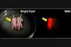
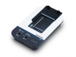
The APEXVIEW™ APX100 benchtop fluorescence microscope makes it fast and simple to acquire expert-quality microscope images. Built with our renowned optics, an intuitive user interface, a powerful AI, and a suite of smart features, the APX100 system combines ease of use with high-quality image data to fit your research needs.
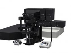
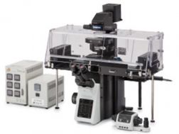
Designed to reduce photobleaching and phototoxicity, the IXplore Live system is optimized for physiological experiments involving live cell and tissue observation. Offering precise environmental control and enhanced rigidity, it supports long-term cell viability and stability for time-lapse imaging applications, such as in cancer, stem cell, and brain research.
Maintain focus accurately and reliably in time-lapse experiments with TruFocus™ Z-drift compensation system
Discover the real morphology of your cells with Olympus silicone immersion optics
Real-time controller helps limit cell disturbance, enabling physiologically relevant data
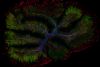
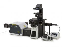
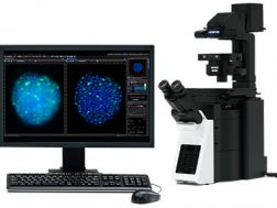
The IXplore Spin system features a spinning disk confocal unit that enables fast 3D image acquisition, a large field of view, and prolonged cell viability in time-lapse experiments. Researchers can use it to perform rapid 3D confocal imaging with high resolution and contrast at greater depths for imaging into thicker samples. The spinning disk also helps to cut down on photobleaching and phototoxicity of samples upon excitation.
Real-time controller (U-RTCE) helps optimize the device’s speed and precision during automated acquisition
TruFocus™ Z-drift compensation system maintains focus for each frame
Precise 3D imaging with improved light collection using X Line™ objectives
Upgrade to the IXplore SpinSR super resolution system as your research progresses
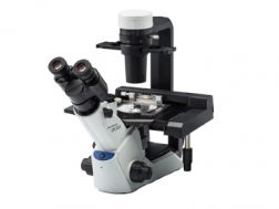
The CKX53 microscope eases the cell and tissue culture workflow, simplifying steps such as live cell observation, cell sampling and handling, image capture, and fluorescence observation. Its integrated phase contrast system, compact, ergonomic design, and stable performance enable simple, efficient cell observation. The universal sample holder and expandable stage accommodate a wide variety of cell culture container types and sizes.
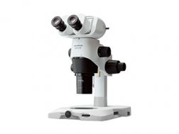
Image macro views of whole organisms to micro views of individual cell structures using the SZX16 research stereomicroscope. Its wide zoom ratio (16.4:1) enables magnifications of 7x–115x with a 1X objective and up to 230x with a 2X objective. Clearly observe fine details owing to the apochromatic optics, reducing chromatic blur for the entire magnification range, and its 0.3 numerical aperture (NA), providing a high 900 line pairs per mm resolution.
Advanced model offering darkfield, brightfield polarization, oblique, and advanced fluorescence observation
Ergonomically designed and provides ample space for manual and automated manipulation and injection tools
2 energy-efficient, long-life LED transmitted light illumination base options
The macro zoom MVX10 microscope’s innovative single-zoom optical path collects light with optimized efficiency at high resolution, offering bright macro fluorescence imaging at all magnifications. Its two-position revolving nosepiece and parfocal objectives enable seamless observation from 4X to 125X with up to 31x zoom.
Maximum fluorescence efficiency plus stereo observation
Optimized high NA optics with minimal autoflourescence
Seamless observation (4x to 125x) with up to 31x zoom
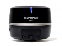
The high-performance DP75 digital microscope camera makes it easy to capture high-resolution brightfield or fluorescence images using a single color camera. It simplifies your microscopy imaging, so you can focus more on your work.
Integrated TruAI denoising maximizes the camera’s image quality in real time
Exceptional color reproduction, making your images as vivid as looking through the microscope oculars
Supports multiple staining combinations and wavelengths up to 1000 nm with a switchable infrared (IR) cut filter
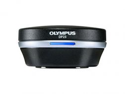
Enabling fast, easy capture of high-quality images that can be clearly observed on a large screen, the DP23 microscope digital camera eases routine life science and clinical research, conferencing, or teaching. Integrate it seamlessly into your microscopy workflow and easily share or stream images.
Share images using the DP23-AOU network solution
Clearly observe live images on a large screen
Fast, high-quality imaging for conferences and teaching
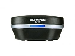
Providing color accuracy and 4K resolution, the DP28 digital microscope camera’s powerful features and wide field of view capture images that enhance tasks such as conferencing, teaching, and clinical research. Integrate it seamlessly into your microscopy workflow for improved work efficiency and image quality.
Providing intuitive operations and a seamless workflow, cellSens software’s user interface is customizable so you control the layout. Offered in a range of packages, cellSens software provides a variety of features optimized for your specific imaging needs. Its Graphic Experiment Manager and Well Navigator features facilitate 5D image acquisition. Achieve improved resolution through TruSight™ deconvolution and share your images using Conference Mode.
Improve experiment efficiency with TruAI™ deep-learning segmentation analysis, providing label-free nuclei detection and cell counting
Modular imaging software platform
Intuitive application-driven user interface
Broad feature set, ranging from simple snapshot to advanced multidimensional real-time experiments
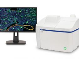
Remotely monitor, analyze, and share your cell cultures’ health, cell count, and confluency using the reliable quantitative data provided by the automated CM30 incubation monitoring system. The system enables label-free observation, reduces the risk of damage to your cultures, and standardizes your culture workflow.
Automatically collects quantitative data on the health and confluency of your cultures
Monitor, analyze, and share your cultures' progress remotely from a PC or tablet
Equipped with oblique epi-illumination for label-free observation