Life Science Solutions
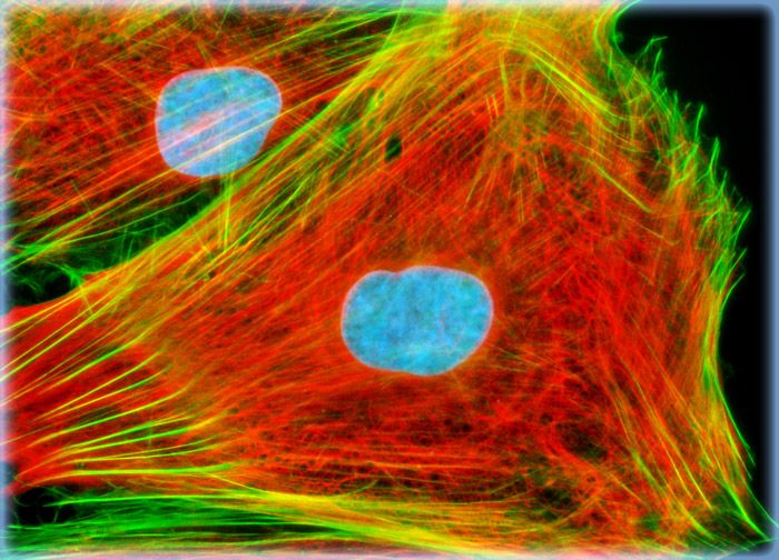
Embryonic Rat Thoracic Aorta Medial Layer Myoblast Cells (A-10 Line)
Focal adhesions were visualized in a log phase adherent monolayer culture of A-10 cells by immunofluorescent treatment with mouse anti-vinculin primary antibodies followed by goat anti-mouse Fab fragments conjugated to the cyanine dye, Cy2. The actin cytoskeletal network was simultaneously imaged with Alexa Fluor 568 conjugated to phalloidin, and nuclei were counterstained with Hoechst 33258.
Sorry, this page is not
available in your country.