Life Science Solutions
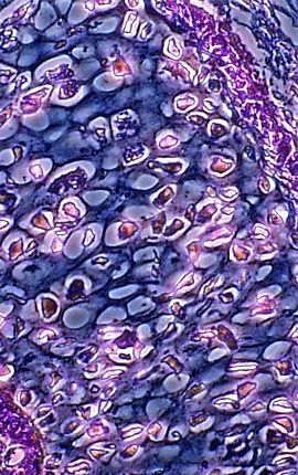
Bird Skin (Epidermis)
A stained thin section of bird skin (epidermis) is illustrated above. As evidenced by this micrograph, combining phase contrast microscopy with classical histological staining techniques often yields enhancement of cellular features.
Sorry, this page is not
available in your country.