Life Science Solutions
Tobacco Mosaic Virus
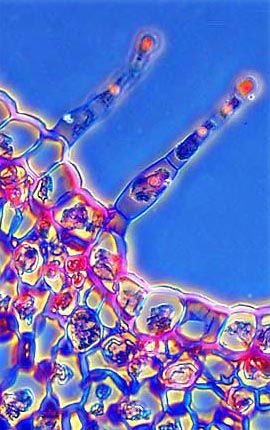
The photomicrograph illustrated above is a medium-magnification phase contrast image of a stained thin section of a tobacco mosaic virus-infected tobacco leaf. Combining phase contrast microscopy with classical histological staining techniques often yields enhancement of cellular features.
对不起,此内容在您的国家不适用。