Not Available in Your Country
Sorry, this page is not
available in your country.
Overview
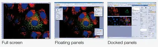
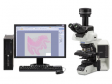
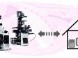
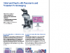
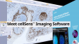
 | Intuitive Operation. Seamless Workflow.The Evident cellSens platform gives you full control over the display and placement of icons, toolbars, and controls, enabling the software to grow and adapt to meet your evolving research needs. |
|---|
cellSens Packages |
cellSens EntrycellSens Entry is the ideal stepping stone for researchers wanting to move into digital image acquisition and documentation, providing all the tools needed for simple image acquisition. |
*cellSens Entry is not available in some areas. |
cellSens StandardThe Evident cellSens Standard software version builds upon the cellSens Entry package, taking acquisition beyond a single image, with advanced image capture processes (e.g. time lapse) and control of motorized and encoded microscope components. The Evident cellSens Standard takes acquisition beyond a single image, with advanced image capture processes (e.g. time lapse) and control of motorized and encoded microscope components. |
|
cellSens DimensionThe most versatile member of the Olympus cellSens family is cellSens Dimension, featuring fully automated image acquisition, powerful analysis tools, and so much more. |
|
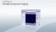
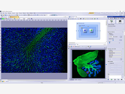 | 5D Experiment AcquisitionAcquire images in five dimensions using tools, like the Graphic Experiment Manager (GEM) and Well Navigator that help you visualize your data acquisition in a user-friendly way. |
|---|
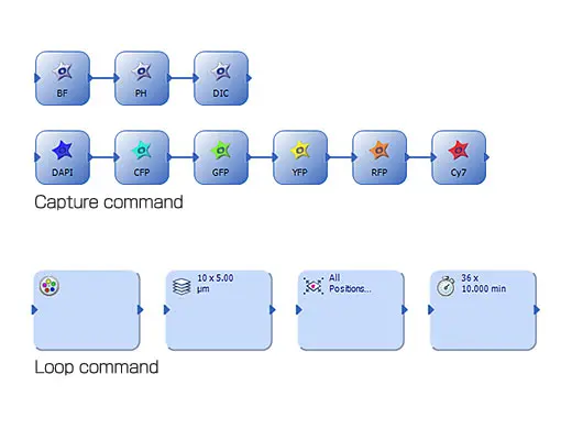
Graphic Experiment Manager (GEM)DimensionThe GEM is a flexible drag-and-drop interface that enables you to build simple or complex experiments within cellSens software. Combine actions within specialized frames to dictate the order and priority of automation and interact with the system during long-term, time-lapse acquisitions without terminating the experiment. To increase efficiency, you can define macro functions, such as executing deconvolution processing in the GEM. |
|
|---|
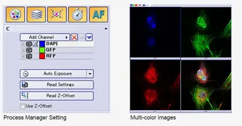
| Process ManagerDimension + StandardThe Process Manager makes it easy to capture multichannel and time-lapse images with just a few clicks. Z-stack imaging is also possible when using a cellSens Dimension license. Dimension + MultipositionStandard + MultipositionUse the optional Multiposition solution to automatically capture multipoint and large-area images when using a motorized stage. You can also navigate around your sample by simply clicking on a point in an image with the Stage Navigation tool. |
|---|
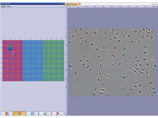
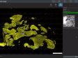
Well Plate NavigationDimension + Multiposition + Well Plate NavigatorThe Well Plate Navigator automatically scans and acquires images from standard and customized well plate formats. All acquired images, sample positions, and user comments can be saved into a structured database for rapid centralized access. Apply unique multidimensional acquisition settings to a single well or multiple selected wells in one step, so you can execute multiple experiments within a well plate in support of complex experiments. |
|
|---|
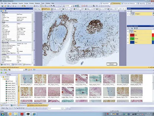 | DatabaseDimension + Database Core or Database ClientStandard + Database Core or Database ClientThe Database Core solution enables you to create user-defined databases with full access control, which can be shared across a network. An interactive query tool makes it easy to find the desired images, associated image properties, user comments, and all related files, like spreadsheets, with automatic preview of the found images. With the Database Client solution, you can conveniently read and write to the shared database from many different stations. Dimension + Multiposition + Well Navigator +Database Core or Database ClientWhen used with the Well Navigator solution, the Database solution makes viewing and analyzing well plate images with a large amount of data more efficient. Simply click on icons for image information, such as the date, file name, or well plate number, and any selection of captured images can be viewed for further analysis. Together, these tools enable you to view captured images and continuously analyze selected images (using Batch Macro commands) using the well plate interface. |
|---|
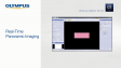
Real-Time Panoramic ImagingDimensionStandard + Manual ProcessCreate stitched images in real time with the Manual Process solution. Manual Process Control makes it simple to move around your sample using a manual stage while the software records and stitches the images in real time, providing a cost-effective alternative to whole slide imaging. Dimension + MultipositionStandard + MultipositionWith cellSens Dimension, and a motorized stage, large-area image acquisition is fully automated with the optional Multiposition solution. When combined with a motorized Z or focus maintenance device, like our TruFocus™ technology, this function can correct for the effects of sample distortion and tilting. |
Related Videos |
|---|
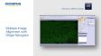
 Original Image (left) versus Extend Focus Image (right) | EFIDimensionStandard + Manual ProcessCreate a single in-focus image from successive image planes as you turn the focus knob using the Extended Focus Imaging (EFI) function. A motorized focus drive fully automates EFI acquisition. EFI composite images can also be created directly from previously captured Z-stacks. |
|---|
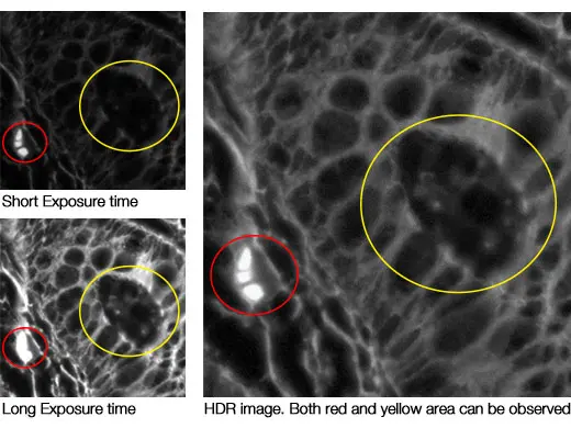
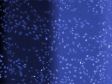
HDR AcquisitionDimensionBy automatically capturing many images at different exposures, the HDR function creates a final image with a higher dynamic range than could be achieved on a single exposure. Low-intensity signals are clearly visible without overexposing the bright areas of the sample. |
|
|---|
 | Image Processing and SharingReveal the true data in your images with TruSight deconvolution and other image processing techniques. Easily share your results with others using Conference Mode or drag and drop data into preconfigured reports. |
|---|
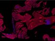
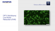
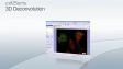
Powerful TruSight™ DeconvolutionDimensioncellSens Dimension includes live 2D deblurring for image preview and acquisition to enable better focusing on thick specimens. Dimension + CI DeconvolutionThe Constrained Iterative Deconvolution solution has the latest algorithms for improved resolution, contrast, and dynamic range with industry-leading speed through GPU processing. Algorithms designed specifically for use with FLUOVIEW™ and Olympus Super Resolution (OSR) images provide clearer, sharper images from high-end systems utilizing ultrafast frame rates and high sensitivities. | Cell line: Human cervical cancer cell line (HeLa)*1 *To learn more about the origin of HeLa cells, visit henriettalacksfoundation.org. |
|---|
| Focus on What's ImportantDimensionObjectively determine the best focus from multidimensional images, including Z-stack and time-lapses, using the Best Focus Extraction tool. This tool is effective in creating T-series images with the best focus possible. Visualize three-dimensional data in a single plane using maximum intensity projections, which combine Z-series planes into a single image with the brightest pixels all in view. |
|---|
Real-Time CollaborationDimensionStandardUse Conference Mode to fill the screen with live or static images for presentation and collaboration. Graphic annotation tools are available at your fingertips for image markup without the need to exit Conference Mode, improving workflow efficiency and saving time. Dimension + NetCamStandard + NetCamUsing standard TCP/IP protocols, the cellSens NetCam Solution facilitates the transfer of live and stored images throughout a network for teaching, mentoring, or supervision. Even when outside of the laboratory, colleagues or supervisors can monitor the work from any point on the network, improving the efficiency of the laboratory. |
Related Videos |
|---|
 | A Different PerspectiveDimensionStandardDisplay images side-by-side for accurate comparison with simultaneous zooming and movement for faster image processing. DimensionTile View mode enables you to view multidimensional datasets all at once, making fine movements or events easier to identify. |
|---|
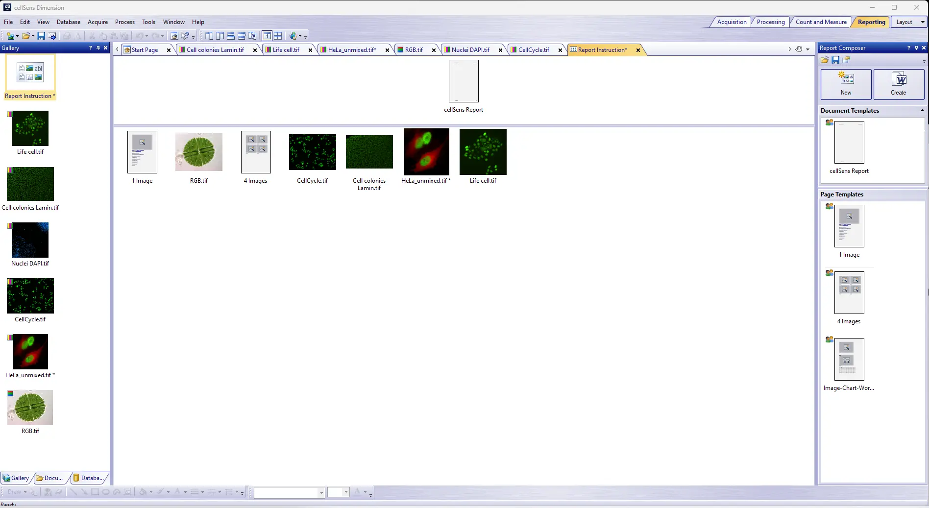
Simple Data ReportingDimensionA convenient Reporting tool combines images and measurement metadata into a report template with easy drag-and-drop operation. These Microsoft Word*2 reports enable you to quickly and easily collaborate with colleagues and communicate results. *2 Requires Microsoft Word version 2010 or later |
|
|---|
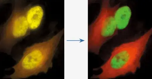
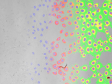
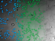
| Spectral UnmixingDimensionWith the linear unmixing algorithm in cellSens Dimension, fluorochromes that overlap in their emission spectra—such as GFP and YFP—can be readily separated to produce crosstalk-free fluorescence images. This linear unmixing tool can also separate autofluorescence-related background signals. |
|---|
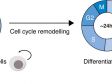
Powerful Analysis ToolsDynamically work with your images to extract the most data for reliable experimental results. The software’s TruAI deep-learning technology offers improved segmentation analysis. Use the Macro Manager to automate entire workflows all the way through image analysis and saving. |
|
|---|
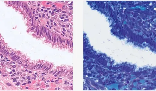
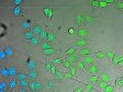
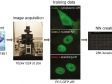
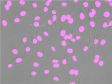
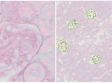
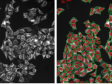
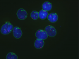
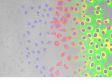
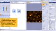
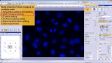
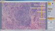
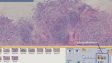
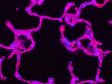
(green) fluorescence image of nuclei, (blue) nuclei detected from brightfield image by TruAI technology Green: You can see that the detection accuracy is low due to unevenness of the GFP label. Blue: Detecting the nuclei with high accuracy despite scratches and dust on vessel | TruAI Deep LearningDimension + Deep LearningStandard + Deep LearningExperience the benefits of cellSens software’s TruAI deep-learning technology to improve your image analysis. From automatic segmentation of complex morphologies without hand labeling to segmentation of cells or organelles using a simple transmitted light image, deep-learning technology offers improved speed and efficiency without the phototoxicity of fluorescence. |
|---|
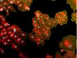
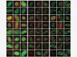
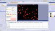
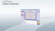
Object Count and MeasureDimensionStandard + Count and MeasureEfficient and precise threshold-based object detection for automated nuclei counting and classification are available. Conveniently export your results to Microsoft Excel for additional analysis. Dimension + Count and MeasureStandard + Count and MeasureExpand on the extensive manual measurements already available in cellSens software with the Count & Measure module. Easily perform automatic object measurement and classification in an interactive interface where recognized objects are always linked with their measurements. Dimension + Count and Measure + Deep LearningStandard + Count and Measure + Deep LearningImprove your count and measurement by detecting objects with deep learning. | 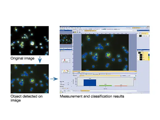 |
|---|
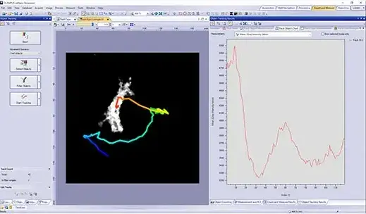
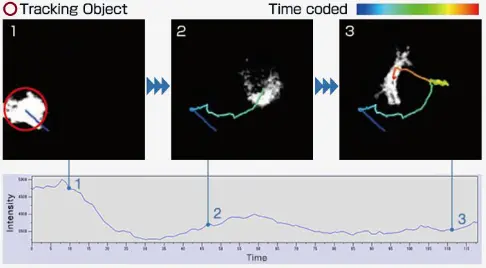
| Live-Cell SolutionsDimensionStandard + Confluency CheckerOur specialized cell identification algorithm enables you to measure cell count and confluency on phase contrast images, including data averaging and total cell count estimation. A cell growth curve can be output by measuring along with time series. Dimension + Count and Measure + TrackingcellSens software's Object Tracking solution provides a powerful and intuitive tool to quantify dynamic processes such as cell movement and division. Moving objects can be automatically detected, tracked, and analyzed over time. |
|---|
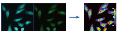
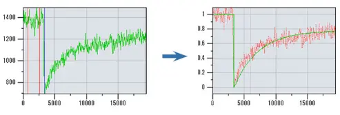
Intensity MeasurementDimensionGraphically depict intensity and ratio values defined by regions of interest (ROIs) and adjust ROI placement to compensate for cell movement. Convert intensity variations to hue and brightness using intensity modulated display (IMD) to visually enhance the fine structures often found within ratio or FRET images. Dimension + Ratio/ FRET or Life Science AnalysisThe Ratio/FRET solution is used to display and analyze real-time ratiometric imaging data. FRET analysis of both sensitized emission and acceptor photobleaching is supported in this user-friendly workflow. Dimension + Life Science AnalysisThe Photo-Manipulation Solution can be used for curve-fitting analysis of FRAP images. | |
|---|---|
|
|
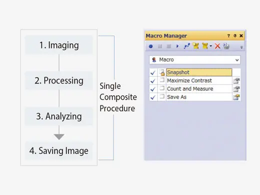 | Simple Macro ExperimentsDimensionSave time and clicks by using the macro manager to automate typical acquisition and data analysis workflows. Batch Macro commands can be applied to multiple images simultaneously and can reduce the time required to complete complex image acquisition and analysis. |
|---|
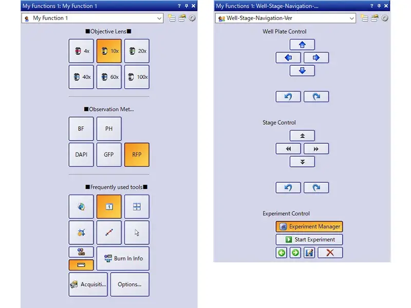
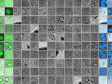
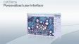
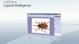
Customizable User InterfaceChoose a recommended layout for image acquisition and analysis or create your own using the My Functions tool set. |
|
|---|
Intuitive at All Skill Levels
Dimension
|
Related Videos |
|---|
 | Build Your Own LayoutDimensionStandardTools and windows can be organized to suit the job at hand, optimizing the layout’s functionality. This enables you to spend less time training and more time imaging. You can create and save custom toolbars for frequently used functions and save them to the My Functions window, where they can be used to improve efficiency when working with your most commonly used tools. |
|---|
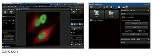
Minimize Monitor GlowDimensionStandardThe Dark Application Skin reduces computer monitor-generated ambient light, helping you adapt to darkened environments, like those required when imaging samples with fluorescence. Icon contrast remains high for easy recognition and quick selection. |
|
|---|
Need assistance? |
The cellSens software package is not for clinical diagnostic use. |
Specifications
cellSens Functions and Optional Solutions |
| Dimension | Standard | Entry | |||
|---|---|---|---|---|---|
| Layout | User experience customization | ✓ | ✓ | ✓ | |
| View | Overlay multiple images | ✓ | ✓ | - | |
| Document groups for side-by-side image comparison | ✓ | ✓ | ✓ | ||
| Movie playback | ✓ | ✓ | ✓ | ||
| Tile view (multiple images in a single data set shown side-by-side) | ✓ | ✓ | ✓ | ||
| Slice view for orthogonal plane viewing of 3D or time-lapse data sets | ✓ | - | - | ||
| Voxel viewer for isosurface and volumetric rendering of 3D and 4D data sets | ✓ | - | - | ||
| Image Acquisition | Snap/movie acquisition | ✓ | ✓ | ✓ | |
| Time-lapse at specified interval | ✓ | ✓ | - | ||
| Automated multiwavelength | ✓ | ✓ | - | ||
| Z-stack | ✓ | - | - | ||
| Multidimensional (XYZT and wavelength) | ✓ | - | - | ||
| Graphical Experiment Manager | ✓ | - | - | ||
| Manual panoramic imaging (Instant MIA and Manual MIA) | ✓ | Manual Process | Manual Process | ||
| Multiposition visitation and stage navigator | Multiposition | Multiposition | - | ||
| Automated panoramic imaging (auto MIA, requires motorized stage) | Multiposition | Multiposition | - | ||
| Instantly create EFI image (manual or motorized Z) | ✓ | Manual Process | Manual Process | ||
| Simultaneous multicolor Imaging (requires two identical cameras** or image splitter) | ✓ | - | - | ||
| Live deblurring | ✓ | - | - | ||
| High dynamic range imaging (HDRI) | ✓ | - | - | ||
| Automated correction collar (ACC) | ✓ | ✓ | - | ||
| Multiwell plate acquisition | Well plate navigator and Multiposition | - | - | ||
| Image Processing | Geometry/combine/filter processing | ✓ | ✓ | - | |
| Fluorescence unmixing | ✓ | - | - | ||
| Brightfield unmixing | Count & Measure | - | - | ||
| Deblurring (No/Nearest Neighbor, Wiener Filter) | ✓ | - | - | ||
| Kymograph | ✓ | - | - | ||
| 2D deconvolution | ✓ | - | - | ||
| 3D deconvolution (constrained iterative deconvolution with GPU process) | CI Deconvolution | - | - | ||
| Image Analysis | Phase analysis | ✓ | - | - | |
| Object measurements and classification | Count & Measure | Count & Measure | - | ||
| Interactive 2D measurements | ✓ | ✓ | ✓* | ||
| Intensity plot over time/z | ✓ | - | - | ||
| Colocalization | ✓ | - | - | ||
| Object counting (manual) | ✓ | ✓ | - | ||
| Object tracking | Tracking and Count & Measure | - | - | ||
| Online ratio and kinetics | Ratio/FRET | - | - | ||
| Ratio analysis (offline) | ✓ | - | - | ||
| FRET analysis | Ratio/FRET or Life Science Analysis | - | - | ||
| FRAP analysis | Photo Manipulation or Life Science Analysis | - | - | ||
| Cell count and confluency measurements | ✓ | Confluency Checker | - | ||
| Deep Learning (TruAI) | Training of neural networks | Deep Learning (TruAI) | Deep Learning (TruAI) | - | |
| Inference using trained neural networks (offline/online) | Deep Learning (TruAI) or Count & Measure | Deep Learning (TruAI) or Count & Measure | - | ||
| Documentation and Collaboration | Automatically compose MS Word reports | ✓ | - | - | |
| Database image and data management solution for microscopy | Database Core | Database Core | - | ||
| Open database and load records/documents from database | Database Client | Database Client | Database Client | ||
| Remoting | Remote live image viewing | NetCam | NetCam | - | |
| * Three-point angle, four-point angle, arbitrary line, closed polygon, polyline, and perpendicular line only. Interactive 2D measurements option is needed to add other measurement tools and make exporting Excel spreadsheets possible.
** Supported cameras: iXon Ultra 897, Zyla 5.5 (USB 3.0), Zyla 4.2 (USB 3.0/CamLink), Neo, iXon Ultra 888, ImagEM X2, ORCA-Flash 4.0 (V2/V3), Prime 95B, Prime BSI, Prime BSI Express, Sona4.2B-11, ORCA Fusion, ORCS-Fusion BT, ORCA-QUEST. |
| Dimension | Standard | |||
|---|---|---|---|---|
| Layout | User experience customization | ✓ | ✓ | |
| Microscope Control | Microscope Control | ✓ | ✓ | |
| View | Slice view for orthogonal plane viewing of 3D or time-lapse data sets | ✓ | - | |
| Voxel viewer for isosurface and volumetric rendering of 3D and 4D data sets | ✓ | - | ||
| Image Acquisition | Automated multiwavelength | ✓ | ✓ | |
| Z-stack | ✓ | - | ||
| Multidimensional (XYZT and wavelength) | ✓ | - | ||
| Instantly create EFI image (manual or motorized Z) | ✓ | Manual Process | ||
| Automated panoramic imaging (auto MIA, requires motorized stage) | Multiposition | Multiposition | ||
| Manual panoramic imaging (Instant MIA and Manual MIA) | ✓ | Manual Process | ||
| Simultaneous multicolor imaging (requires two identical cameras or image splitter)*1 | ✓ | - | ||
| Live deblurring | ✓ | - | ||
| High dynamic range imaging (HDR) | ✓ | - | ||
| Automated correction collar (ACC) | ✓ | ✓ | ||
| Multiwell plate acquisition | Well Plate Navigator and Multiposition | - | ||
| Image Processing | MIA | ✓ | ✓ | |
| Geometry/combine/filter processing | ✓ | ✓ | ||
| Morphological filter | Count & Measure | Count & Measure | ||
| Fluorescence unmixing | ✓ | - | ||
| Brightfield unmixing | Count & Measure | - | ||
| Kymograph | ✓ | - | ||
| 2D deconvolution | ✓ | - | ||
| 3D deconvolution (constrained iterative deconvolution) | CI Deconvolution | - | ||
| Image Analysis | Interactive 2D measurements | ✓ | ✓ | |
| Object counting (manual) | ✓ | ✓ | ||
| Colocalization | ✓ | - | ||
| Object measurements and classification | Count & Measure | Count & Measure | ||
| Object tracking | Tracking and Count & Measure | - | ||
| Online ratio and kinetics | Ratio/FRET | - | ||
| Ratio analys (offline) | ✓ | - | ||
| FRET analysis | Ratio/FRET or Life Science Analysis | - | ||
| FRAP analysis | Life Science Analysis | - | ||
| Cell count and confluency measurements | ✓ | Confluency Checker | ||
| Deep Learning (TruAI) | Training of neural networks | Deep Learning (TruAI) | Deep Learning (TruAI) | |
| Inference using trained neural networks (offline/online) | Deep Learning (TruAI) or Count & Measure | Deep Learning (TruAI) or Count & Measure | ||
| Report | Report function (Microsoft Word is needed) | ✓ | - | |
| Documentation and Collaboration | Database image and data management solution for microscopy | Database Core | Database Core | |
| Open database and load records/documents from database | Database Client | Database Client | ||
* Supported cameras: iXon ultra 897, Zyla 5.5 (USB 3.0), Zyla 4.2 (USB 3.0/CamLink), Neo, iXon Ultra 888, ImagEM X2, ORCA-Flash 4.0 (V3), Prime 95B, Prime BSI, Prime BSI Express, Sona4.2B-11, ORCA-Fusion, ORCA-Fusion BT, ORCA-QUEST. |
cellSens Solutions■ Included □ Optional |
| Dimension | Standard | Entry | |||
|---|---|---|---|---|---|
| Manual Process | Easily create high-resolution composite images (Instant MIA) by simply moving the manual stage. You can also acquire a focused image (EFI) over the entire surface by manually shifting the Z direction. | ■ | □ | □ | |
| Encoded Device | Encoded devices (objectives, light intensity, etc.) make it easy to recall settings. | ■ | ■ | □ | |
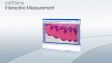
| Interactive Measurement | Draw a polyline, rectangle, or circle on top of your image to obtain exportable measurement data. Measurement results can be exported to Excel. | ■ | ■ | □ | |
| Database Client | Access to the database created with the Database Core option. | □ | □ | □ | |
| Database Core | Make data management and browsing more efficient by creating a database that can easily search and sort acquired images based on data, such as imaging conditions and acquisition date. | □ | □ | ||
| Confluency Checker | Determine the confluency of unstained live cells in culture dishes through quantitative measurements for reliable data. | ■ | □ | ||
| Multiposition | Multipoint and stitched images can be acquired using the motorized stage. When combined with the motorized Z, a focus map can be created from multiple points of focus, and you can obtain stitched images with little focus deviation by removing sample tilt and distortion. | □ | □ | ||
| Count & Measure | Define the morphology of an object, and the software will identify all similar objects and present segmentation analysis results in a chart. | □ | □ | ||
| NetCam | Facilitates the transfer of live and stored images through a network for teaching, mentoring, or supervision. | □ | □ | ||
| Deep Learning | Efficient segmentation analysis powered by deep learning enables challenging target detection, such as label-free nucleus detection. | □ | □ | ||
| Well Plate Navigator*1 | Easily set the capture settings for each well. The well position and name can be tagged to images, making data management easier and well plate screening more efficient. | □ | |||
| CI Deconvolution | Access to GPU-based deconvolution as well as popular and custom TruSight deconvolution algorithms to improve the sharpness, contrast, and dynamic range of reconstructed images. | □ | |||
| Ratio/FRET | Obtain ratio measurements from your images as they are being acquired. | □ | |||
| Tracking*2 | Measure and analyze the luminance and speed of individual cells that move and divide over time. | □ | |||
| Life Science Analysis | FRAP/FRET analysis can be performed on the acquired image. | □ | |||
| Photo Manipulation | Enables cell frap module control and FRAP analysis. | □ | |||
| Laser Control | Enables NI USB-6343 BNC to control external devices. | □ | |||
| Automated Correction Collar (ACC) | Operating automated correction collar. | □ | □ | ||
Super Resolution for cellSens*3 | Renewed super resolution (online and offline) | □ | |||
| *1 Requires Multiposition option
*2 Requires Count Measure option *3 Also for Desktop |
Products with Confirmed Functionality |
| Dimension | Standard | Entry | |||
|---|---|---|---|---|---|
| Olympus | Camera | DP23, DP23M, DP28, DP74, DP75, DP80, XM10, UC90, LC20, LC30, LC35, SC50, SC180 | ✓ | ✓ | ✓ |
| Micoscope | BX43, BX53, BX63, BX61, BX61WI, IX83, IX85, IX73, IX81, SZX16A | ✓ | ✓ | - | |
| IX81-ZDC, IX81-ZDC2 | ✓ | - | - | ||
| Peripherals | BX-DSU, IX3-DSU, IX3-ZDC, IX3-ZDC2, IX2-DSU, U-CBF, cellTIRF (multiline, single line), USB-ODB converter, Real Time Controller (U-RTCE, U-XRTC), IX5-ZDC | ✓ | - | - | |
| Light Source | U-LGPS | ✓ | ✓ | - | |
| Hamamatsu | Camera | ImagEMX2, ORCA-Flash 4.0 V3, ORCA-Flash 4.0 LT PLUS, ORCA-Flash 4.0 LT3, ORCA-Fusion, ORCA-Fusion BT, ORCA-QUEST | ✓ | - | - |
| ORCA-spark | ✓ | ✓ | - | ||
| Image Splitter | W-View Gemini | ✓ | - | - | |
| Q-Imaging | Camera | Retiga 6000 | ✓ | - | - |
| Photometrics | Camera | Prime (PCI-Express), Prime 95B, Prime BSI, Prime BSI Express, Moment | ✓ | - | - |
| Image Splitter | Dual View DV2 / QuadView QV2 | ✓ | - | - | |
| Andor | Camera | iXon Ultra 897, iXon Ultra 888, iXon Life 888, iXon Life 897, Sona4.2B-11,Zyla4.2/Zyla4.2 PLUS (Camera-link,USB3.0), Zyla5.5 (Camera-link 10tap,USB3.0), ZL41 Cell 4.2 (Camera-link,USB3.0), Neo5.5 | ✓ | - | - |
| Vincent Associates | Shutter | Uniblitz shutter (VCM-D1, VMM-D1, VMM-D3) | ✓ | ✓ | - |
| CoolLED | Light Source | pE-1, pE-2, pE800, pE-4000 | ✓ | - | - |
| pE-300white, pE-300ultra, pE-340fura | ✓ | ✓ | - | ||
| Excelitas | Light Source | X-Cite120LED, X-Cite XYLIS, X-Cite TURBO | ✓ | - | - |
| Lumencor | Light Source | SOLA SEII, SEII 365, Spectra X | ✓ | - | - |
| Sutter | Shutter, FW | Lambda 10-3/10-B | ✓ | - | - |
| Prior | Motorized XY Stage | ProScan III, Optiscan III | Multiposition | - | - |
| Shutter, FW, Z-drive | ProScan (I, II, III) , Optiscan III | ✓ | - | - | |
| Piezo Z (Control via Real Time Controller) | NanoScanZ NZ100 | ✓ | - | - | |
| Ludl | Motorized XY Stage | Mac 6000 | Multiposition | - | - |
| Shutter, FW, Z-drive | Mac 6000 | ✓ | - | - | |
| Märzhäuser | Motorized XY Stage | Tango, Pilot Stage | Multiposition | - | - |
Z-drive Controller | Tango | ✓ | - | - | |
| Physik Instrumente | Piezo Z (Control via Real Time Controller) | PIFOC P-721 | ✓ | - | - |
| Applied Scientific Instrumetation | Motorized XY Stage | MS-2000 | Multiposition | - | - |
| Z-drive Controller | MS-2000 | ✓ | - | - | |
| National Instruments | Digital TTL Device | NI USB-6501 | ✓ | - | - |
| NI USB-6343 BNC | Laser Control | - | - | ||
| Yokogawa | CSU | CSU-X1, CSU-W1 | ✓ | - | - |
| For details on Windows OS compatibility, please contact your Evident sales representative. |
| Dimension | Standard | |||
|---|---|---|---|---|
| Olympus | Camera | DP22, DP23, DP23M, DP27, DP28, DP74, DP75, DP80, XM10, UC90, LC20, LC30, LC35, SC50, SC180 | ✓ | ✓ |
| Micoscope | BX43, BX53, BX63, BX61, BX61WI, IX83, IX85, IX73, IX81, SZX16A | ✓ | ✓ | |
| IX81-ZDC, IX81-ZDC2 | ✓ | - | ||
| Peripherals | BX-DSU, IX3-DSU, IX3-ZDC, IX3-ZDC2, IX2-DSU, IX2-ZDC, IX2ZDC2, U-CBF, cellTIRF (multiline, single line), USB-ODB converter, Real Time Controller (U-RTCE) | ✓ | - | |
| Light Source | U-LGPS | ✓ | ✓ | |
| Hamamatsu | Camera | ImagEMX2, ORCA-Flash 4.0 V3, ORCA-Flash 4.0 LT PLUS, ORCA-Flash 4.0 LT3, ORCA-Fusion, ORCA-Fusion BT, ORCA-QUEST, ORCA-Halo* | ✓ | - |
| ORCA-spark | ✓ | ✓ | ||
| Image Splitter | W-View Gemini | ✓ | - | |
| Q-Imaging | Camera | Retiga 6000 | ✓ | - |
| Photometrics | Camera | Prime (PCI-Express), Prime 95B, Prime BSI, Prime BSI Express, Moment, Kinetix, Kinetix22 | ✓ | - |
| Image Splitter | Dual View DV2 /QuadView QV2 | ✓ | - | |
| Andor | Camera | iXon Ultra 897, iXon Ultra 888, iXon Life 888, iXon Life 897, Sona4.2B-11, Zyla4.2/Zyla4.2 PLUS (Camera-link,USB3.0), Zyla5.5 (Camera-link 10tap,USB3.0), ZL41 Cell 4.2 (Camera-link,USB3.0), Neo5.5 | ✓ | - |
| Vincent Associates | Shutter | Uniblitz shutter (VCM-D1, VMM-D1, VMM-D3) | ✓ | ✓ |
| Ludl | Motorized XY Stage | Mac 6000 | Multiposition | - |
| Shutter, FW, Z-drive | Mac 6000 | ✓ | - | |
| Prior | Motorized XY Stage | ProScan III, Optiscan III | Multiposition | - |
| CoolLED | Light Source | pE-1, pE-2, pE800, pE-4000, pE-400 max | ✓ | - |
| pE-300white, pE-300ultra, pE-340fura | ✓ | ✓ | ||
| Excelitas | Light Source | X-Cite120LED, X-Cite XYLIS, X-Cite TURBO, X-Cite NOVEM | ✓ | - |
| Lumencor | Light Source | SOLA SEII, SEII 365, Spectra X | ✓ | - |
| Sutter | Shutter, FW | Lambda 10-3/10-B | ✓ | - |
| National Instruments | Digital TTL Device | NI USB-6501 | ✓ | - |
| NI USB-6343 BNC | Laser Control | - | ||
| Yokogawa | CSU | CSU-W1 | ✓ | - |
| For details on Windows OS compatibility, please contact your Evident sales representative. |
Compatible image formats |
| Read and write | JPEG, JPEG2000, TIFF, BMP, AVI, PNG, VSI, PSD(Adobe Photoshop), Big TIFF, OIR | ||||
|---|---|---|---|---|---|
| Read only | GIF, OIF/OIB(FLUOVIEW format), Cell, STK (MetaMorph), MRC (Medical Research Council) | ||||
System requirements |
| OS | Microsoft Windows 10 Professional (64-bit) (22H2), Microsoft Windows 11 Pro (64-bit)(24H2) | ||||
|---|---|---|---|---|---|
| OS Language | English, Simplified Chinese, Japanese, German and Italian (Entry and Standard) | ||||
| CPU | Intel Core i5, Intel Core i7, Intel Core i9, Intel Xeon Recommended for high-speed image acquisition: QuadCore | ||||
| RAM | 8 GB for general applications, 16 GB or more is recommended for high-speed image acquisition (for DP23/DP28/DP23M cameras, dual memory is recommended for high frame rate imaging), 32 GB or more is recommended for deep learning | ||||
| HDD |
5 GB for installation
Recommended for high-speed image acquisition: solid state drive (SSD) | ||||
Software version update
A version update is available for the next version following the version written on the license card (excludes updating sub-minor versions).
|


![cellSens [ver.4.3] User Manual](https://lifescience.evidentscientific.com.cn/modules/imageresizer/e65/c22/b4f84f0c0c/100x75p50x71.png)
![cellSens [ver.4.3] Database Manual](https://lifescience.evidentscientific.com.cn/modules/imageresizer/9ae/6e1/f5b888459f/100x75p50x72.png)
![cellSens [ver.4.3] Installation Manual](https://lifescience.evidentscientific.com.cn/modules/imageresizer/b73/b72/4f7800c26d/100x75p50x74.png)
![cellSens [ver.4.3] Hardware Manual](https://lifescience.evidentscientific.com.cn/modules/imageresizer/285/481/93b8c65b3c/100x75p50x74.png)
![cellSens [ver.4.2.1] User Manual](https://lifescience.evidentscientific.com.cn/modules/imageresizer/5d1/62e/06aacbbaeb/112x84p74x50.jpg)
![cellSens [ver.4.2.1] Installation Manual](https://lifescience.evidentscientific.com.cn/modules/imageresizer/078/418/8acae569e9/112x84p63x50.jpg)
![cellSens [ver.4.2.1] Hardware Manual](https://lifescience.evidentscientific.com.cn/modules/imageresizer/f03/fa0/b089a67de1/112x84p69x50.jpg)
![cellSens [ver.4.2.1] Database Manual](https://lifescience.evidentscientific.com.cn/modules/imageresizer/71b/33b/d3fd3100b2/112x84p67x50.jpg)
![cellSens [ver.4.2] Database Manual](https://lifescience.evidentscientific.com.cn/modules/imageresizer/59f/e2f/3d66fc4eed/112x84p63x50.jpg)
![cellSens [ver.4.2] Installation Manual](https://lifescience.evidentscientific.com.cn/modules/imageresizer/a98/7cf/61c3347ad8/112x84p69x50.jpg)
![cellSens [ver.4.2] Hardware Manual](https://lifescience.evidentscientific.com.cn/modules/imageresizer/b13/8ca/f52be69c09/112x84p67x50.jpg)
![cellSens [ver.4.2] User Manual](https://lifescience.evidentscientific.com.cn/modules/imageresizer/aca/a27/563aa88d26/112x84p71x50.jpg)
![cellSens [ver.4.1] Installation Manual](https://lifescience.evidentscientific.com.cn/modules/imageresizer/771/3f4/5736605aef/112x84p61x50.jpg)
![cellSens [ver.4.1] Hardware Manual](https://lifescience.evidentscientific.com.cn/modules/imageresizer/20d/b5f/2c79201869/112x84p68x50.jpg)

![cellSens [ver.4.1] Database Manual](https://lifescience.evidentscientific.com.cn/modules/imageresizer/cda/5d9/4122b59f26/112x84p63x50.jpg)
![cellSens [ver.4.1] User Manual](https://lifescience.evidentscientific.com.cn/modules/imageresizer/8b7/530/b58b358776/112x84p61x50.jpg)


Downloads
Installers and Version Checker |
cellSens V4.4.1 64bit Installer | cellSens V4.3 64bit Installer | cellSens V4.2.1 64bit Installer |
cellSens V4.2 64bit Installer | cellSens V4.1.1 64bit Installer | cellSens V3.2 64bit Installer |
cellSens V2.3 32bit InstallercellSens V2.3 64bit Installer | cellSens V1.18 32bit InstallercellSens V1.18 64bit Installer | cellSens V1.16 32bit InstallercellSens V1.16 64bit Installer |
VERSION 1.7 OR LATER | Upgrading to a Windows 10 PC | |
Release Note
Version 4.4.1New Hardware Support
New Functions and Improvements
Version 4.3New function/improvement
Version 4.2.1New hardware support
New function/improvement
Version 4.2New hardware support
New function/improvement
Version 4.1.1New hardware support
Minor bug improvements
Version 4.1New hardware support
New function/improvement
Version 3.2New hardware support
New function/improvement
Version 2.3New hardware support
New function/improvement
Version 2.2New hardware support
New function/improvement
Version 2.1New hardware support
Product portfolio changes
New function/improvement
|