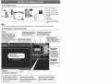
Not Available in Your Country
Sorry, this page is not
available in your country.
- Overview
- Applied Technologies
- Experiment Data Management and Analysis
- Specifications
- Resources
- Specifications
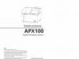
Overview
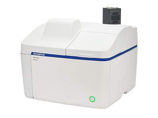
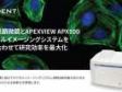
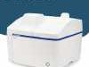
 | Benchtop Fluorescence MicroscopeExceptional Imaging Made EasyThe APEXVIEW APX100 benchtop fluorescence microscope makes it fast and simple to acquire expert-quality microscope images. Built with renowned Olympus optics, an intuitive user interface, a powerful AI, and a suite of smart features, the APX100 system combines the ease of use of an all-in-one-microscope with high-quality image data to fit your research needs. |
|---|
 Application Image GalleryThe APX100 microscope supports a wide range of research imaging applications for slides, dishes, and well plates. Use the included imaging methods such as multichannel, stitching, time-lapse, and Z-stack acquisition in any combination to fit your research protocol. |
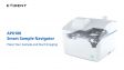
Stay Focused on Your ResearchThe APX100 all-in-one microscope’s smart sample navigator and fast autofocus enable you to spend less time searching for samples and more time collecting data. | 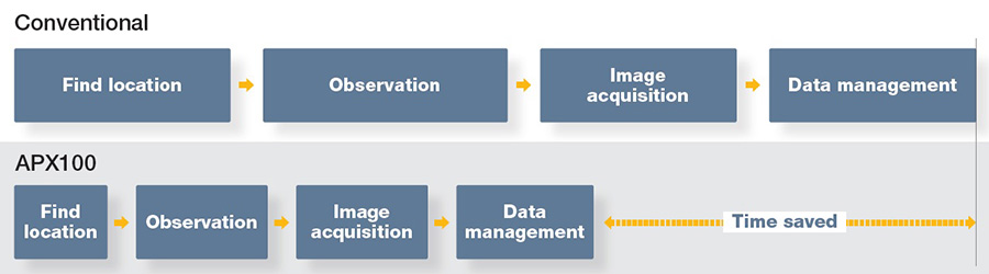 |
|---|
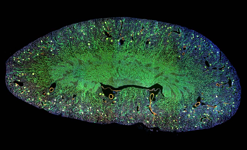
Mouse kidney. Alexa Fluor 488 WGA, Alexa Fluor 568 Phalloidin, DAPI. | Publication-Quality Images in a Few ClicksThe APX100 microscope is built with the same quality optics and technology as our high-end systems, so you can be confident in the quality of your images. |
|---|
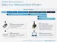
Fast, Efficient Microscope Data ManagementThe system keeps your data organized and acquisition parameters saved for future reference. |
|
|---|
Testimonials |
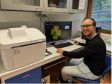
| ”I have been genuinely pleased with the APX100 performance so far in a busy, diverse laboratory. It is feeding data to multiple in-house projects, as well as helping us set up capabilities for new academic collaborations. The options for upgrades are very good, with the advantage of a lower capital expenditure investment.”
Mark Taylor, PhD
| |
Need assistance? |
Applied Technologies
Automatic Sample DetectionPlace your sample in the sample holder, and the smart sample navigator automatically acquires a macro image. The AI then locates your sample on the slide and automatically positions it above the objective, so that you can choose the observation method and immediately begin capturing images. | Related Videos |
Related Videos | Simple Layout and Workflow Maximize EfficiencyDesigned for simplicity, using the APX100 microscope is as easy as loading your sample, closing the lid, and pushing a button.
|
|---|
High-Speed AutofocusWith an autofocus up to twelve times faster than conventional autofocus algorithms, you can locate the imaging plane quickly. | Related Videos |
|---|
 | Advanced Optical TechnologyOur advanced optical technology gives you the flexibility to capture high-quality images in many research applications.
|
|---|
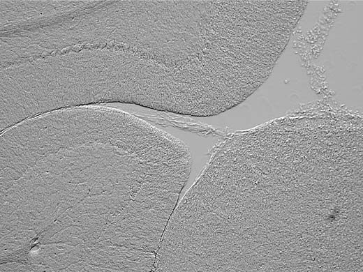
Gradient ContrastAcquiring sharp images at the edges of a well in a multi-well plate can be challenging. The contrast is poor even when using oblique contrast, and DIC does not work well with plastic containers. Using our unique gradient contrast method, you can capture sharp images in a wider area and with higher contrast than the conventional method.
|
Rat brain acquired by Gradient Contrast |
Mouse Kidney acquired by Gradient Contrast |
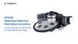
 Rat brain slice Left: Spherical aberration is causing blur / Right: Spherical aberration corrected with motorized correction collar | Clearer Images with Motorized Spherical Aberration CorrectionAccount for the thickness of your cover glass quickly and simply with the system’s motorized spherical aberration correction.
Watch a video to see how the motorized spherical aberration correction works |
|---|
Tools for Clearer ImagesPhotobleaching Prevention ModeThe APX100 system’s photobleaching prevention mode minimizes light exposure and phototoxicity. The fluorescence illuminator only emits excitation light for the time it takes to acquire the image. The static image is then displayed on the screen, providing time to take measurements or evaluate captures. |
|
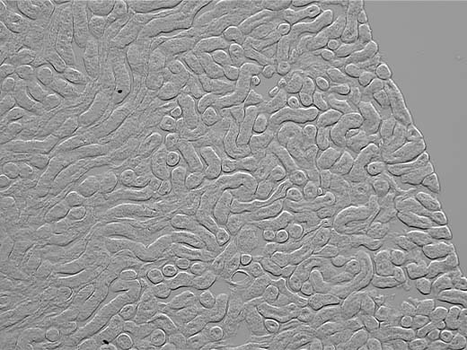
Reduces the Halo Effect in Phase Contrast ImagingThe system can reduce the halo-like artifacts that are sometimes visible in phase contrast observations, making sample structures easier to observe. | Neuro 2a cells Left: Original image / Right: Halo-like artifacts reduced by the software |
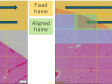
Automated Correction for Uneven SamplesThe built-in focus map allows you to set different focal planes for imaging while also correcting for sample tilt. Obtain sharp stitched images with no focus shift, even for thick, uneven samples. |
|
A Line 20X Objective for Plastic ContainersThe A Line 20X phase contrast objective (UCPLFLN20XPH) with a numerical aperture of 0.7 enables bright, high-resolution imaging of cells in plastic dishes. | 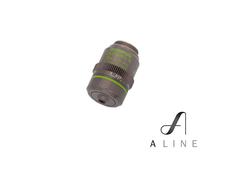 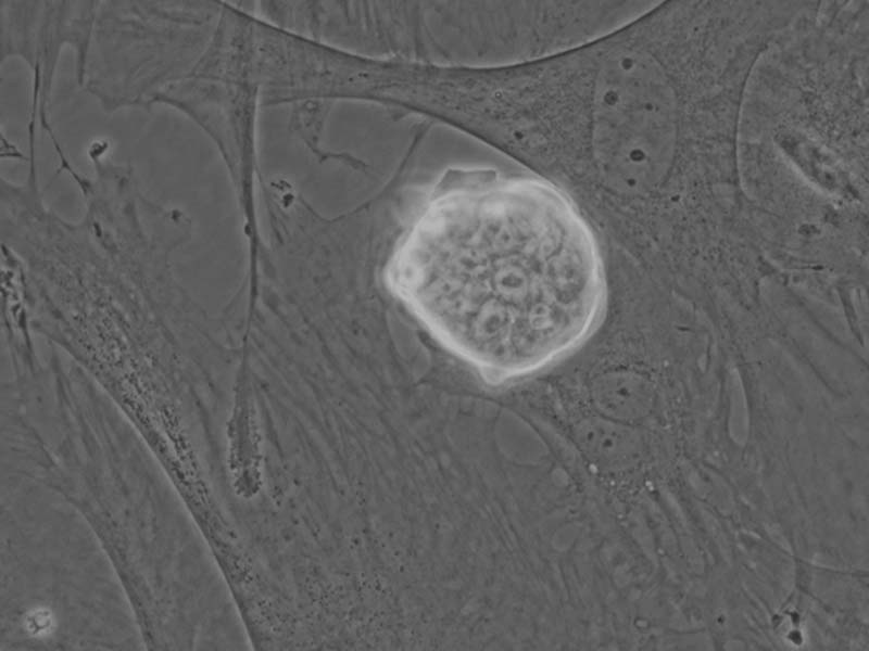  |
Need assistance? |
Experiment Data Management and Analysis
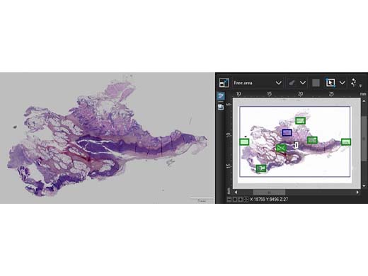
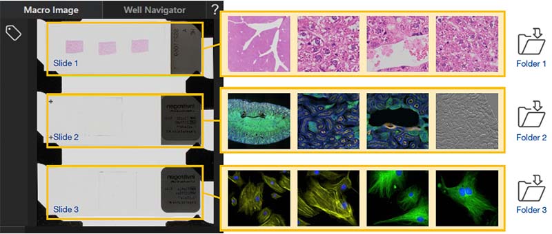
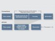
Images Are Organized and Easy to FindThe APX100 microscope has a dedicated system to organize and store your data. When you acquire an image, the software automatically creates folders for each sample and saves the data into the correct one. The consistent indexing keeps your data organized and easy to locate. |
|
Recall Image Acquisition SettingsThe APX100 system saves all important acquisition settings with the image data, making it easy to recall experiment conditions. |
|
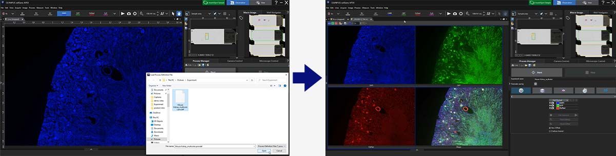
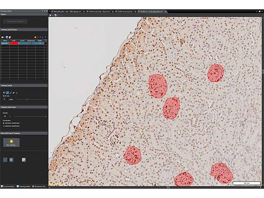
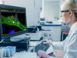
Image Processing and Analysis Functions that Accelerate ResearchOur advanced cellSens image processing and analysis software works seamlessly with the APX100 system, providing you the tools to process and analyze your data quickly and efficiently. |
cellSens Image AnalysisAn optional cellSens software license provides powerful analysis capabilities.
|
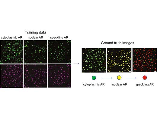
TruAI Deep-Learning Applications | |
|
|
TruSight DeconvolutionFluorescence blur is a challenge when imaging thick specimens with widefield fluorescent microscopes. Olympus’ TruSight deconvolution algorithms improve image quality, enabling you to obtain a higher signal-to-noise ratio, enhance resolution, and see deeper in thick specimens. |
Rat brain, stain: Hoechst, RPCA-Iba1, MCA-7D5, CPCA-GFAP Left: Original image / Right: Processed with TruSight deconvolution | Rat brain, stain: Hoechst, RPCA-Iba1, MCA-7D5, CPCA-GFAP Left: Original image / Right: Processed with TruSight online deblur |
Need assistance? |
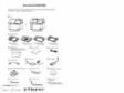
Specifications
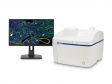
| The APX100 system is available in two frame options—the standard unit configuration (APX100-SU) has a cost-effective, built-in monochrome camera to capture fluorescence image data while the high-end camera unit configuration (APX100-HCU) contains an open port on the monochrome camera path that supports wider FOV camera options. |
APX100-SU
| APX100-HCU
| ||
|---|---|---|---|
| Microscope | Observation method | Brightfield, fluorescence, phase contrast, gradient contrast | |
| Sample holder | Glass slides (3 slides), 35 mm dish (3 dishes), microplate, general | ||
| Objectives |
Choose from 25 available objectives (4X–100X)
Six-position motorized revolving nosepiece | ||
| Motorized aberration correction | One motorized position, five standard positions for objectives | ||
| Transmitted illuminator |
Built-in Köhler illumination for transmitted light, high color rendering LED
Condenser: WD 45 mm, including PHL, PH1, PH2, and PH3 | ||
| Stage | Motorized XY stage with automatic control | ||
| Focusing | Motorized focusing with automatic control | ||
| Magnification changer | Color/monochrome: 0.5x fixed |
Color: 0.5x fixed
Monochrome: 1x, 2x | |
| Fluorescence |
Fluorescence illuminator with fly-eye lens
Choose from 18 mirror cubes; eight-position motorized mirror turret High brightness light guide light source (U-LGPS) Fluorescence LED illuminator (X-Cite XYLIS) Motorized ND filter changer (100%, 25%, and 6%) | ||
| Macro optical system | Built-in, 0.07x macro optics | ||
| Anti-vibration mechanism | Built-in | ||
| Camera | Color camera | 6.41 megapixels, 1/1.8 color CMOS | |
| Monochrome camera | 6.41 megapixels, 1/1.8 monochrome CMOS |
Hamamatsu ORCA-Fusion (USB 3.0)
Teledyne Photometorics Prime95B (USB 3.0) | |
| Software | Operation software |
cellSens APEX, cellSens APEX Entry
(Optional software is not available for cellSens APEX Entry) | |
| Optional software |
Time-Lapse CS-APS-TL-VF
Well Plate Navigator CS-S-WN-VF Full Count and Measure CS-S-CM-VF CI Deconvolution CS-S-DE-VF | ||
| Optional units | Stage-top incubator | Tokai Hit STX series (APX dedicated model) | |
| Solution administration system | Tokai Hit KSX-Type2 | ||
| Environment | Weight | 34.6 kg (76.3 lb) | 35.3 kg (77.8 lb) |
| Power consumption |
70 W
(APX100-SU/HCU only) | ||
| Power supply ratings |
Input: AC 90–264 V, 50–60 Hz
Output: DC 24 V/5 A | ||
System Requirements |
| OS | Microsoft Windows 10 Pro (64-bit), Microsoft Windows 11 Pro (64-bit) |
|---|---|
| CPU | Equivalent to Intel® Core™ processor (3.3 GHz, 6 cores, 18 MB 3200 MHz) or Intel® Core™ processor (2.7 GHz, 14 cores, 24 MB 4800 MHz) |
| RAM |
16 GB or more
DDR4 SDRAM (ECC/Unbuffered/16 GB) or DDR5 SDRAM (4400 MHz/ECC/Unbuffered/16 GB) |
| HDD | OS: 256 GB or more, data: 2 TB or more |
| I/F |
USB3 ×4 ports or more (or USB3.1 Gen1×2), RS-232C × 1 port or more*
*When using CPU; Intel® Core™ processor (2.7 GHz, 14 cores, 24 MB 4800 MHz), the RS232C serial card requires the following: FIFO buffer memory: 64 bytes or more |
| Display |
More than full HD 1920 × 1080
(fixed to full HD setting) |
Resources


Specifications
APX100 Standard | APX100 Extra Sensitive | APX100 Live | ||
|---|---|---|---|---|
| 显微镜 | 观察方式 | 明场(彩色/单色模式),相衬,Gradient contrast,荧光 | ||
| 标本托架 | 玻片(3 片),35 mm 培养皿(3 皿),多孔板,通用型 | |||
| 物镜 |
XAPO(20,40)
UPLFLN10X2PH;UCPLFLN20XPH | XAPO(10,20,40,60O) |
XAPO(10,20,40)
SAPO(30S) | |
|
共25枚物镜可选(4X–100X)
6孔位电动物镜转盘 | ||||
| 电动球差校正 | 支持单孔位电动球差校正,另外5孔位为标准物镜孔位 | |||
| 透射光照明 |
内置科勒照明,高显色性LED透射光光源
聚光镜: 工作距离 45 mm, 包含相差环板 PHL,PH1,PH2,和PH3 | |||
| 载物台 | 自动控制的电动XY载物台 | |||
| 对焦机构 | 自动控制的电动Z轴 | |||
| 变倍装置 | 彩色/单色相机: 固定0.5x |
彩色相机: 固定0.5x
单色相机:1x,2x | ||
| 荧光照明 |
荧光复眼照明
18种荧光激发块可选;8孔位电动荧光激发块转盘 高亮度光导式荧光光源 (U-LGPS),电动ND调节 (100%,25%,和6%) | |||
| 宏观光学系统 | 内置,0.07x 宏观物镜 | |||
| 减震机构 | 内置 | |||
| 相机 | 彩色相机 | 640.1万像素,1/1.8 彩色CMOS | ||
| 单色相机 | 640.1万像素,1/1.8 单色CMOS | 高灵敏度sCMOS | ||
| 功能 | 智能样品导航 | ● | ● | ● |
| 全电动多色荧光 | ● | ● | ● | |
| 多位点和大图拼接 | ● | ● | ● | |
| Z-Stack | ● | ● | ● | |
| 受限迭代消卷积 | 选配 | ● | ● | |
| 延时拍摄 | 选配 | 选配 | ● | |
| 活细胞培养装置 | 选配 | 选配 | ● | |
| 其他可选功能 | 孔板导航 CS-S-WN-VF,全功能计数测量 CS-S-CM-VF | |||
| 运行环境 | 重量 (主单元) | 34.6kg | 35.3kg | |
| 能耗 | 70 W | |||
| 供电 |
Input : AC 90–264 V, 50–60 Hz
Output : DC 24 V/5 A | |||
系统要求 |
| 操作系统 | Microsoft Windows 10 Pro(64位) |
|---|---|
| CPU(中央处理器) | 相当于Intel Xeon处理器W-1250(3.3 GHz,6核,12 MB 2666 MHz) |
| RAM(随机存取存储器) | 16 GB或以上DDR4 SDRAM(ECC/无缓冲/16 GB) |
| 硬盘 | OS:256 GB或以上,数据:2 TB或以上 |
| I/F | 4个或以上USB 3(或2个USB 3.1 Gen1)端口,1个或以上RS-232C端口 |
| 显示器 | 超全高清1920×1080 *固定为全高清设置 |