Brightfield illumination has been one of the most widely used observation modes in optical microscopy for the past 300 years. The technique is best suited for utilization with fixed, stained specimens or other kinds of samples that naturally absorb significant amounts of visible light. Images produced with brightfield illumination appear dark and/or highly colored against a bright, often light gray or white, background. This digital image gallery explores a variety of stained specimens captured with an Olympus BX51 microscope coupled to a 12-bit QImaging Retiga camera system and a three-color liquid crystal tunable filter.
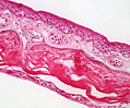 Amphibian Skin
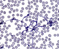 Bacteria
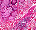 Bald Scalp
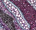 Bracken Fern
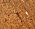 Cerebrum
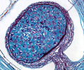 Lycopodium
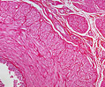 Ductus Deferens
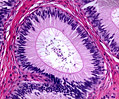 Epididymis
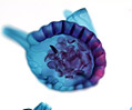 Fern Spores
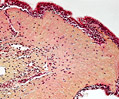 Frog Epithelium
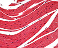 Frog Muscle Tissue
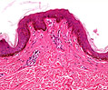 Pigmented Skin
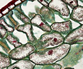 Hemlock Leaf
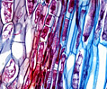 Horsetail Strobilus
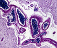 Cerebral Cortex
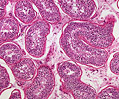 Immature Testes
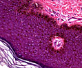 Keloid Scar Tissue
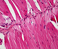 Cardiac Muscle
 Cerebellum
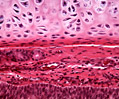 Hyaline Cartilage
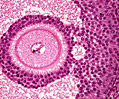 Graafian Follicle
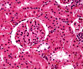 Mammalian Kidney
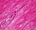 Smooth Muscle
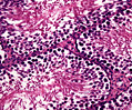 Mammalian Testes
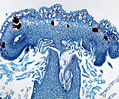 Marchantia Liverwort
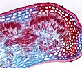 Oleander Leaf
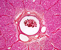 Optic Nerve
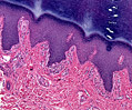 Palmar Skin
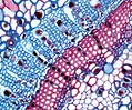 Pine Needle
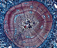 Pine Stem
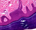 Plantar Skin
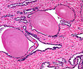 Prostate Gland Old
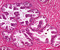 Prostate Gland Young
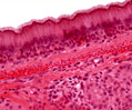 Columnar Epithelium
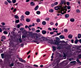 Salamander Liver
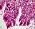 Columnar Epithelium
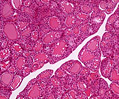 Thyroid Gland
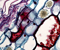 Zamia Stem