Thin unstained, transparent specimens are excellent candidates for imaging with classical differential interference (DIC) microscopy techniques over a relatively narrow range (plus or minus one-quarter wavelength) of bias retardation. The digital images presented in this gallery represent a wide spectrum of specimens, which vary from unstained cells, tissues, and whole organisms to both lightly and heavily stained thin and thick sections. In addition, several specimens exhibiting birefringent character are included to demonstrate the kaleidoscopic display of color that arises when anisotropic substances are imaged with this technique.
 American Dog Tick
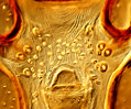
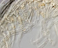 Amphipods
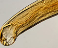 Hookworm
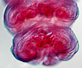 Aurelia Jellyfish
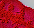 Canine Tapeworm
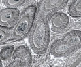 Cat Testes Stained
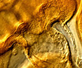 Chicken Embryo
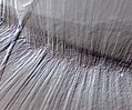 Chicken Embryo Lens
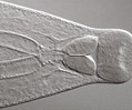 Chinese Liver Fluke
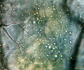 Chironomid Fly Larva
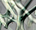 Sponge Fibers
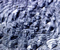 Ctenoid Fish Scale
 Cucumber Tapeworm
 Cycloid Fish Scale
 Deer Tick
 Desmid Algae
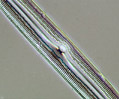 Diatom Frustule
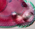 Digenetic Trematode
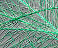 Down Feathers
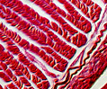 Earthworm Muscles
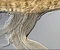 Earthworm Nerves
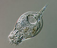 Euchlanis Rotifer
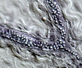 Adipose Tissue
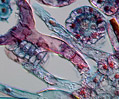 Fern Leaves
 Frog Heart Muscle
 Frog Testes
 Fungus Fruiting Bodies
 Human Cerebrum
 Cheek Epithelial Cells
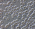 Human Erythrocytes
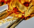 Human Flea
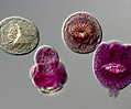 Hydatid Cysts
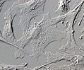 Muntjac Deer Skin
 Intestinal Fluke
 Intestine Thin Section
 Jellyfish Sensors
 Kapok Fibers
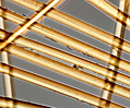 Kevlar Fibers
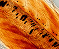 Lancelets
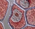 Lancelet
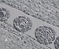 Lily Flower Buds
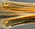 Lone Star Tick
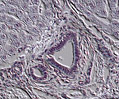 Mammalian Liver
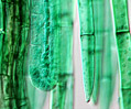 Moss Antheridia
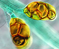 Moss Bulbils
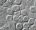 Mouse Kidney
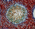 Mushroom Fungus
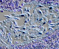 Nucleic Acid Stains
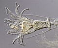 Obelia Hydroid
 Pennaria Hydrozoa
 Pine Tree Pollen
 Planaria Cross Section
 Polypropylene Fibers
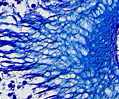 Man-of-War Tentacles
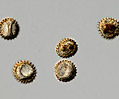 Ragweed Pollen
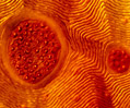 Rocky Mtn. Wood Tick
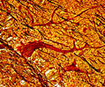 Human Cerebellum
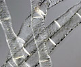 Spirogyra Algae
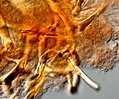 Sponge Skeleton
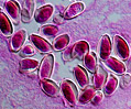 Liver Fluke Eggs
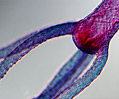 Stained Hydra
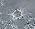 Sun Animalcules
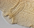 Taenia Tapeworm
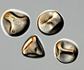 Timothy Grass Pollen
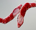 Trematode Rediae
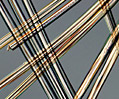 Triacetate Fibers
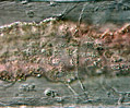 Tubifex Worms
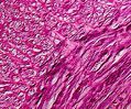 Vas Deferens
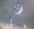 Vorticella Ciliates
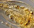 Water Flea
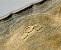 Whipworm Eggs
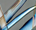 Wild Silk Fibers
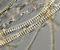 Zygnema Algae