Life Science Solutions
Uterine Adenomyosis
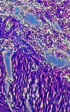
A stained thin section of human uterine tissue exhibiting damage from uterine adenomyosis is illustrated above. As evidenced by the micrograph, combining phase contrast microscopy with classical histological staining techniques in pathological research often yields enhancement of cellular features.
对不起,此内容在您的国家不适用。