TruResolution™ Automated Spherical Aberration Correction for High-Resolution Deep Tissue Imaging
In recent years, microscopy technologies in the life sciences have advanced rapidly, with growing demand for high-resolution imaging of deep tissue structures. However, deep imaging is often affected by optical aberrations—particularly spherical aberration, which can significantly degrade image quality. Precise correction of these aberrations is therefore essential.
This white paper provides a detailed overview of the technological innovations and benefits brought by the TruResolution™ automated spherical aberration correction system in the FLUOVIEW™ FV5000 confocal and FV5000MPE multiphoton laser scanning microscopes. The discussion highlights how the TruResolution system addresses conventional challenges in spherical aberration correction through automation and intelligent optimization.
An Overview of Manual Spherical Aberration Correction and Its Challenges
Objective lenses are composed of multiple lens elements and manufactured through precise design and engineering processes to enable high-accuracy observation of fine structures, such as cells and organelles. These lenses are designed to suppress aberrations (errors in image formation), allowing for extremely clear specimen images under ideal conditions.
However, when the refractive index of the medium, such as the specimen or cover glass, differs from that of the immersion liquid, light refraction causes a discrepancy in the focal depth between rays entering through the center and those entering through the periphery of the objective lens. This phenomenon, known as spherical aberration, leads to reduced resolution and fluorescence intensity.
To correct this aberration, adjusting the correction collar built into the objective lens is effective. By rotating the correction collar, the focal depth difference between central and peripheral rays can be compensated, resulting in optimal imaging performance.
For example, when focusing into water using a water immersion objective lens (Figure 1a), setting the correction collar to 0 yields optimal imaging. In contrast, when observing tissue through a cover glass (Figure 1b), spherical aberration occurs if the correction collar is not adjusted, causing the focal spot to spread and reducing resolution and fluorescence intensity. Proper adjustment of the correction collar (Figure 1c) can effectively correct this aberration.
However, in laser scanning microscopy, observations are typically conducted in a darkroom environment, making manual adjustment of the correction collar during image acquisition difficult. Moreover, adjusting the correction collar alters the focal position, requiring experience and skill to achieve optimal settings.
Additionally, in deep tissue imaging, even if the correction collar is adjusted near the surface, spherical aberration reappears when the observation depth increases (Figure 1d). Therefore, it is extremely challenging to set the optimal correction value at every depth when acquiring Z-stack images.
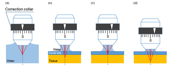
Figure 1. Schematic figures of spherical aberration caused by cover glass or tissue and the effect of correction collar adjustment.
a) Focusing under ideal conditions. When observing a specimen immersed in water using a water-immersion objective lens, light rays from both the center and periphery of the lens converge at the same depth, resulting in no spherical aberration.
b) Focusing with spherical aberration. When observing tissue through a cover glass with water as the immersion medium, refraction at the glass interface causes a discrepancy in focal depth between central and peripheral rays, leading to spherical aberration.
c) Focusing after spherical aberration correction. By properly adjusting the correction collar, the focal depth difference between central and peripheral rays is compensated, achieving ideal focusing conditions.
d) Focusing at deeper positions after surface-level correction. When the correction collar is set for surface observation and the focal plane is moved deeper, spherical aberration reoccurs, causing the focal spot to spread.
Optimizing Spherical Aberration Correction Using a Motorized Collar and Automated Algorithms
The TruResolution system addresses conventional challenges in spherical aberration correction by enabling intuitive and precise adjustment of the correction collar.
First, by equipping the objective lens with a motorized correction collar, users can control the collar via software without physically touching the lens—even in darkroom environments—greatly improving operability. Additionally, the system includes a mechanism that automatically adjusts the Z-position of the objective lens in response to the collar’s rotation angle. This ensures that the focal position remains stable during collar adjustment, maintaining accurate focus at all times (Figure 2a).
Furthermore, the TruResolution system incorporates an intelligent algorithm that automatically determines the optimal correction collar position. By acquiring multiple images at different collar settings and analyzing their contrast curves, the system identifies the peak contrast point to calculate the ideal correction value with high precision (Figure 2b). This enables users to apply the optimal correction with a single click from the software interface.
During Z-stack image acquisition, users can pre-register the appropriate collar positions for each depth. The correction collar then rotates automatically during imaging, ensuring optimal image quality at every focal plane. With the TruResolution system, it is now possible to perform deep tissue Z-stack imaging with precise spherical aberration correction, resulting in consistently bright and high-resolution images across all depths.
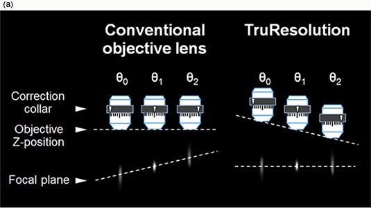
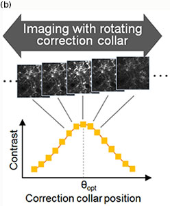
Figure 2. Schematic figures of correction collar control and the optimization algorithm in the TruResolution system.
a) When conventional objectives collars are rotated, the focal plane also changes (left). TruResolution objectives maintain the focal plane by automatically changing the Z-position of the objective according to the rotation angle (right).
b) Finding the optimal correction collar angle (θopt): A contrast curve is determined by calculating the contrast value of each acquired image at different correction collar angles. The optimal correction collar position is calculated by determining the peak of this contrast curve.
Deep Tissue Imaging in Multiphoton Microscopy with the TruResolution System
The TruResolution system has enabled precise correction collar adjustments during deep Z-stack imaging, which was previously difficult. As a result, bright and high-resolution images can now be acquired consistently at all depths.
This is particularly impactful in multiphoton excitation microscopy, where resolution heavily depends on the size of the excitation spot due to the absence of confocal pinholes or cameras. In deep tissue imaging, fluorescence signals tend to attenuate due to light scattering. However, maintaining a small excitation spot size increases excitation density, helping to compensate for signal loss. Therefore, spherical aberration correction is critically important for both resolution and fluorescence intensity.
Figure 3 shows the results of imaging fluorescent beads embedded in a gel that simulates the refractive index and scattering properties of mouse brain tissue. With the TruResolution system, the excitation spot size remains consistent across varying depths, and image brightness is stable. In contrast, when the correction collar is fixed at the surface setting, the spot size increases with depth, resulting in reduced brightness.
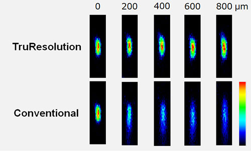
Figure 3. Effect of the TruResolution system on deep imaging of fluorescent microbeads in brain-mimicking gel. Fluorescent microbeads (diameter = 200 nm) in a gel with optical characteristics similar to live mouse brain (refractive index: 1.36, light scattering coefficient: 43 cm-1) excited at 960 nm with constant laser power used for all images.
Upper row: Microbead XZ images acquired at different depths using the TruResolution system for automated spherical aberration compensation.
Lower row: Microbead XZ images acquired at different depths using a fixed correction collar initially adjusted for optimal imaging at the surface of the gel. Image brightness scales are normalized at each depth. All the images were acquired with the FV30-AC25W objective lens.
Next, Figure 4 shows in vivo imaging of neuronal dendrites in the mouse brain at a depth of 400 µm. Using the TruResolution system, brighter and clearer images are obtained even under identical excitation conditions.
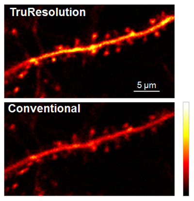
Figure 4. In vivo imaging of neuronal dendrites in live mouse brain (Thy1-YFP-H mouse, sensory cortex). Images were acquired at a depth of 400 µm using an excitation wavelength of 960 nm and the FV30-AC25W objective lens, with identical excitation intensity.
Top image: Automated spherical aberration compensation by the FV30-AC25W TruResolution objective yields sharp, high-contrast images of dendritic spines.
Bottom image: For comparison, the same field of view was captured with the correction collar optimized for the sample surface, as is typical with conventional correction collars.
The TruResolution system is also effective with cleared tissue samples. In cleared tissue samples, the refractive index can vary significantly depending on the reagents used, potentially affecting optical performance. For example, the XLPLN10XSVMP objective lens supports a refractive index range of 1.33 to 1.52. However, if the correction collar is not properly adjusted, aberrations may occur, leading to degraded image quality.
With the TruResolution system, the correction collar can be automatically adjusted even for cleared samples, enabling consistently bright and high-resolution imaging.
Figure 5a shows an XZ image of a Z-stack acquired from a mouse brain cleared with ScaleA2, observed to a depth of approximately 4 mm. Using the TruResolution system, bright and uniform images were obtained even in deep regions. Notably, when the refractive index of the immersion medium matches that of the sample, only a single correction collar setting is required.
Figure 5b presents an XY image at a depth of 2.7 mm, where the image acquired with the TruResolution system is noticeably brighter and clearer compared to that obtained with a fixed correction collar setting.
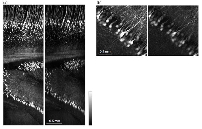
Figure 5. TruResolution system for cleared mouse brain samples.
Mouse brain samples were cleared using the ScaleA2. Left image was acquired with automated correction collar adjustment using the TruResolution system, and the right image was acquired with manual correction collar adjustment based on the refractive index of CUBIC-clearing reagent. Excitation intensity was identical in both conditions. Excitation wavelength: 960 nm; Objective lens: FV30-AC10SV.
a) Maximum intensity projection of a 250 µm-thick section along the Y-axis after Z-stack acquisition.
b) Maximum intensity projection of a 100 µm-thick section at a depth of 2.7 mm along the Z-axis.
High-Resolution Imaging in Confocal Laser Scanning Microscopy Using the TruResolution System
In confocal laser scanning microscopy, variations in cover glass thickness can cause spherical aberration, making it difficult to acquire high-resolution images. If the correction collar is not properly adjusted, images may appear blurred and dim, significantly reducing observation accuracy.
The IXplore™ IX85 inverted microscope platform is equipped with a motorized correction collar drive unit, compatible with most Evident objectives that are equipped with a correction collar and designed for inverted microscopes. By switching objective lenses according to the observation purpose, the system can be flexibly operated as part of the TruResolution system.
Figure 6 shows XYZ images acquired from Z-stack imaging of a mouse brain slice cleared with RapiClear. When using the TruResolution system, both XY- and Z-axis resolution are improved, resulting in brighter and sharper images.
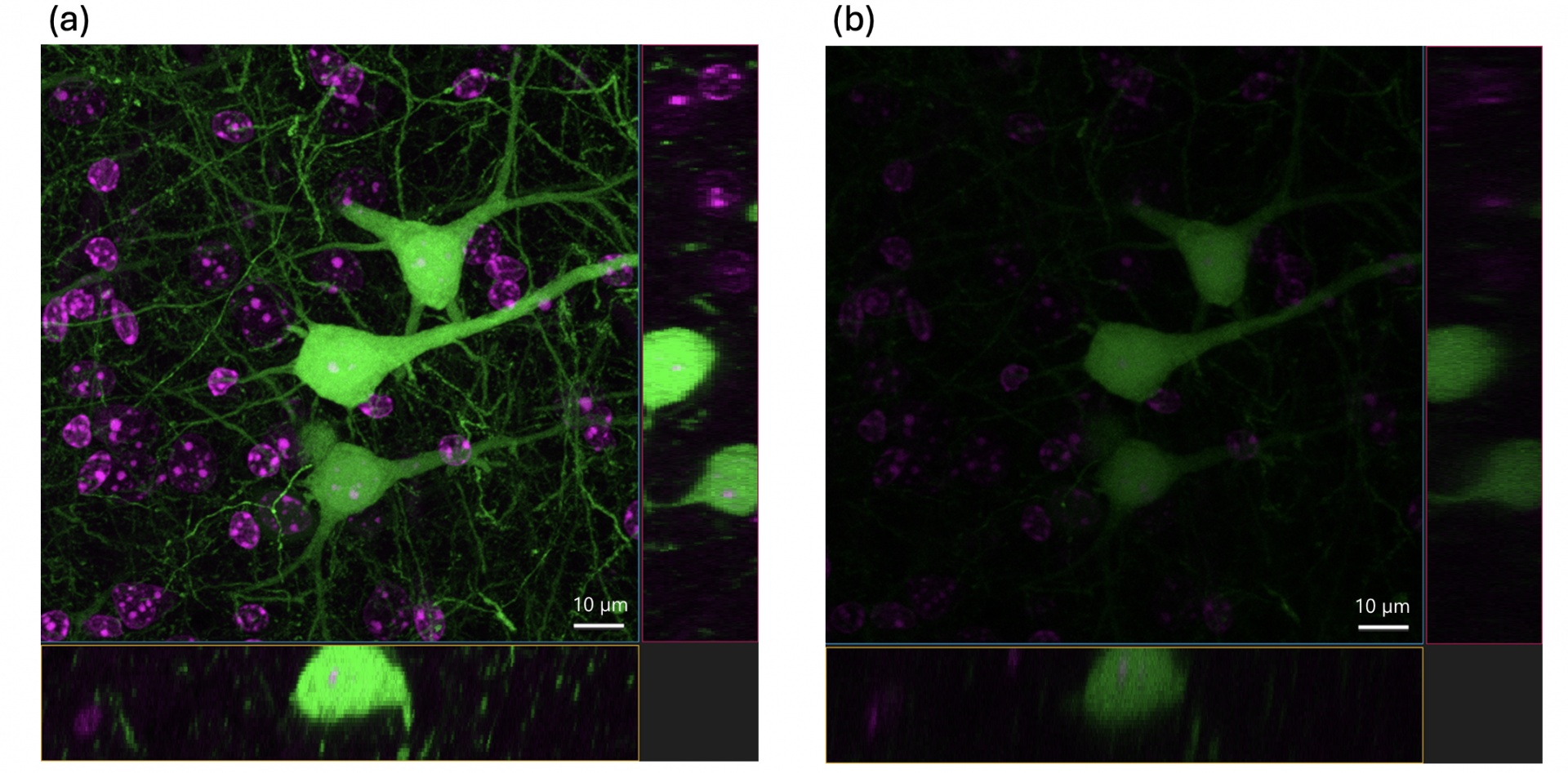
Figure 6. Confocal imaging of a mouse brain slice cleared with RapiClear 2.
a) Image acquired with automated correction collar adjustment using the TruResolution system.
b) The image was captured with the correction collar rotated fully toward the direction of lowest refractive index. This condition corresponds to the scenario in which the correction collar is held and rotated while attaching the objective lens to the revolving nosepiece.
These results confirm that automatic correction collar adjustment with the TruResolution system enables acquisition of brighter, higher resolution images.
Conclusion
This white paper detailed the automated spherical aberration correction technology provided by the TruResolution system, along with its demonstrated effectiveness. Traditionally, adjusting the correction collar during deep tissue imaging or when working with cleared samples has posed significant challenges, often resulting in inconsistent image quality and complex operation.
The TruResolution system fundamentally resolves these issues through a suite of integrated features: motorized control of the correction collar, automated maintenance of the focal position, intelligent determination of optimal correction settings, and full compatibility with Z-stack imaging.
As the demand for deeper imaging and adaptation to diverse sample conditions continues to grow, the TruResolution system is expected to serve as a core technology that supports high-precision and highly reproducible imaging—greatly expanding the possibilities of microscopic observation.
Author
Hiromi Utsunomiya
Life Science High-End Imaging, Product Management, Evident
Acknowledgments
Application images were acquired at the RIKEN CBS-EVIDENT Open Collaboration Center, courtesy of Dr. Hiromu Monai, Dr. Hajime Hirase, and Dr. Atsushi Miyawaki.
References
For more details on the studies mentioned in this white paper, please refer to the following article:
1. Ue, Y., Monai, H., Higuchi, K., et al. “A Spherical Aberration-Free Microscopy System for Live Brain Imaging.”Biochemical and Biophysical Research Communications, 2018, Vol. 500, 236–241.
Sorry, this page is not
available in your country.