FORMERLY
Get in Touch
Get in Touch
Products
Applications
Blog
Resources
Learn
Support
Shop
Careers
FORMERLY
What is EVIDENT?
Search
Shopping Cart
English (English)
|
简体中文 (Simplified Chinese)
Life Science Solutions
Application Notes
Home
/
Application Notes
アプリケーション資料
Isolation of Circulating Tumor Cells: A Model Workflow for Single-Cell Analysis
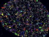
Light Up Your Research: Imaging Microbes at the Nanoscale
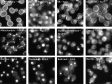
Light Up Your Research: Imaging DNA Replication Sites at the Nanoscale
How to Track Single Molecules in Living Cells
How to Immobilize Non-adherent Cells for Single Molecule Imaging
How to Obtain Simultaneous Multicolor Nanoscopy with One Laser
How to Analyze Clusters in SMLM Data
Accelerate Hematological Analysis with AI-Powered Digital Pathology
SLIDEVIEW VS200 for Digital Pathology
SLIDEVIEW VS200 for Life Science Research
HPF Counting Remains a Core Tool in Clinical Microscopy
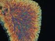
Polarized Microscopy and What It Can Teach Us About the Materials That Make Up Our Skeletal Tissue
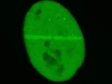
Identification of Viable Oocytes in Phase Contrast Images Using Deep Learning
Yeast Protein Localization Classified Using TruAI™ Deep-Learning Technology
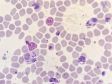
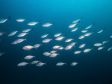
Facilitating Malaria Research with a Battery-Powered Microscope Illumination Solution
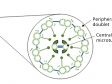
Revealing the Regulatory Mechanisms for the Proper Assembly of Motile Multicilia Using High-Speed Live-Cell Imaging
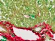
Identifying Cirrhosis in Liver Tissue Using a Research Slide Scanner
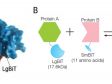
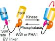
Imaging Intracellular Localization of Protein-to-Protein Interactions Using NanoBiT® Technology
Automating Migration Assay Imaging and Quantification Using an Incubation Monitoring System
Monitoring Spheroid Formation in U-Bottom Multiwell Plates Using an Incubation Monitoring System
Remotely Monitoring the Primary Culture of Bone Marrow-Derived Mesenchymal Stem Cells
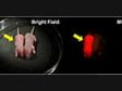
Verifying the Tumor Labeling of a Novel Fluorescent Nanoprobe Using NIR Imaging
Monitoring the Effect of Medium Components on the Growth and Differentiation of Human iPS Cell-Derived Liver Bud Organoids Using the Olympus Provi™ CM20 Incubation Monitoring System
5 Basic Microscope Adjustments for ART Research
Advanced Live-Cell Analysis Using AI-Driven High-Content Screening Systems
Advantages of Using the SLIDEVIEW™ VS200 Slide Scanner’s Motorized Condenser to Adjust Specimen Contrast
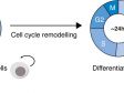
Monitoring Cell Cycle Dynamics During Stem Cell Differentiation Using the scanR System’s TruAI™ Deep-Learning Technology
Quantification of C-Fos-Positive Neurons in Mouse Brain Sections Using TruAI™ Deep-Learning Technology
Monitoring the Whole Process of Human iPS Cell-Derived Liver Bud Organoid Differentiation Using the CM20 Incubation Monitoring System
Observation of Neural Structures between the Cortex and Thalamus in the Marmoset Brain Using FLUOVIEW FV3000
Extended Time-Lapse Imaging of Cell Motility and Proliferation Using the Olympus FLUOVIEW FV3000 and TruFocus Z-Drift Compensation System
Determining Cell Proliferation and Cytotoxicity with Increased Accuracy and Ease Using the CM20 Incubation Monitoring System
How Old Is the Fish?—Using a SLIDEVIEW™ VS200 Research Slide Scanner to Overcome the Challenges of Inspecting Fish Otoliths
Comparing Human iPS Cell Lines using the CM20 Incubation Monitoring System Part 3: Reducing Differentiation Efficiency Variations in Liver Bud Organoids Among iPS Cell Lines
Predicting Multi-Class Nuclei Phenotypes for Drug Testing Using Deep Learning
Rapid Automated Detection and Segmentation of Glomeruli Using Self-Learning AI Technology
Support Biopsy Efficiency with the Simplified Operation of the IX73 Inverted Microscope
Using Intravital Multiphoton Microscopy to Visualize Dynamic Interactions between Neutrophils and Monocytes/Macrophages in an Influenza-Infected Mouse Airway
Automated Analysis of Label-Free Organoid Imaging Data
Total Support for Assisted Reproductive Technology (ART) Research
Imaging of Drug Response in Drug Efficacy Assessment
Comparing Human iPS Cell Lines using the CM20 Incubation Monitoring System Part 2: Variations in the Efficiency of Liver Bud Organoid Differentiation among iPS Cell Lines
Accelerating and Optimizing the Segmentation and Analysis of Pancreatic Islets with the VS200 Research Slide Scanner and TruAI Deep-Learning Solution
Optimizing Your Oblique Observation Using Our Flexible Contrast Solution
ライブセルイメージング・細胞生物・分子生物学分野での発光イメージング活用事例
光受容体(オプシン5)活性化の発光Ca2+イメージング(蛍光Ca2+イメージングとの比較)
ショウジョウバエの胚発生過程におけるプロモーターアッセイの発光イメージング
Comparing Human iPS Cell Lines using the CM20 Incubation Monitoring System Part 1: Observing Differentiation into iPS Cell-Derived Liver Bud Organoids
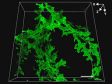
Dynamic Volumetric Imaging with the FV3000RS Confocal Microscope: 3D Reconstruction of Actin Dynamics in a Colletotrichum graminicola Spore
Digitizing Very Thick Fluorescent Samples with the SLIDEVIEW VS200 Research Slide Scanner
Label-Free Transmigration Assay Using scanR TruAI for Self-Learning Microscopy
Observing a Vascularized Tumor Spheroid on a Chip with a Confocal Microscope
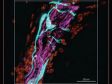
Distinguishing Brain and Tumor Blood Vessels in Deep Tissues Using Multiphoton Microscopy
Cell Quality Evaluation with the CM20 Incubation Monitoring System : Improve the Reproducibility of Experiments through the Characterization of Cells
Perform Accurate and Efficient Microscopy Image Analysis Using TruAI based on Deep Learning
Mapping Aortic Valve Cells Using the FLUOVIEW FV3000 Microscope
High-Resolution Imaging of Bone Cell Interactions Using the FLUOVIEW FV3000 Confocal Microscope and an X Line 40X Oil Immersion Objective
Selective Isolation of Adherent Single Cells
Selective Isolation of Living Cells for Omics Analysis
Connectome Imaging Using the FLUOVIEW FV3000 Confocal Microscope and X Line Objectives
Digitizing Slides Using a Manual Microscope and Digital Camera
Multiplexing with the FLUOVIEW FV3000 Confocal Microscope
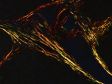
Discovering Fine Neurovascular Structures in Tibial Epiphysis Using the FLUOVIEW FV3000 Microscope
3D Observation of Cleared Mouse Liver Using the FLUOVIEW FV3000 Microscope
Visualizing DNA Repair Proteins with the FLUOVIEW FV3000 Confocal Microscope
Identifying Collagen Fiber Types I and III Stained with Picrosirius Red Using the BX53 Microscope Equipped with Olympus’ High Luminosity and High Color Rendering LED
The Inner Focus Articulating Nosepiece and FVMPE-RS® Multiphoton Microscope Enable Fast Volumetric and Off-Angle Brain Imaging
3D Time-Lapse Imaging of Spheroids with the FLUOVIEW FV3000 Confocal Microscope: 48-Hour Continuous Observation of Antibody-Dependent Cell-Mediated Cytotoxicity (ADCC)
Development of a New Fucci(CA) Application: A Fluorescent Probe for Visualizing Cell Cycles
Routine Microscopy: Improving Productivity Through Better Ergonomics
Experience the Clarity of 4K Microscopy
Polarizing Observation Technique for Bone Histomorphometry
Fluorescence, phase-contrast, and bioluminescence imaging of live cells incubated in plastic bottom dishes using a 20X high numerical aperture (NA) phase contrast objective lens
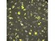
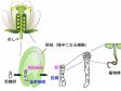
Application of silicone immersion objectives to long-term live-cell imaging of plant zygote embryogenesis
Application of silicone immersion objectives to long-term 3D live-cell imaging of mouse embryo during development
Application of the Z-Drift Compensation System IX-ZDC to multidimensional cell-based assay at the single cell level
Use of Low Chromatic Aberration Objective PLAPON60XOSC for Quadruple Immunofluorescence of Brain Tissue
Using silicone oil immersion objectives with a confocal laser scanning microscope for deep tissue observation in cleared specimens
TIRF Imaging of Changes in Membrane Morphology and Molecular Dynamics
Single-Molecule Fluorescence Imaging on the Cell Membrane
Tackling diabetes with confocal microscopy
Good sausage, bad sausage: Analysis with VS-Analysis
Show more
このページはお住まいの地域ではご覧いただくことはできません。
メールでのお問い合わせ
メールマガジンの登録
Print
Cancel
Redirecting
You are being redirected to our local site.
Attention: Please enable JavaScript
Sorry, this page is not available in your country