Not Available in Your Country
Sorry, this page is not
available in your country.
Overview
 |
Clearer Insights,
|
|---|
Powering Discovery Across Applications |
Live-cell imaging and dynamic processes
| Screening and large-scale acquisition
| 3D models and complex samples
| Fluorescence and multiplex imaging
|
Expand Your View, Enhance Your VisionSee and capture more—and reduce your acquisition times—with an unmatched field of view (FOV) in the industry and an array of advanced end-to-end imaging features that set a new standard in clarity and precision, including groundbreaking new objective technology Unmatched 26.5mm FOV View more of your sample at once with an industry-leading field number (FN) of 26.5mm on two integrated imaging ports. Cover larger areas in fewer images, get more data in each image, minimize the need for image stitching, and speed up your workflows. |
Comparison of how many cells you can acquire in different FN. |

Need assistance? |
Clearer Insights
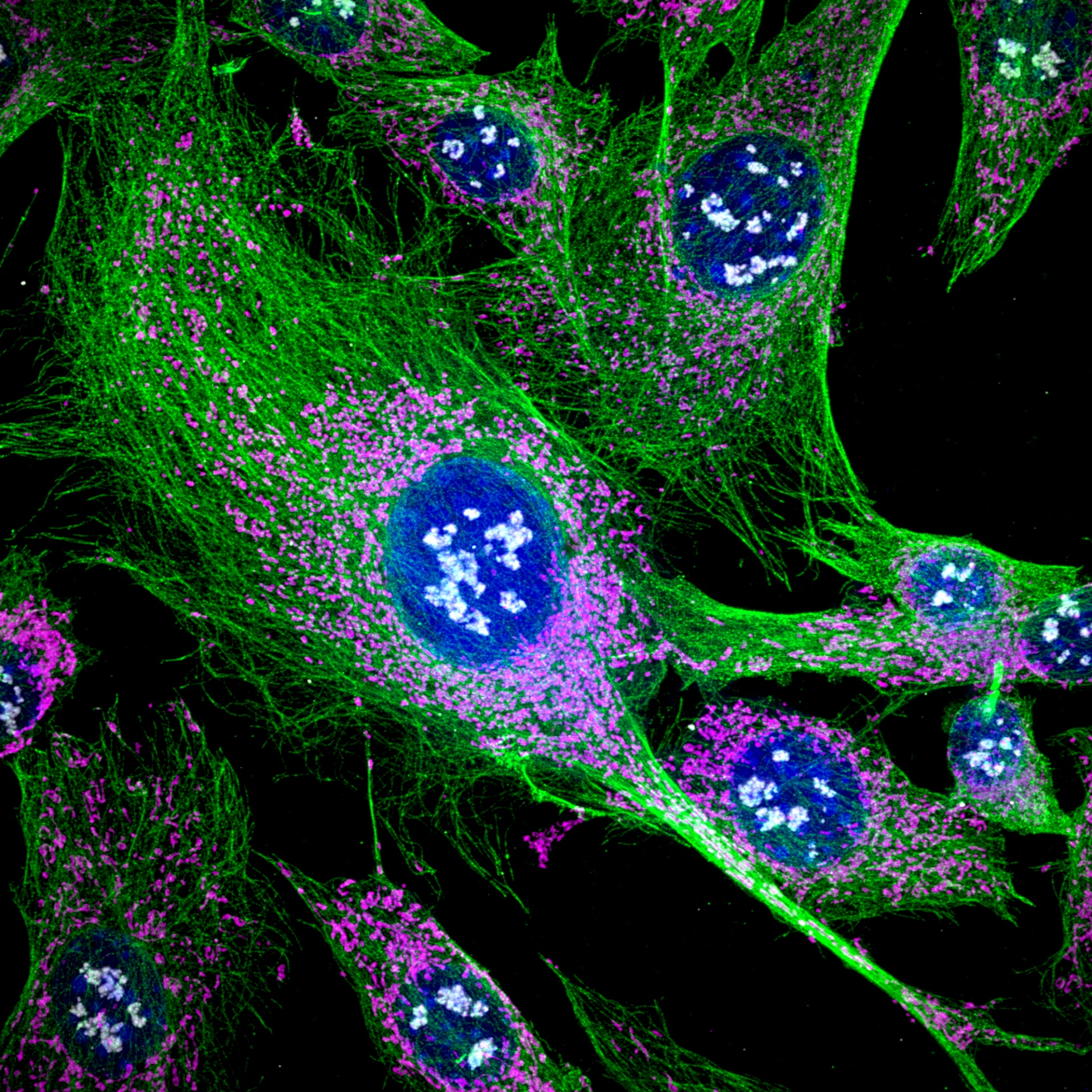
Cultured NIH 3T3 cells. Blue: nuclei, Green: tubulin, Red: HSP60, Gray: fibrillarin. Sample provided by EnCor Biotechnology Inc. | Experience New Levels of Precision ClarityWith an innovative, strategic selection of advanced beginning-to-end imaging features, the IXplore™ IX85 is setting a new standard in precision and clarity—enabling you to see more fine details than ever before and easily obtain impactful, publication-ready images. |
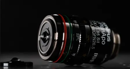
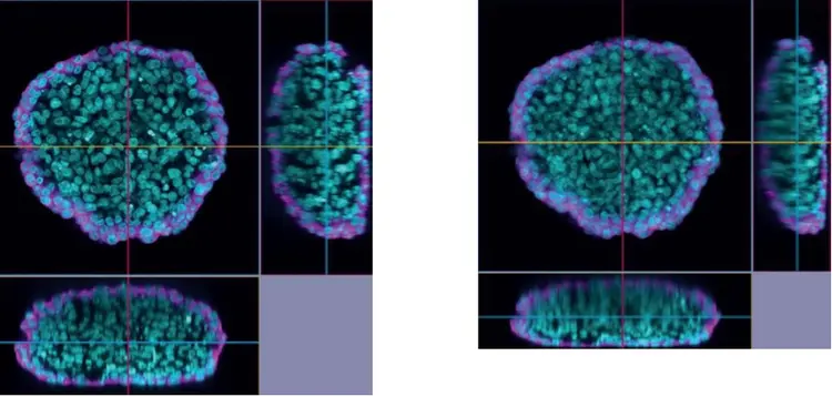
See More with Deeper ImagingSee deeper into your samples, catch more signal, and image the real shape of cells over time—all without the hassle of immersion liquid. Pair the IXplore IX85 with the world’s first multi-immersion silicone gel objective lens (LUPLAPO25XS). Featuring groundbreaking silicone gel pad technology, LUPLAPO25XS delivers the image quality of silicone immersion with the ease of use of a dry objective.
|
XYZ image comparison. Left: LUPLAPO25XS silicone gel pad objective, NA 0.85. Right: UPLXAPO20X dry objective, NA 0.8. Cleared HeLa* 1 cell spheroid (cyan: nuclei, magenta: microtubules). Captured on the IXplore IX85 Spin. *To learn more about the origin of HeLa cells, visit henriettalacksfoundation.org. |
|---|
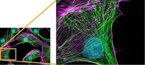
Images acquired using an X Line UPLXAPO60XO objective lens. | Consistent QualityEvident’s expansive range of X Line™ and A Line™ objectives can meet any research need, pushing your system further. Taking full advantage of the IXplore IX85’s 26.5 mm FOV, X Line objectives deliver exceptional correction of spherical and chromatic aberrations throughout a wide field of view. With high numerical apertures (NA) and outstanding transmission, they provide sharp, consistent images. |
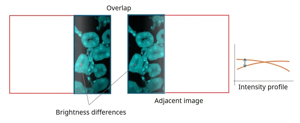
Edge-to-Edge Clarity and PrecisionIntelligent Shading Correction automatically compensates for uneven illumination across the field of view to generate stitched images without manual adjustment, improving efficiency and consistency in large-area imaging. |
Left: Without Intelligent Shading Correction. Right: With Intelligent Shading Correction. |
|---|
Stunning 3D ImagesUse TruSight technology to create 3D images with confocal-like sharpness at the speed of a widefield microscope. Explore 3D samples in real time with advanced software solutions that help you see more, faster. |
|---|
Need assistance? |
End-to-End Automation
Optimize Your Workflow, Accelerate New DiscoveriesThe IXplore IX85’s automated acquisition features, customizable interfaces, and task management software help you work efficiently while staying confident in your results. |
Automated Spherical Aberration CorrectionThe automated correction collar automatically fine-tunes your objective settings and optimizes image quality by reducing spherical aberrations caused by variations in cover glass thickness.
| |
 Automated correction collar on the IXplore IX85 platform. | 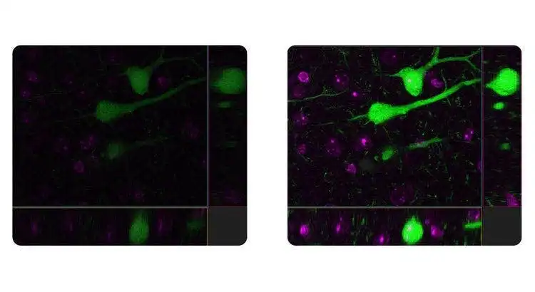 The automated correction collar automatically fine-tunes your objective settings and optimizes image quality. Left: Without auto correction collar. Right: With auto correction collar. |
Real-Time Image Acquisition AnalysisFurther enhance productivity with real-time image processing and analysis. Advanced imaging tools support consistent, accurate results.
|  |
Your Live Cells in FocusThe IXplore IX85 platform offers enhanced rigidity to reduce the effects of vibration and temperature on your microscope. This facilitates reliable live-cell and time-lapse imaging by helping maintain the desired focus position on the Z-axis. Pair the IXplore IX85 with our TruFocus™ Z-drift compensator to capture cellular dynamics through high-precision, multipoint time-lapse images that are aligned and in focus. |
Close Monitoring of Cell Migration GrowthUse cellSens™ Object Tracking and Count and Measure solutions to analyze the movement and division of live cells in time-lapse or Z-stack image sets. Confluency Checker tools are a proven way for you to measure confluency on phase contrast images as well as fluorescence. | 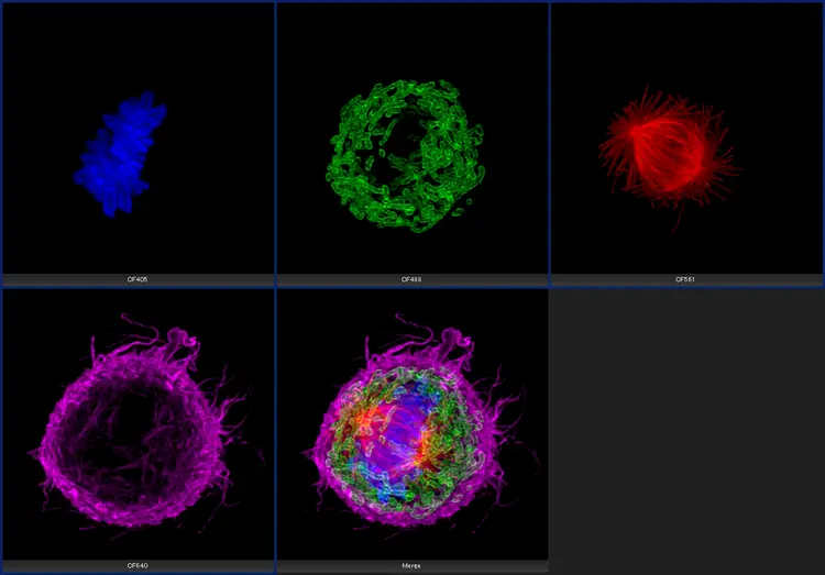 Cultured Cos 7 cell. |
 Left: Without TruSight / Right: With TruSight | Improve Experiment Efficiency with Advanced DeconvolutionWith cellSens Dimension software, you can use live 2D deblurring for preview and acquisition to enable exceptional focusing on thicker specimens.
|
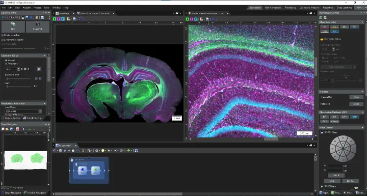
Fully Automated EfficiencyThe Graphical Experimental Manager (GEM) of cellSens Dimension software enables fully automated multidimensional observation (X, Y, Z, T, wavelength, and positions) to make experiment setup even easier. | 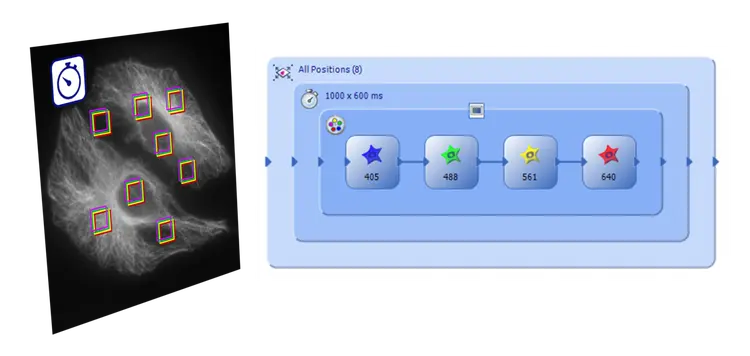 |
Need assistance? |
Unmatched Customizability
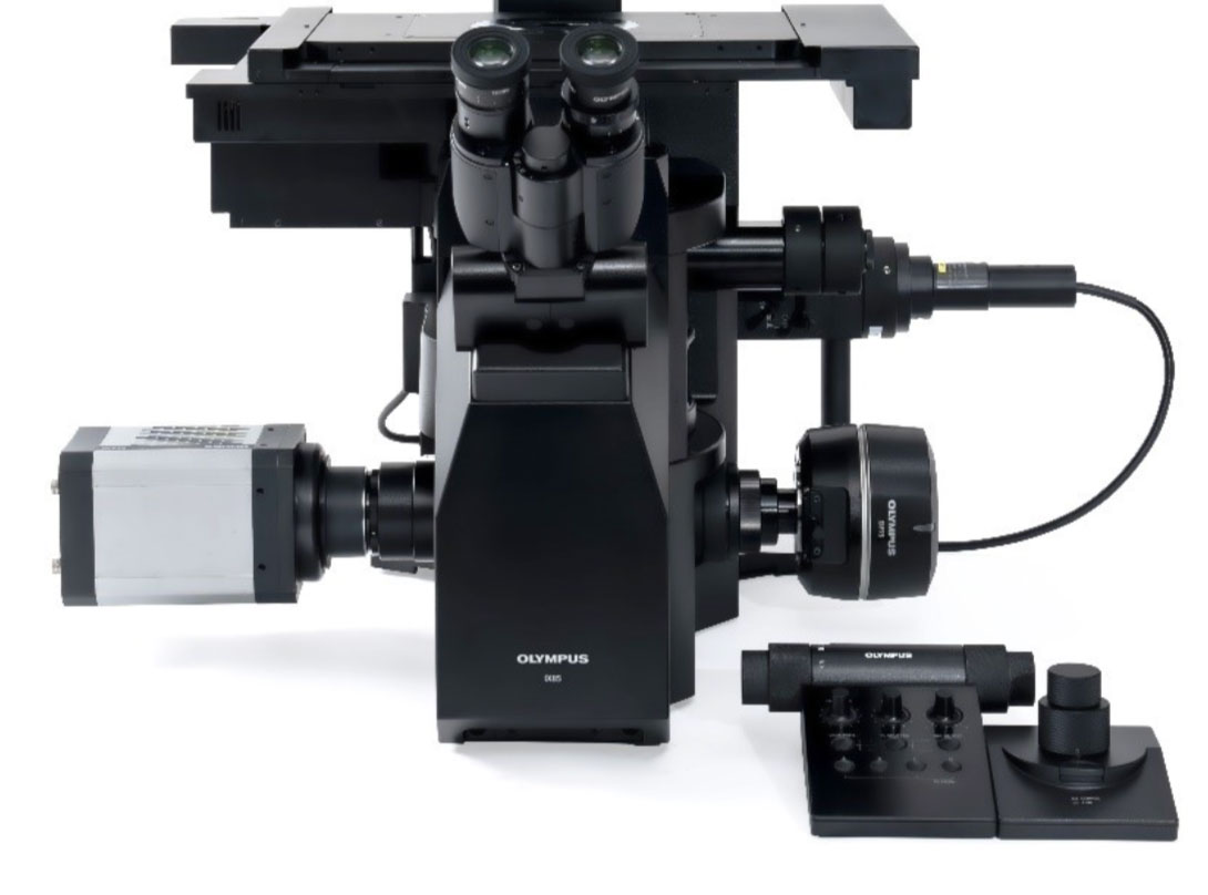
One Platform, Infinite PossibilitiesStay at the forefront of technology with the IXplore IX85 Pro’s unmatched customizability. Now you can support diverse imaging modalities across a variety of applications with no sacrifice in performance—and empower your lab to stay prepared for whatever the future brings. | |
| New Built-In Ports Two built-in camera ports (left and right) and an optional third port enable enhanced functionality and the capacity to create a multi-camera configuration setup. Four optical paths are available: left (100%), right (100%), binocular (100%), and BI 50%/L 50%. |
|---|---|
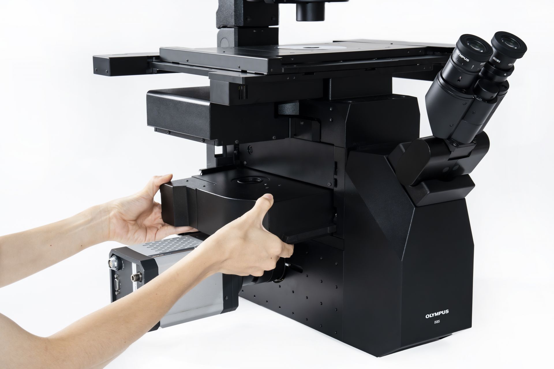
Versatile Deck Structure Customize your microscope setup for your own experiments. An open frame design makes it simple to add or swap modules to customize functions as needed. You can also upgrade from one to two decks as research needs change. |
|
| New Built-In Threaded Holes Built into the frame, threaded holes around the camera ports turn the IXplore IX85 Pro into a canvas for innovation, whether you’re building a custom system or integrating the frame into your product. |
Future-Proof ReliabilityIdeal for busy lab settings, long-term imaging projects, and live-cell imaging, the IXplore IX85 platform provides durability and performance backed by exceptional service and support. Confidence in Every UseThe IXplore IX85’s durable frame design is built to withstand a multitude of uses, ensuring consistent performance and reducing downtime. The platform provides maximum stability, minimizing small movements that could disrupt delicate long-duration imaging experiments. With the same service and support that you’ve come to know and expect from Evident, you can be confident that your IXplore IX85 will perform as intended for years to come. Upgrade and Expand Your Imaging Technology as Needs EvolveThe IXplore IX85 lets you upgrade and expand your imaging platform as research needs evolve by adding features such as TruFocus Z-drift compensation, environmental control, and imaging modalities like confocal laser scanning, spinning disk, and photomanipulation. For high-content screening, integration with scanR analysis software streamlines data processing and quantitative evaluation. Third-party integration is easy with support for incubation systems, motorized stages, and more. |

Need assistance? |
Specifications
| IX85P1ZF | IX85P2ZF | |||
|---|---|---|---|---|
| Microscope frame | Optical system | UIS2 optical system | ||
| Revolving nosepiece |
Motorized 6-position revolving nosepiece (DIC slider attachable),
One position for Automated Correction Collar Simple water proof structure | |||
| Focus |
Stroke: 10.5 mm
Minimum increment: 0.01 um, Maximum nosepiece movement speed: 3mm/s | |||
| Intermediate Magnification Changer |
3 positions (Coded)
1X / 1.6X / 2X | |||
| Light path selection |
Motorized 4 positions
Eyepiece 100%, left 100%, right 100%, eyepiece 50%/left 50% | |||
| Deck insert layer | 1 layer | 2 layers | ||
| Maximum port field number |
Left/Right side port: FN26.5, BI port: FN22
Deck right side port: FN18 |
Left/Right side port: FN18, BI port: FN22
Deck right side port: FN18 | ||
| Focus compensator |
TruFocus
Z drift compensator |
Offset method (Focus search, one-shot focus, continuous focus),
Class 1 laser product, laser wavelength: 830nm | ||
| Transmitted light illuminator |
Pillar tilt mechanism (30 ° inclination angle, with vibration reducing mechanism),
Condenser holder (with with 88 mm stroke, refocusing mechanism), Field iris diaphragm adjustable, 4 filter holders Light source: High power LED light source | |||
| Observation tube | Widefiled (FN22) |
• U-TBI90BK Wide field tilting binocular
• U-BI90 Wide field binocular • U-TR30-2/U-TR30NIR Wide field trinocular | ||
| Stage | Motorized stage |
• IX5-SSA: Stage stroke: X: 116mm x Y: 78mm, maximum stage movement speed: 40mm/s, Knob controller
• 3rd party motorized stage | ||
| Mechanical stage with right handle | Stage stroke: X: 116mm x Y: 78mm, | |||
| Plain stage | 232 mm (X) x 240 mm (Y) stage size, stage insert plate exchangeable (ø110 mm) | |||
| Condenser |
Motorized long working
distance condenser |
W.D. 27 mm, NA 0.55, motorized turret with 7 position slots for optical devices
(3 positions for ø30 mm and 4 positions for ø38 mm), motorized aperture and polarizer | ||
|
Long working distance
universal condenser | NA 0.55, W.D. 27 mm 5 positions for optical devices (3 positions for ø30 mm and 2 position for ø38 mm) | |||
| Ultra long working distance | NA 0.3, W.D. 73.3 mm, 4 positions for optical devices (for ø29 mm) | |||
| Fluorescence illuminator |
L-shape-fluorescence
illuminator | L-shaped design with exchangeable FS and AS modules, slider shutter and ND filter poket | ||
| Fluorescence mirror turret |
Motorized fluorescence
mirror turret | Motorized turret with 8 positions, built-in shutter, simple waterproof structurer | ||
| Fluorescence light source |
• U-LGPS: LED and LDP light source, Class 1 laser product
• 3rd party LED light source | |||
| Control unit (IX5-MCZ) | Nosepiece position, light path selection, filter turret position, FL shutter ON/OFF, DIA LED power, DIA LED ON/OFF, 4 customizable button | |||
| Control box (IX5-CBH) | PC interface | USB (Type-C), RS-232C | ||
| Operating enviornment |
• Indoor use
• Ambient temperature: 5 ºto 40 ºC (41 º to 104 ºF) • Maximum relative humidity: 80% for temperatures up to 31 ºC (88 ºF), decreasing linearly through 70% at 34 ºC (93 ºF), 60% at 37 º C (99 ºF), to 50% relative humidity at 40 ºC (104 ºF) • Supply voltage fluctuations: Not to exceed ±10% of the normal voltage | |||