

Olympus Image of the Year Award 2019Image of the Year Award 2019 has come to a close. Have a look at below to see the beautiful images we received this year. For the latest information and submission details, click here! |
|
The Global Winner
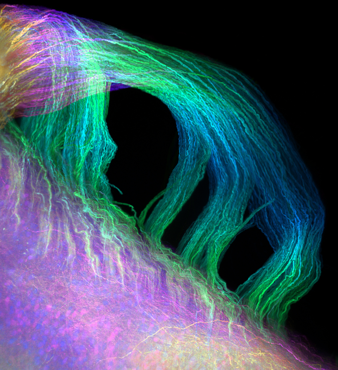
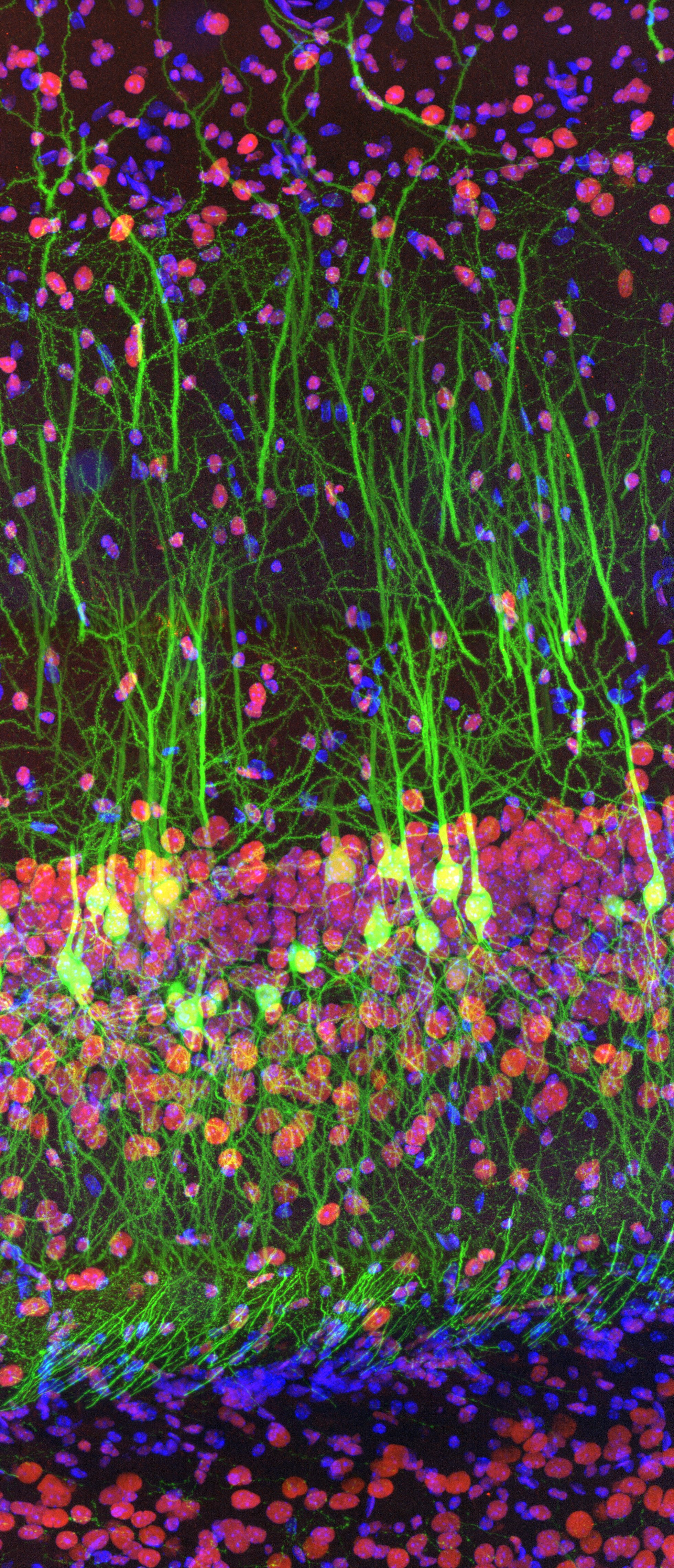 | The winning image was taken by Ainara Pintor (Spain). The stunning fluorescence image shows the immunostaining of Thy1-EGFP mouse brain slice with two fluorophores. In green, the excitatory hippocampal neurons, which express Green Fluorescent Protein under Thy1 promoter. In red, Fat mass and obesity-associated (FTO) protein revealed with Alexa Fluor 594 antibody. In blue, cell nuclei labelled with DAPI. Captured with a super resolution confocal microscope system. |
Regional Winners
Americas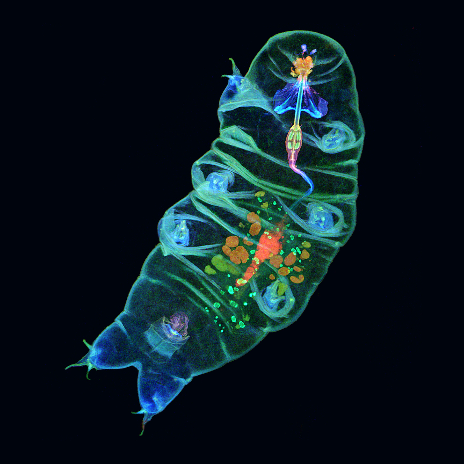 | Europe, the Middle East and Africa | Asia-Pacific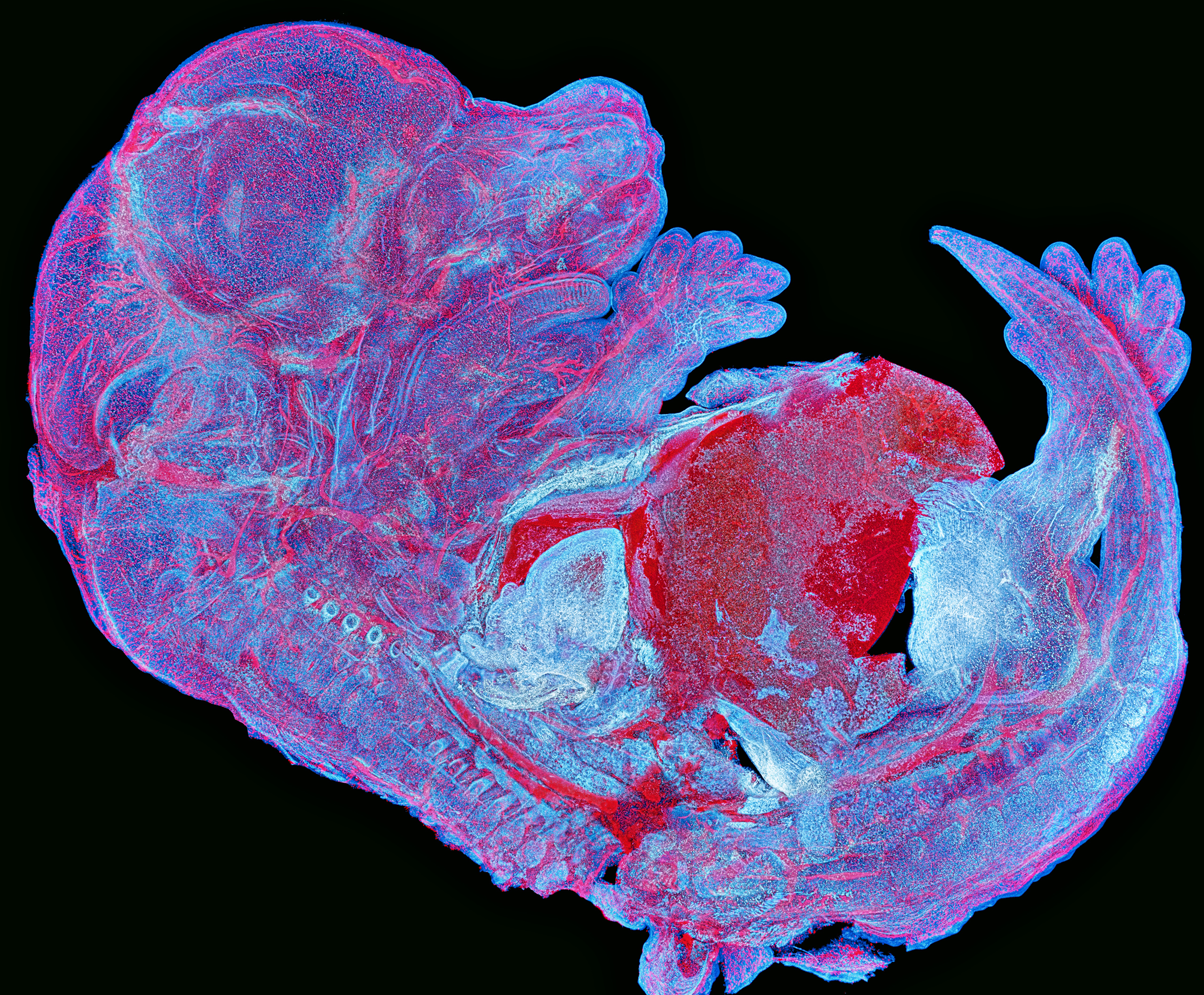 |
The winning image was taken by Tagide deCarvalho (U.S.A.). The beautiful fluorescence image shows the inside of a tardigrade with colorful details. | Alan Prescott (U.K.) 's image won the EMEA regional prize. As titled "The Mouse's Whiskers" by Alan, this fascinating shape was captured from a frozen section of a mouse using multiple fluorescent labels. | Asia-Pacific regional prize was awarded to Howard Vindin (Australia). This remarkable image shows the autofluorescence of a mouse embryo with 950 tiles stitched together. |
Honorable Mentions
 This is the ovary of a gall-inducing wasp anselmella miltoni girault showing their eggs captured with a confocal microscope. |  Inflorescence of arabidopsis thaliana with young, developing flower buds expressing a fluorescent reporters. | 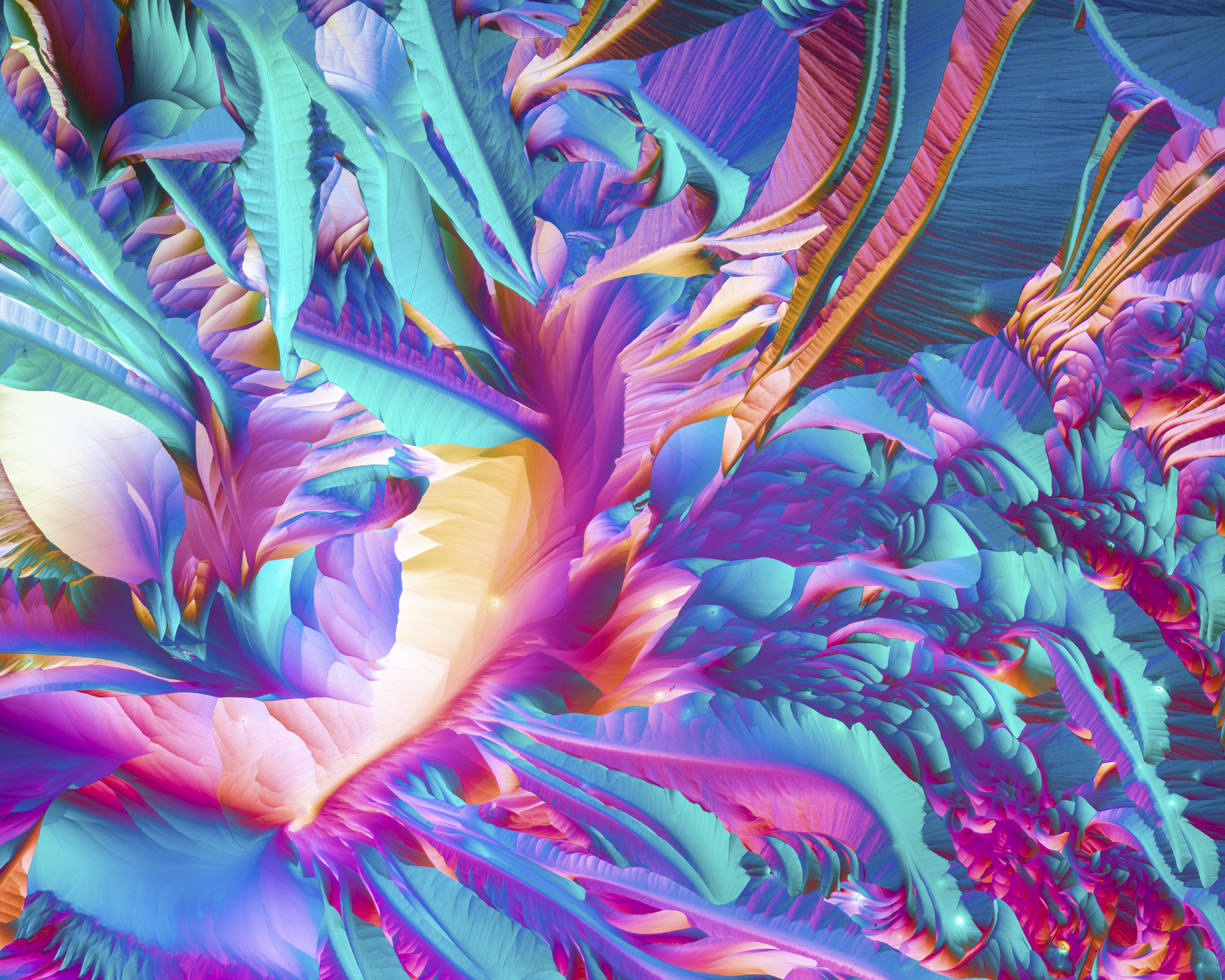 A preparation of amino acids L glutamine and beta alanine crystalized out of an ethanol solution and photographed at 50X using polarizing filters. |
 Mouse spinal cord with a GFP expression and cleared with the CLARITY method. |  Image of 3D depth: color-coded reconstruction of confocal images of tubulin (microtubuli) in mitotic COS7 monkey fibroblast cells. |  Captured a part of foldable wings of an insect, and named as “a road in the sky”, because veins look like roads and spines on wing membrane are like stars. |
 Different photonic crystals in insects on the elytron of the longhorned beetle Sternotomis pulchra. | 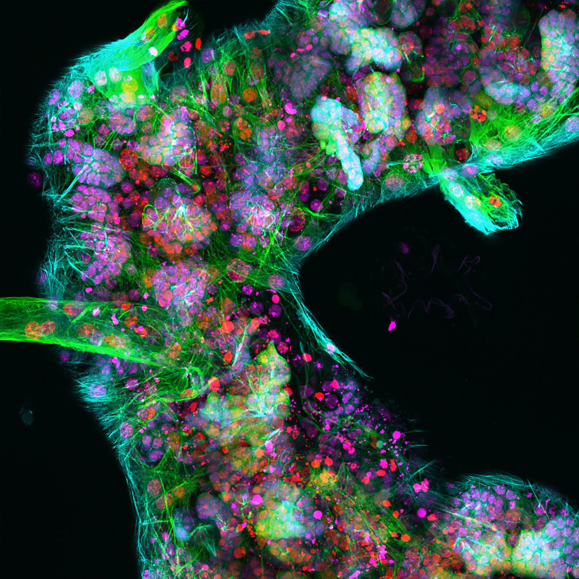 Nuclei (red) and microtubules (green) highlight the neurons of the brain. | 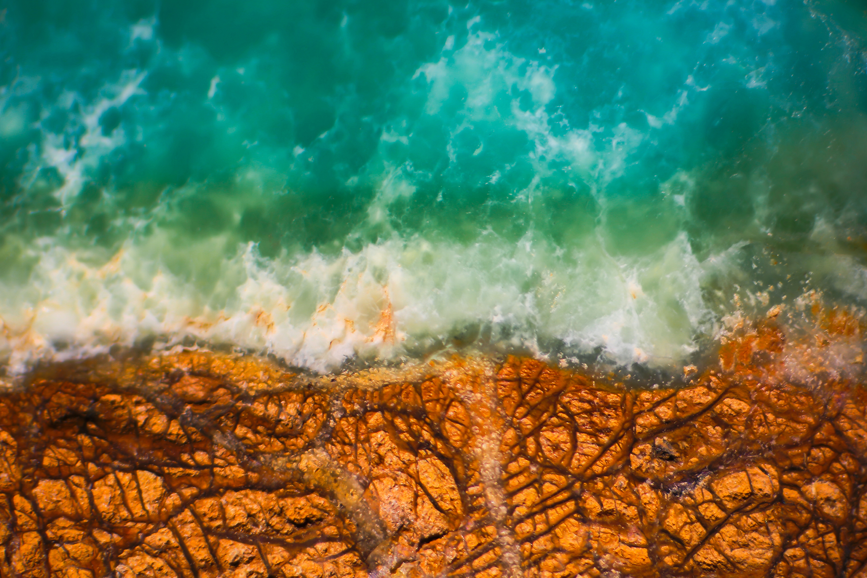 This image shows the green gem material, prase opal, which when magnified through a microscope remarkable resembles an aerial view of a coastline. |
Download Wallpapers for Your Screen
Download the Image of the Year wallpaper package now for free and beautify your screen!
Sorry, this page is not
available in your country.
Sorry, this page is not
available in your country.