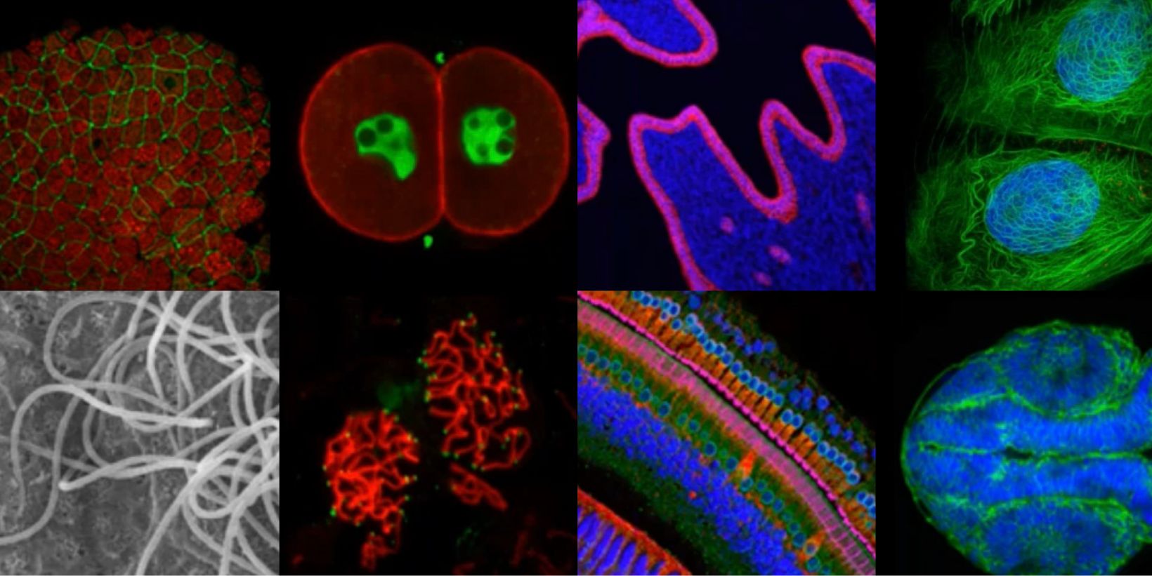
Biologists have a significant toolbox at their disposal when it comes to microscopically imaging 3D samples, such as organoids. From widefield microscopy to confocal, superresolution, multiphoton and lightsheet, each have their own set of pros and cons that must be carefully considered before making an informed choice on the most suitable to address your biological question. Often a correlative approach is required, applying several techniques to address the question from different perspectives. It is also crucial to consider the method of sample preparation and optimise each of the potential steps which can include fixation, permeabilisation, labelling and mounting. Further, the images generated by all techniques can be enhanced with post-processing techniques, such as deconvolution, which can enable or help to improve subsequent image analysis and interpretation.
This presentation aims to introduce each of the microscopy techniques that may be applicable for imaging 3D samples and highlight their relative performance attributes in terms of sample viability, speed, resolution, contrast and depth penetration – highlighting that each technique and instrument will come with trade-offs between these parameters1.
1 Jonkman, Brown, Wright, Anderson & North (2020) Tutorial: guidance for quantitative confocal microscopy. Nature Protocols 15:1585–1611

