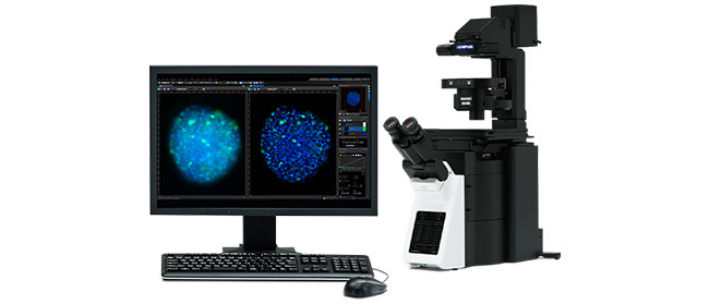
Hardware development in microscopy has enabled scientists to collect more and more image data in a very efficient and automated way. But to gain quantitative information of this data, images need to be analyzed in a robust way.
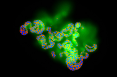
Image segmentation and classification lies at the heart of image analysis. The better the scientist can segment and classify objects of interest in their images, the better quantitative information they can gain, improving the quality of the results.
In 2019 Olympus launched TruAI technology, an image analysis approach based on deep neural networks with a focus in segmentation and classification. TruAI technology makes segmentation and classification easy and accessible by non-experts, even in the most difficult scenarios, like in low contrast brightfield images, low signal-to-noise fluorescence images, or in situations of highly dense cell confluency. When installed in the microscope that is acquiring the images, it speeds up the research as soon as analysis results can be available, shortly after the acquisition has concluded, or even at the same time as the acquisition.
In this tech talk, through a collection of examples measured with our live cell imaging systems, high-content screening station, and whole slide scanner, you will see what TruAI technology can do for your research and get a preview of what is coming next.
Presenter: Manoel Veiga, PhD
Application Specialist, Olympus Soft Imaging Solutions
Manoel Veiga earned his PhD in Physical Chemistry in the University Santiago de Compostela, Spain. Following two postdocs in the Universities Complutense of Madrid and WWU Münster, he joined PicoQuant GmbH. After five years supporting customers worldwide in the fields of FLIM and time-resolved spectroscopy, he joined Olympus Soft Imaging Solutions GmbH in 2017, where he works as Global Application Specialist with a focus on high-content analysis and deep learning.


