Evident LogoOlympus Logo

咨询专家

Advances in 3D Optical Imaging Technologies: An Overview 

With rapid development in fluorescent proteins, synthetic fluorochromes, and digital imaging, advanced 3D imaging technologies are now available to investigators to provide critical insights into the fundamental nature of cellular and tissue functions. 3D and 4D imaging systems have become very common tools among biologists. However, there are several technical challenges and limitations in performing successful 3D and 4D imaging. Olympus has developed a wide range of 3D imaging microscopes to overcome these challenges and to satisfy the requirements of researchers across different disciplines.

In this talk, we will be taking you through the different imaging modalities that Olympus offers including widefield based fluorescence microscope like the Olympus IXplore and how in combination with image processing tools like Olympus TruSight can render usable 3D data sets. 

However, more sophisticated 3D imaging systems are required for imaging samples such as organoids and spheroids. We will therefore illustrate the importance of laser-based systems that have been effectively used for deeper imaging into samples, like the Olympus FV3000 laser scanning confocal microscope, IXplore SPIN spinning disk confocal microscope and finally the FVMPE-RS multiphoton-excitation microscope. Along with these, we will stress the importance of high-quality objective lenses optimised for 3D imaging.

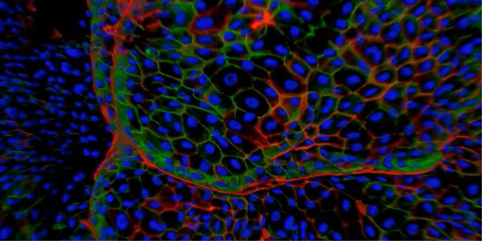
Advances in 3D Optical Imaging Technologies: An Overview

With rapid development in fluorescent proteins, synthetic fluorochromes, and digital imaging, advanced 3D imaging technologies are now available to investigators to provide critical insights into the fundamental nature of cellular and tissue functions. 3D and 4D imaging systems have become very common tools among biologists. However, there are several technical challenges and limitations in performing successful 3D and 4D imaging. Olympus has developed a wide range of 3D imaging microscopes to overcome these challenges and to satisfy the requirements of researchers across different disciplines.

In this talk, we will be taking you through the different imaging modalities that Olympus offers including widefield based fluorescence microscope like the Olympus IXplore and how in combination with image processing tools like Olympus TruSight can render usable 3D data sets. 

However, more sophisticated 3D imaging systems are required for imaging samples such as organoids and spheroids. We will therefore illustrate the importance of laser-based systems that have been effectively used for deeper imaging into samples, like the Olympus FV3000 laser scanning confocal microscope, IXplore SPIN spinning disk confocal microscope and finally the FVMPE-RS multiphoton-excitation microscope. Along with these, we will stress the importance of high-quality objective lenses optimised for 3D imaging.

Experts
Mr. Srivats Hariharan
Manager, Applications and Marketing
Olympus Singapore

Mr. Srivats Hariharan is an Applications & Marketing Manager in Olympus life science team in the Asia Pacific region. He holds a bachelor’s degree in Mechanical Engineering from Nanyang Technological University, Singapore and has experience working in biomedical research labs and A*STAR Microscopy Core Facility where he supported researchers on confocal and live cell imaging technologies and help setup single molecule super-resolution and light sheet microscopes. He joined the life-science team of Olympus Singapore in 2011 as Product Manager and in-charge of supporting research customers and business partners in South-East Asia and Taiwan.

Advances in 3D Optical Imaging Technologies: An Overview2024年11月25日
Sorry, this page is not available in your country
Ask the Expert Sign-up

By clicking subscribe you are agreeing to our privacy policy which can be found here.

对不起,此内容在您的国家不适用。

Sorry, this page is not available in your country