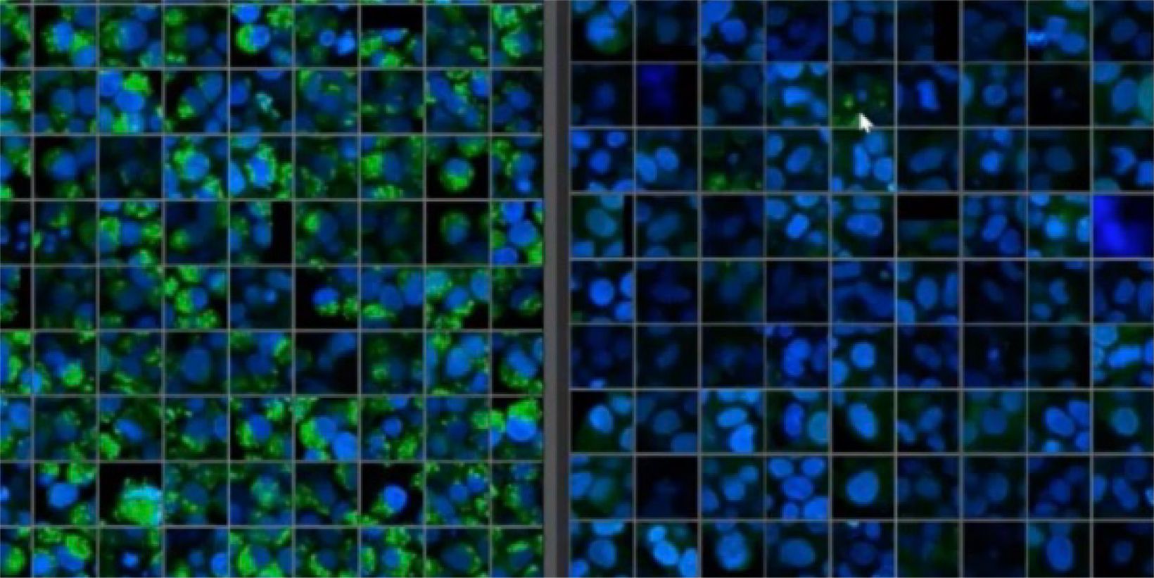
Three-dimensional cell culture models such as patient-derived organoids (PDO) and spheroids have increased in popularity because they can provide a 3D microenvironment that more closely reproduces in vivo conditions compared to 2D monolayer culture. Phenotypic and functional heterogeneity arise among cancer cells within the same tumor because of genetic change, environmental differences and reversible changes in cell properties. Therefore, evaluation of cell-specific responses is important for accurate prediction of drug efficacy and kinetics in vivo.
Microscopic imaging technique such as confocal microscopy is promising to monitor the cell-specific responses at higher spatial resolution. Here, we introduce a novel 3D cell analysis software “NoviSight”. “NoviSight” recognizes 3D objects based on fluorescence intensity and analyzes them based on various information such as their sizes, the cell number, viability, and subcellular features contained in the recognized objects. In this webinar, we will introduce a case study of the analysis of patient-derived cancer organoids and spheroids using NoviSight. Then, we will demonstrate NoviSight software.
Three-dimensional high-throughput cell analysis platform represents the first step toward the development of tools for understanding the pharmacological mechanisms of drugs or drug targets in the preclinical tissue models. We hope that our platform will provide invaluable information for the study of basic research, drug efficacy, dosage, pharmacology and ADMET before clinical phases of drug development.

