对不起,此内容在您的国家不适用。
- Overview
- The Ellison Institute and the TIC
- Leadership
- Olympus and Precision Medicine Research
- Resources
- Contact Us
Overview
Ellison Institute-Olympus Innovation PartnershipThe Lawrence J. Ellison Institute for Transformative Medicine (Ellison Institute)-Olympus Innovation Partnership in Multiscale Bioimaging is working to demonstrate the clinical application of new technologies that combine the workflow of a surgical biopsy and primary diagnosis with microscopic tumor characterization. The result: precision medicine and individualized treatment plans with the aim of improving health outcomes and driving patient-centered healthcare worldwide. Transforming Drug Discovery ResearchOlympus supports the Ellison Institute’s precision medicine research with our advanced imaging systems that enable the application of emerging technologies, such as our deep-learning artificial intelligence (AI) software. Our imaging experts collaborate closely with the Institute’s scientists in their studies, exploring how AI image recognition can be used to improve cancer treatment. 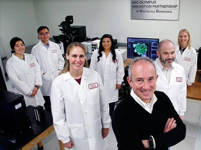      |
The Ellison Institute and the TIC
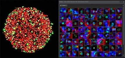
Creation of the PartnershipThe co-development agreement between the Ellison Institute and Olympus, which originated as a collaboration between the University of Southern California (USC) and Olympus, is a multiscale partnership designed to advance research into the prevention, diagnosis, and treatment of cancer through specialized precision medicine. The signing of the partnership was overseen by the scientific leadership of two internationally recognized trailblazers at USC, medical oncologist and researcher, David Agus, MD (Ellison Institute) and bioimaging technologist and researcher Scott Fraser, PhD (Translational Imaging Center (TIC)). This agreement equipped these labs with the latest Olympus microscopy technology, enabling their researchers to perform multiscale bioimaging of single cells, live cells, tumor microenvironments, organ systems, and the whole patient. Watch this video to hear Dr. Agus and Dr. Fraser explain the partnership’s benefits: Related VideosRedefining Cancer Research: The Innovation Partnership TodayAs its name suggests, part of the Lawrence J. Ellison Institute for Transformative Medicine’s mission is to uncover new and groundbreaking ways to treat cancer. Scientists at the Ellison Institute are using Olympus’ latest microscopy imaging solutions to study ways to improve and accelerate oncology drug discovery research. Challenges of Oncology Drug Treatment TestingIn the approval stages preceding clinical trials of cancer drugs, researchers perform extensive testing on human tumor cells in the lab to validate whether a drug molecule stops or slows cancer cell growth. Once these treatments reach the clinical trial stage, there is an unfortunately high failure rate for cancer drugs, despite the demonstrated efficacy in the lab. Innovating Drug Discovery Solutions Using Novel MethodsTo help improve the success rate of cancer drugs at the clinical stage, scientists at the Ellison Institute are innovating methods to optimize the precision of drug efficacy testing. These studies involve using Olympus’ advanced imaging solutions, including our high-content screening systems, TruAI™ deep-learning technology, and 3D modeling software, which are being applied to 3D biomimetic preclinical cancer models, such as patient-derived organoids.    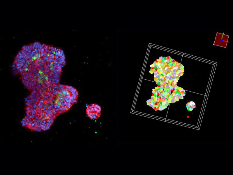  These images were captured using an Olympus FLUOVIEW™ FV3000 system after whole mount immunostaining and tissue clearing. Source: Seungil Kim, Ph.D., USC Ellison Institute Leveraging these emerging technologies, Ellison Institute researchers are conducting novel experiments to validate their potential to improve the precision of drug testing in the lab, thereby increasing the potential for success in subsequent clinical trials. Improving the approval rates for cancer drugs at the clinical trial stage could have far-reaching impacts for cancer patients and their physicians, offering more treatment options for precision oncology.
Olympus’ microscopy solutions experts collaborate closely with the researchers, providing technical support and assistance with image acquisition and analysis as needed. Discover the Lawrence J. Ellison Institute for Transformative MedicineThe Ellison Institute unites international experts from diverse disciplines and employs novel technologies to spark innovation and drive transdisciplinary research with the goal of redefining cancer and wellness in the 21st century. By harnessing the expertise of leading physicians, scientists, and thought leaders from around the world, the Ellison Institute advances cancer research, deepening our understanding of the complex biological processes that take place within a tumor and patient. |
Leadership
LeadershipBrought to fruition by leaders of the Ellison Institute, TIC, and Olympus, the Ellison Institute-Olympus Innovation Partnership supports the efforts of multidisciplinary scientists by providing them the advanced microscopy tools they need to overcome challenges and achieve breakthroughs in cancer research, diagnostics, and patient care.
|
Olympus and Precision Medicine Research
What is precision medicine?
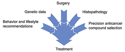
What is pharmacogenomics?Pharmacogenomics is the study of how genes affect a person’s response to particular drugs and is a key part of precision medicine. This relatively new field combines pharmacology (the science of drugs) and genomics (the study of genes and their functions) to develop effective, safe medications and doses that are tailored to variations in a person’s genes. What is precision oncology or precision cancer medicine?The terms precision oncology and precision cancer medicine can be used interchangeably. Precision oncology involves identifying the best possible treatment and prevention strategies for individual patients and using and sharing this knowledge to advance precision oncology research for the population at large. The capacity to start the right treatment at the right time for the right duration can save patients from unnecessary treatments, as well as ensure that an optimal sequence of treatments is being employed from the outset. Researchers use patients’ unique molecular data, combined with their clinical data and environment, to achieve precision cancer medicine. How does Olympus support precision medicine?Olympus provides a range of tools to assist in precision oncology, including the following:
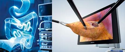
3D and AI imaging and analysis solutionsUsing 3D images acquired by our scanning systems, Olympus’ 3D analysis software can provide statistical information about tumor spheroids and organoids. Our deep-learning technology enables researchers to train the scanning system’s neural network to perform rapid label-free object identification and classification. The Ellison Institute, for example, uses Olympus’ FV3000 microscope and cellSens software’s TruAI deep-learning module for automated high-speed drug screening. They also configured an automated high-content screening system with the FV3000 microscope, exploiting our software’s macro-micro scanning function for 3D organoid detection and targeted imaging. Tumor localization and primary diagnosisMinimally invasive, multimodal endoscopes enable physicians to precisely locate the tumor and collect samples for biopsy with minimal disruption of surrounding tissue. Histology and 3D/4D cellular and molecular characterizationIn traditional histology, the biopsied tissue is stained, enabling a trained pathologist to examine it using a microscope or for the tissue to be scanned by a whole slide imaging microscope. New 3D/4D cellular and molecular characterization methods are providing physician-scientists with more information, including the capacity to compare changes in live-cell imaging over time—the 4th dimension. Live tumor tissue is biopsied from the patient in the clinic and transported to the research lab, where the tumor tissue is dissected into single cells and placed in a series of mini-well microplates, where they grow into spheroids. Because each microwell is self-contained, researchers can test different compounds to determine which combination might be the most effective weapon in fighting an individual patient's tumor. Scientists can also capture high-resolution 3D/4D images of the organoids to obtain critical information about the individual patient’s tumor, including the number and type of cells, morphology, cell growth or death rates, and then ultimately, use this information to help monitor the effectiveness of the latest anti-cancer compounds.  Slide scanning histology |
Resources
ArticlesSelectScience—Incubation monitoring for cancer cell line quality control and banking Microscopy Today—Towards Precision Medicine: The Ellison Institute and Olympus Partnership is Working to Change the Future of Cancer Medicine Application NotesAutomated Analysis of Label-Free Organoid Imaging Data TalksSeungil Kim, PhD, Ellison Institute Staff Scientist, spoke at a SLAS 2021 special interest group session on the topic of “High content imaging approaches to organs-on-chips: recent advancements and future prospects” (Jan 26, 2021) Shannon Mumenthaler, PhD, Ellison Institute Lab Director, spoke at the Olympus Discovery Summit on the topic of “High-Content Imaging of 3D Cancer Models” (Apr 27, 2021): |
Contact Us
联系我们 |


