Life Science Solutions
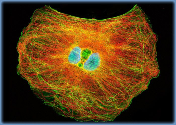
Distribution of Actin and Microtubules in OK Cells
Immunofluorescence with mouse anti-alpha-tubulin was employed to visualize distribution of the microtubule network in a log phase monolayer culture of opossum kidney cells. The secondary antibody (goat anti-mouse IgG) was conjugated to Alexa Fluor 546 and mixed with Alexa Fluor 488 conjugated to phalloidin to simultaneously image tubulin and the actin cytoskeleton.
对不起,此内容在您的国家不适用。